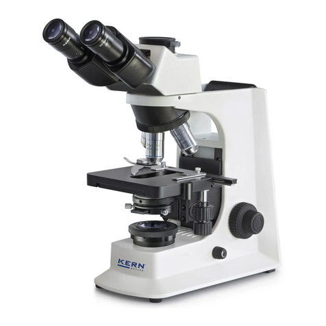
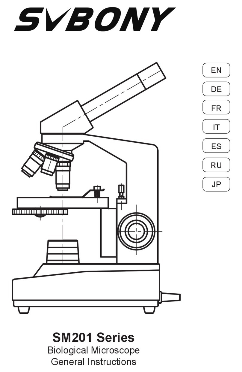
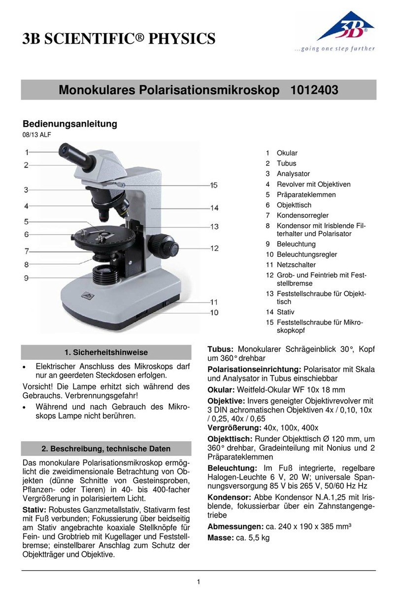
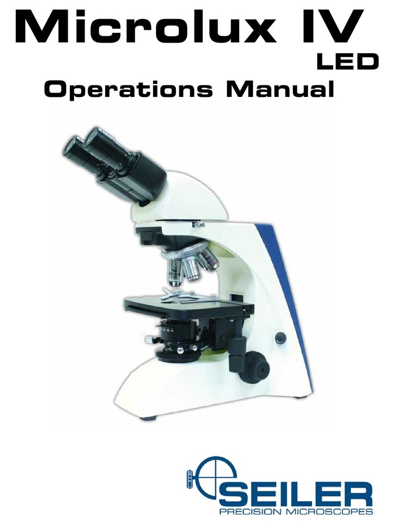
Fisher Science Education S71000 User manual

Microscope
Instruction Manual

2
INTRODUCTION
Thank you for your purchase of a Fisher Science Education microscope. Your new microscope
is a precision instrument carefully checked to assure that it reaches you in good condition. It is
designed for ease of operation and years of carefree use. The information in this manual probably far
exceeds what you will need to know in order to operate, troubleshoot and maintain your microscope.
However, it is provided to answer questions that might arise, and to help you avoid any maintenance
expense that may be unnecessary.
TYPES OF MICROSCOPES COVERED IN THIS MANUAL
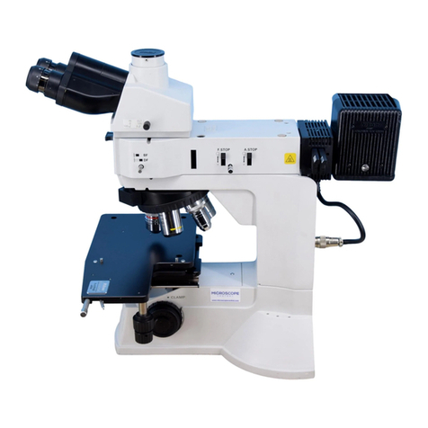
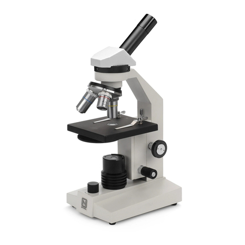
The compound microscope combines two optical lens systems. The lens closest to the specimen
slide, the objective, magnies the primary image and the top lens, called the eyepiece, further
magnies the image. Magnication of the objective times the magnication of the eyepiece is the
total magnication produced by this combination of lenses. The image produced by the compound
microscope is upside down and reversed. Compounds are used for viewing standard 1” by 3” 1mm
thick transparent specimen slides with cover slips. Prepared specimen slides can be purchased to t
the classroom subject matter or user can make his own specimen slides.
The stereo microscope is an instrument that incorporates two separate optical system aligned to
produce three-dimensional images. Primary uses of the stereo microscope are the inspection and
assembly of small parts, examining plants and insects, dissecting of biological specimen. Stereo
microscopes provide an upright, unreversed image that permits easy manipulation of the object being
viewed while looking through the microscope. Stereos are designed for viewing solid objects at low
magnication, but they will also permit viewing of some transparent specimen slides.
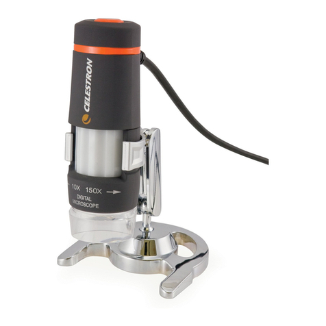
Digital microscopes have all of the features of a compound microscope or stereo microscope but
are enhanced with the addition of a built-in digital camera. With software included with these digital
microscopes, they become real time learning tools.

3
FISHER SCIENCE EDUCATION MICROSCOPES
TABLE OF CONTENTS
Microscope Terminology 4-5
Care and Maintenance 6-11
Troubleshooting 12-13
General Instructions for Compound Microscopes 14-15
Model S71000, S71001A and S7001B Instructions 16
Model S71000C, S71000D, S71000E and S71000F Instructions 17
Model S71001, S71001A, S71001B, S71001C, S71001D, S71001E,
S71001F and S71001G Instructions 18
Model S71002, S71002A and S71002B Instructions 19
Model S71002C, S71002D, S71002E, S71002F and S71002G Instructions 20
Model S71003, S71003A, S71003B and S71003C Instructions 21
Model S71003D, S71003E, S71003F, S71003G, S71003H
and S71003J Instructions 22
General Instructions for Stereo Microscopes 23-25
Model S71004 and S71004A Instructions 26
Model S71005, S71005A, S71005B and S71005C Instructions 27
Model S71006, S71006A, S71006B, S71007 and S71007A Instructions 28
Model S71008 and S71008A Instructions 29
Model S71009, S71009A and S71010 Instructions 30
General Instructions for Digital Microscopes and Model S71011 31
Model S71012 Instructions 32
Model S71012A Instructions 33
Model S71012B Instructions 34
Model S71013 Instructions 35
General Use of WiFi and Model S71014 Instructions 36
Model S71014A and S71014B Instructions 37
WiFi Camera Operation 38
Fisher Science Education Warranty 39

4
MICROSCOPE TERMINOLOGY
• ABBE CONDENSER: The 1.25 N. A. Abbe condenser lens positioned under center of stage is
requited when using 100x objective lenses. In addition the Abbe condenser is focusable by one
of two methods, a spiral mount condenser with lever to lower or raise the assembly or else a rack
and pinion mount with a knob to provide movement in the up and down direction
• ARM: Main support for microscope components.
• BASE: Housing and platform of the instrument to which the arm is attached in addition it usually
contains an illumination system for the microscope.
• BODY TUBE: On simple vertical tube models it holds the eyepiece tube and objective, on
advanced models it holds nosepiece, objectives and head.
• COAXIAL FOCUSING KNOBS: Coaxial focusing system combines both the coarse and ne
focus into one set of knobs located on the same axis. The control is designed for a continuous
operation over the range of stage movement.
• COARSE FOCUSING KNOBS: Large knobs located on each side of arm, raise or lower stage to
bring specimen image into focus.
• CONDENSER LENS (0.65): Condenses light rays from sub stage illumination and lls the back
element of objective lens to improve image resolution. A 0.65 condenser lens xed in center of
stage is provided on microscopes with objectives up to 40x (400 times magnication).
• DIOPTER ADJUSTMENT: Adjustable eyepiece diopter permits focusing adjustment of image for
any difference in vision between users eyes.
• DISC DIAPHRAGM: Disc located below stage with holes of various apertures, designed to help
achieve optimum resolution of the objective lens. Larger apertures used for higher magnications,
and smaller apertures used for lower magnications.
• EYEPIECE (ocular lens): Lens closest to the eye magnies the primary image formed by the
objective lens.
• EYEPIECE TUBE: This is the component that holds the eyepieces in place. Elementary, student
and high school models have set screws in the eyepiece tube used to lock eyepieces in place.
• FIELD OF VIEW: View area that is seen through the lens system of the microscope.
• FILTER: Daylight blue lter designed to make incandescent illumination appear white.
• FILTER HOLDER: Attached to bottom of iris diaphragm that swings out allowing user to insert
lter of choice. If a built in neutral lter is provided it should be removed from the optical path
when using 40x and 100x objectives.
• FINE FOCUSING KNOBS: Smaller knobs, located close to the coarse focusing knobs, permit
more precise adjustment of the image.
• FOCUSING EYEPIECE TUBE BINOCULAR HEAD: Focusing eyepiece tube models, (two
diopters) used to adjusting parfocality and focusing adjustment of image for any difference in
vision between eyes.
• HEAD: Upper portion of the microscope which contain prisms and eyepiece tube or tubes.
• INCIDENTAL ILLUMINATION: Primarily used on stereo type microscopes to provide illumination
from above the specimen.
• INTERPUPILLARY DISTANCE: Interpupillary distance (IPD) is the distance between the center
of the pupils of the two eyes. Interpupillary distance is critical for the design of binocular viewing
heads so that the left and right image can blend into one image.

5
• IRIS DIAPHRAGM: Iris Diaphragm, opening and closing of iris is controlled by lever. It is
designed to help achieve optimum resolution of the objective lens. Larger apertures used for
higher magnications, and smaller apertures used for lower magnications.
• MAGNIFICATION: Total magnication obtained with each objective lens is determined by
multiplying the magnication of the eyepiece times the magnication of the objective. Keep
in mind that as magnication increases, eld of view (area of the specimen seen when looking
through microscope) decreases.
• MECHANICAL STAGE: Permits precise, mechanical manipulation of the specimen slide.
• MECHANICAL TUBE LENGTH: Distance between the top of eyepiece tube to mounting face of
nosepiece. (On more advanced models, above elementary type, the tube length is 160mm).
• NEUTRAL DENSITY FILTER: Neutral colored frosted lter designed to soften illumination hot
spots.
• NUMERICAL APERTURE (NA): Mathematical formula devised by Ernst Abbe for the direct
comparison of objective lens to resolving power.
• NOSEPIECE (REVOLVING TURRET): Designed to hold objective lenses permitting changes of
magnication by rotating different powered objective lenses into optical path. Forward facing
position used on elementary and high school models. Reverse facing nosepiece position used on
more advanced models permits easier access to stage when positioning specimen slides.
• OBJECTIVE LENS: Lens closest to the object being viewed, forms rst image of the specimen.
• OIL IMMERSION LENS: High power (100x) objective lens which requires a medium of immersion
oil between the lens and the slide
• RESOLVING POWER: Ability of the optical system to distinguish and separate ne structural
details in a specimen. The resolving power is limited by the NA of the objective, and it also
depends upon the working NA of the sub-stage condenser, the higher the effective NA of the
system the greater will be the resolving power.
• RHEOSTAT: Variable potentiometer that adjusts the light intensity of the illuminator.
• SEIDENTOPF BINOCULAR HEAD: Seidentopf heads remain parfocality when changing
interpupillary distances and are supplied with one adjustable diopter adjustment to adjust image
for any difference in vision between left and right eyes.
• SPECIMEN SLIDE: Typically a 3 by 1 inch by 1mm thick glass with a specimen held for
observation covered by a .01mm thick cover glass.
• STAGE CLIPS: Two locked-on clips hold specimen slide in place on stage.
• STAGE: Platform of the microscope where the specimen slide is placed.
• SAFETY RACK STOP: Prevents higher power objectives from breaking specimen slides, and
prevents damage to objective lens. Stop has been pre-adjusted at the factory.
• TENSION ADJUSTMENT COLLAR: The tension collar is used to adjust and control “body tube”
or “stage” drift (Instrument not remaining in focus).
• TRANSMITTED ILLUMINATION: Used to illumination both compound and stereo microscopes
providing illumination from below specimen.
• WORKING DISTANCE: Distance between the top of specimen slide and the front of the objective
lens.

6
CARE AND MAINTENANCE OF MICROSCOPES
I. WARNING: For your own safety, turn switch off and remove plug from power source before
maintaining your microscope. If the power-cord, AC power converter, recharger, or any of the
supply cords are worn, cut or damaged in any way, have it replaced immediately to avoid shock or
re hazard.
II. OPTICAL MAINTENANCE
A. Do not attempt Do not attempt to disassemble any lens components. Consult a microscope
service technician when any repairs not covered by instructions are needed. Prior to cleaning
any lens surface, brush dust or dirt off lens surfaces using a camel hair- brush. Or use air to
blow dust and lint off surfaces. Use of compressed air in a can, available at any computer
supply store, is a good source of clean air.
B. Do not remove eyepieces or objective lenses to clean. Clean only the outer lens surface.
Breath on lens to dampen surface, then wipe with lens paper or tissue or use a cotton swab
moistened with distilled water. Wipe lenses with a circular motion, applying as little pressure
as possible. Avoid wiping dry lens surface as lenses are scratched easily. If excessive dirt
or grease gets on lens surfaces, a small amount of distilled water or Eyeglass lens cleaner
can be used on a lens tissue. To clean objective lenses, do not remove objectives from
microscope. Clean front lens element only, following same procedure. NOTE: Fingerprints or
other matter on the front lens element of the objective lens is the single most common reason
that you will have difculty in focusing the microscope. Before having costly servicing done,
or before returning for “warranty repair”, make certain to examine the front lens element with
a magnifying glass or eye loupe for the presence of such contaminants. If a microscope is
returned for warranty repair, and it is determined that such contaminants are the problem, this
is not covered under warranty and an estimate for cleaning will be submitted for cleaning.
III. MECHANICAL MAINTENANCE
A. Rack Stop (compound microscopes only): Rack stop screw has been pre-adjusted at the
factory and should not require re-adjustment. However, if you do attempt re-adjustment of
rack stop use the following procedure. Locate rack stop as shown on drawing of your particular
model. Loosen locking nut by turning counter clockwise that should allow you to loosen rack
stop screw by rotating counter clockwise.
a. Models with ne focus adjustment require setting it at mid-range, then using coarse focus
knobs focus on standard slide until sharp image is obtained.
B. Rotate rack stop screw in clockwise direction until tight. Lock into position with the locking
nut.
IV. VIEWING BODY OR STAGE DRIFT: Coarse focus tension adjustment collar prevents the viewing
body or stage from drifting down from its own weight and causing the image to move out of focus.
This has been adjusted at the factory, but over the course of time it may require adjustment. It is
recommenced that you leave the tension adjustment as loose as possible for ease of focusing, yet
not so loose that it permits viewing body or stage to drift out of focus on its own weight.
A. Mid range focusing Models S71000A and S71000B do not require adjustments for stage drift.
B. Coarse focus only and coarse focus with ne focus models have a tension adjustment collar
located between arm and coarse focus knob on left side of microscope. Use a small jewelers
screwdriver or use supplied 0.90 “L” hex wrench, loosen the setscrew located in only one of
the four holes on tension adjustment collar. Turn collar clockwise to tighten tension, counter-
clockwise to loosen tension. Use of a wide rubber band will provide a better grip on the tension
adjustment collar. After adjusting, tighten the setscrew to lock collar in place.
C. Coaxial coarse and ne focus models have the tension adjustment knob located between
stand and coarse focus knob of microscope (on right side with arm facing user). Tighten
tension of coarse focus knobs by turning tension adjustment knob in a counter clockwise
direction.

7
V. METAL PARTS
A. Clean metal parts using a damp cloth to remove dust and dirt from all metal and painted
surfaces, followed by a dry cloth.
VI. ELECTRICAL MAINTENANCE: The extent of electrical maintenance, by other than a qualied
technician should be bulb, battery or fuse. Be certain to turn switches to the off position and
remove power plug from power source outlet before changing bulbs or fuses.
A. Replacing Incandescent Lamps on Elementary Model S71000A:
a. Replace the lamp by laying microscope on side to reveal base plate located on bottom of
illuminator base.
b. Locate screws securing rubber feet. Using a screwdriver, remove the rubber feet and
base plate to expose bulb.
c. Remove bulb by depressing lamp slightly and rotating in a counter-clockwise direction
until it pops up.
d. Using Fisher Science Education replacement lamp no. S74015 (15 watt 120 volt double
contact base) insert new bulb, gently depress into socket and rotate clockwise. Wipe
bulb to insure that it is clean and free of all ngerprints.
e. Replace base plate and rubber feet.
B. Replacing Incandescent Lamp on Elementary Models S71000C and S71000E:
a. Replace the incandescent lamp by locating the locking screw on bottom of illuminator
housing shaft and remove screw. Pull shaft with lamp out of main illuminator housing.
b. Remove by depressing lamp slightly; rotate in counter-clockwise direction until it pops up.
c. Using Fisher Science Education replacement lamp no. S74015 (15watt 120volt double
contact base) gently depress lamp into socket and rotate clockwise. Wipe bulb to insure
that it is clean and free of ngerprints.
d. Replace lamp socket assembly into main lamp housing and replace locking screw.
C. Replacing Incandescent Lamps on Middle/High School Models S71001 and S71001A:
a. Make sure that the illuminator eld lens housing and lamp are cool before attempting to
remove. Take off illuminator eld lens housing by rotating in a counter-clockwise direction.
This housing is secured tightly, to prevent easy removal by students, so a very rm grip
and some strength will be required.
b. Remove lamp by depressing lamp slightly and rotate in a counter-clockwise direction until
it pops up slightly.
c. Using Fisher Science Education replacement lamp no. S74017 (20watt 120 volt double
contact base) replace lamp by gently depressing lamp into socket and rotate clockwise.
Wipe bulb to insure that it is clean and free of all ngerprints.
d. Replace illuminator eld lens housing by rotating in a clockwise direction and tighten.
D. Replacing LED bulbs on Models S7100B, S71001B, S71001C, S71001D, S71001E, S71001F,
S71001G, S71002, S71002A, S71002B, S71002C, S71002D, S71002E, S71002F, S71002G,
S71011, S71014, S71014A, and S71014B:
a. To replace LED bulb remove the illuminator eld lens housing by loosening the hex screws
located on the bottom edge of housing (Use the 0.90 “L” type hex key wrench supplied
with your microscope). Remove lens housing to expose LED bulb.
b. Remove LED bulb by grasping the plastic base of bulb and gently pulling straight up.
c. Using Fisher Science Education replacement lamp no S74018 (LED bulb with lamp base)
insert the new LED “bulb” making sure to align the lamp base with lamp socket. Wipe
bulb to insure that it is clean and free of ngerprints.
d. Replace illuminator eld lens housing and tighten hex screw to secure lens housing to
illuminator base.
E. Replacing Led bulbs on Elementary Models S71000D and S71000F:
a. Do not attempt to replace LED bulb on these two models. LED bulb is not replaceable
by end user.

8
F. Replacing the Incidental (Top) Illuminator LED bulb on Middle/High School Stereo Models
S71005A and S71005C:
a. To replace top light replace top light, remove two chrome crosshead screws securing top
lighthouse to light bracket and the two chrome crosshead screws securing coiled cord
clamp to arm.
b. Remove front lens from lighthouse by rotating in a counter clockwise direction. Holding
top lighthouse in one hand, remove silver spacer then feed coiled cord through bottom of
lighthouse, pushing LED lamp assembly out of lighthouse.
c. Unplug connector located on bottom of LED lamp mount and remove from housing.
d. Using Fisher Science Education replacement lamp no S74022 (LED bulb with special
base) plug the connector to supply cord. Wipe bulb to insure that it is clean and free of
ngerprints.
e. Reassemble lamp assembly in reverse order
G. Replacing the Transmitted (Bottom) Illuminator LED bulb on Middle/High School Stereo Models
S71005A and S71005C:
a. To replace transmitted (bottom light), loosen setscrew located at side of base and remove
stage plate. Removing stage plate will expose the bottom LED bulb.
b. Grasp LED bulb and pull straight up and out of socket.
c. Using Fisher Science Education replacement lamp no S74023 (LED bulb with special
base) insert the new LED bulb making sure to align the lamp base with lamp socket. Wipe
bulb to insure that it is clean and free of ngerprints.
d. Replace the stage plate and tighten locking setscrew.
H. Replacing Halogen Lamps on College/Laboratory Models S71003, S71003A, S71003B,
S71003C, S71003D, S71003E, S71003F, S71003G, S71003H, S71003J, S71012 and S71012A:
a. Carefully lay instrument on its side, taking care to avoid damage to specimen slide holder
located on top of mechanical stage.
b. Loosen large chrome screw located on hinged door of illuminator base.
c. Swing door open to expose the halogen lamp.
d. Using a tissue or cloth, gently grasp the halogen bulb and pull lamp straight out of lamp
socket.
e. Using Fisher Science Education replacement lamp no S74020 hold lamp with tissue paper
and insert bulb base pins straight into lamp socket. Wipe bulb to insure that it is clean and
free of ngerprints.
f. Make sure to use the proper 12-volt bulb in order to prevent serious damage to the
electrical system of the microscope.
g. Close hinged door and tighten the chrome locking screw
I. Replacing the Transmitted (Bottom) Lamp on High School/College Stereo Models S71007 and
S71007A:
a. Gently lay microscope on its side to reveal base plate on bottom of illuminator base.
a. Observe screws located in rubber feet. Using a screwdriver, remove the rubber feet and
base plate to expose bulb.
b. Remove bulb by depressing lamp slightly and rotating in a counter-clockwise direction
until it pops up.
c. Using Fisher Science Education replacement lamp no S74015 (120volt 15watt double
contact base) insert new lamp by gently pressing into socket and rotate in a clockwise
direction until it snaps into place. Wipe bulb to insure that it is clean and free of all
ngerprints.
d. Replace base plate and secure rubber feet.
J. Replacing the Incidental (Top) Lamp on High School/College Stereo Models S71007 and
S71007A:
a. Remove top lamp housing by grasping it and rotating in a counter-clockwise direction to
expose the lamp.

9
b. Remove light bulb by rmly grasping and depressing lamp slightly and rotating in a
counter-clockwise direction until it pops up.
c. Using Fisher Science Education replacement lamp no S74024 (12volt 10watt double
contact base) insert new lamp by gently pushing into socket and rotate in a clockwise
direction until it pops into place. Wipe bulb to insure that it is clean and free of all
ngerprints.
d. Replace light housing by rotating in a clock-wise direction until tight.
K. Replacing the Incidental (Top) Lamp on Middle/High School Stereo Models S71004, S71004A,
S71005, S1005B, S71006, S71006A and S71006B:
a. Remove the knurled light housing locking screw located on side of rectangular shaped
lamp housing.
b. Remove the rectangular shaped light housing and expose top bulb.
c. Gently grasp bulb and pull bulb from two tension clips, which hold it in place.
d. Using Fisher Science Education replacement lamp no S74021 (12volt 10watt) holding new
bulb with tissue (to avoid getting body oil on surface of bulb), push bulb into tension clips
until it slips into position.
e. Replace light housing and lock in place with knurled locking screw.
L. Replacing the Transmitted (Bottom) Lamp on Middle/High School Stereo Models S71004,
S71000A, S71005, S71005B, S71006, S71006A, S71006B and S71012B:
a. To replace transmitted (bottom light), loosen setscrew located at side of base and remove
stage plate. Removing stage plate will expose the bottom bulb.
b. Gently grasp bulb and pull bulb from two tension clips, which hold it in place.
c. Using Fisher Science Education replacement lamp no S74021 (12volt 10watt) holding new
bulb with tissue (to avoid getting body oil on surface of bulb), push bulb into tension clips
until it slips into position.
d. Replace stage plate and secure with locking setscrew.
M. Replacing the Incidental (Top) Illuminator Halogen Bulb on Digital Stereo Model S71012B:
a. Remove front lens housing from microscope by rotating in a counter clockwise direction.
b. Remove light bulb by rmly grasping and pulling straight out from bi-pin socket
c. Using Fisher Science Education replacement lamp no S74027 (12 volt 10 watt bi pin
halogen) orientate the two pins with the socket and rmly push pins into socket.
d. Replace top lens housing by rotating in a clock-wise direction until tight.
N. Replacing the Transmitted (Bottom) Lamp on Digital Stereo Model S71013:
a. Gently lay microscope on its side to reveal base plate on bottom of illuminator base.
b. Observe screws located in rubber feet. Using a screwdriver, remove the rubber feet and
base plate to expose bulb.
c. Remove light bulb by rmly grasping and pulling straight out from bi-pin socket
d. Using Fisher Science Education replacement lamp no S74028 (12volt 10watt Halogen bi-
pin base) orientate the two pins with the socket and rmly push pins into socket.
e. Replace base plate and secure rubber feet.
O. Replacing the Transmitted (Bottom) Lamp on College Stereo Models S71008, S71008A,
S71009, S71009A and S71010:
a. Gently lay microscope on its side to reveal base plate on bottom of illuminator base.
b. Observe screws located in rubber feet. Using a screwdriver, remove the rubber feet and
base plate to expose bulb.
c. Grasp uorescent lamp and pull bulb from socket.
d. Using Fisher Science Education replacement lamp no S74019 (5watt uorescent) holding
push bulb into tension clips until it slips into position.
e. Replace base plate and secure rubber feet.
P. Replacing the Incidental (Top) Lamp on College Stereo Models S71008, S71008A, S71009,
S71009A and S71013:
a. Remove top lampshade by grasping it and rotating in a counter clockwise direction

10
exposing top lens housing.
b. Remove top lamp lens housing by grasping it and rotating in a counter-clockwise direction
to expose the lamp.
c. Remove light bulb by rmly grasping and pulling straight out from bi-pin socket.
d. Using Fisher Science Education replacement lamp no S74025 (12volt 15watt Halogen
bi-pin base) hold lamp with tissue paper, orientate the two pins with the socket and rmly
push into socket. Wipe bulb to insure that it is clean and free of all ngerprints.
e. Replace top lens housing by rotating in a clock-wise direction until tight.
f. Replace top lampshade by rotating in a clock-wise direction until tight.
Q. Replacing the Incidental (Top) Lamp on College Stereo Model S71010:
a. Remove top lampshade by grasping it and rotating in a counter clockwise direction
exposing halogen reector lamp.
b. Remove light bulb by rmly grasping and pulling straight out from bi-pin socket.
c. Using Fisher Science Education replacement lamp no S74026 (12volt 10watt Halogen
Reector) orientate the two pins with the socket and rmly push pins into socket.
d. Replace top lens housing by rotating in a clock-wise direction until tight.
R. Replacing Fuses on Models Requiring a Fuse:
a. Fuse is located at the rear side of microscope base.
b. To remove fuse from holder, insert a 6mm screwdriver blade into slot located in rear of
fuse holder cap. Slightly depress and rotate screwdriver ¼ turn in direction of arrow,
release pressure on screwdriver to release fuse.
c. Pull cap and fuse out of fuse holder.
d. Insert proper fuse into fuse cap. Insert fuse cap into fuse holder.
e. Using screwdriver, rotate fuse cap assembly in opposite direction of arrow until slot
engages, depress fuse cap and rotate ¼ turn to lock into fuse holder.
S. Recharging Batteries on Cordless Models:
a. Must use the supplied Automatic Switching Recharger when charging batteries.
b. It is recommended that you charge the batteries before initial use and after prolonged
storage as the batteries may have discharged.
c. Plug output cord from battery charger into DC recharging socket located on LED
illuminator. Your automatic switching recharger operates on 100 to 240 volts AC 50/60
Hz. Plug recharger into your AC wall outlet.
d. Battery recharger is also equipped with an automatic “trickle charge” feature; the red
LED indicator lamp located on recharger will be illuminated when batteries are receiving
maximum charge. After batteries are charged, the red LED indicator lamp will turn to
green and charger automatically switches to “trickle charge”.
e. The charger can be left plugged in, but for safety reasons it is a good idea to disconnect
the charger from the AC wall outlet and the output cord from recharging socket after 12
hours. Batteries and charger may feel warm when charging, and unplugging the recharger
is a safety precaution.
T. Replacing Batteries on All Rechargeable LED Microscopes, except Models S71000C and
S71000F:
a. Gently lay microscope on its side. Remove the rubber feet located on bottom of base
and remove base plate. Observe battery compartment inside of illuminator base.
b. Remove the screw securing battery cover to bottom of illuminator. Slide cover back
to expose and remove batteries. Remove “ALL” 3 batteries and replace with new
rechargeable AA nickel hydride batteries, insert with correct polarity according to
markings on battery holder.
c. Replace battery cover and secure screw.
d. Replace base plate and the rubber feet.
e. Recharge batteries as described in Care and Maintenance of Microscopes located on
page 10 (S).

11
U. Replacing Batteries on Models S71000C and S71000F:
a. Do not use regular AA alkaline batteries. Use of other Use of other than rechargeable AA
nickel metal hydride batteries could result in batteries exploding during recharging.
b. Carefully lay the microscope on its side.
c. Locate battery compartment cover on bottom of illuminator. Carefully remove the two
screws securing battery cover to bottom of illuminator. Slide cover back to expose
batteries.
d. Remove all 3 batteries and replace with new rechargeable AA nickel hydride batteries,
making certain to insert with correct polarity according to markings on battery holder.
Replace cover and secure screws.
e. Recharge batteries as described in Care and Maintenance of microscopes located on
page 10 (S).

12
ELECTRICAL
PROBLEM REASON FOR PROBLEM SOLUTION
Lights fail to operate
AC power cord not connected
Outlet inoperative
Lamp burned out
Fuse is blown
Connect outlet plug to outlet
Have qualied service repair outlet
Replace lamp
Replace fuse
Fuse burns out too soon
Improper fuse
Fuse blows instantly when
replaced
Replace with proper fuse
Short in electrical system - have
qualied technician repair
Light bulb burns out too soon
or immediately
Incorrect bulb, voltage or lamp
base used
Replace with specied lamp
Light ickers
Lamp not properly inserted into
socket
Loose connection at AC outlet
Electrical short
Properly insert lamp
Have qualied service technician
repair outlet
Properly install fuse holder
TROUBLESHOOTING
FOCUSING
PROBLEM REASON FOR PROBLEM SOLUTION
Image does not remain in
focus
Stage or body of microscope
drops from its own weight
Adjust tension control
Image will not focus
(compound models)
Rack stop not set at proper
position
Slide is upside down
Slide cover slip is too thick
Adjust rack stop
Place slide on stage with cover slip
up
Use 0.17mm thick cover slip

13
IMAGE CONCERNS
PROBLEM REASON FOR PROBLEM SOLUTION
No image Nosepiece not indexed properly Move revolving nosepiece until
objective lens clicks into position
Poor resolution
(Image not sharp)
Diaphragm improperly adjusted
Too much light
Objective lenses dirty
Eyepiece lens dirty
Washed out image
Specimen slide dirty
Spots on eld of view
(Eyepiece or condenser lens
dirty)
No immersion oil used on 100X
objective lens
Bubbles (air) in immersion oils
Adjust disc diaphragm or iris
diaphragm
Adjust light intensity control to a
lower position
Clean objective lenses
Clean eyepiece lenses
Adjust disc or iris diaphragm
Clean slide
Have qualied service technician
clean inside of lens
Use small amount of immersion
oil between the objective and the
slide
Remove bubbles by carefully
moving nosepiece back and forth

14
GENERAL INSTRUCTIONS FOR COMPOUND MICROSCOPES
I. Remove microscope, vinyl dustcover and any components from Styrofoam lined carton. Retain
the container, and use it for extended storage, for transporting, or in case the microscope ever
needs to be shipped. Always carry microscope by grasping arm with one hand and placing other
hand under base. Place the microscope directly in front of you in a manner, which permits you
to comfortably look into the eyepiece. On models with a rotating head, note that the head of the
microscope rotates 360°, permitting you to operate the microscope from the front or the back,
whichever is most convenient. Most users will position the microscope with the arm facing you so
that focusing knobs are most convenient to reach.
II. Turn illuminator on in order to illuminate the specimen.
A. Models supplied with a variable rheostat knob or dial for controlling light levels.
B. Before operating microscope, adjust intensity (rheostat) control knob or dial to its minimum
position. (This will help extend the life of the light bulb).
C. Turn illuminator switch to the “on” position.
D. Rotate intensity knob until image is illuminated.
E. As different magnications are selected it will be necessary to adjust the intensity of light to
match requirements of objective and specimen slide.
III. Focusing your compound microscopes.
IV. Rotate coarse focus knobs in a direction that stage moves “away” from the objective lenses as far
as possible.
V. Positioning specimen slide.
A. Stages with standard stage clips. Place specimen slide cover slip facing up, under the two
stage clips located on stage. Move specimen slide until it is centered over lens in center
condenser lens located in middle of stage.
B. Mechanical Stage Models: Swing movable nger on holder outward, place specimen slide
(cover slip facing up) on top of slide against xed side of slide holder. Slowly release movable
nger until it makes contact with specimen slide. Using mechanical X and Y controls move
specimen slide until it is centered over the condenser lens.
VI. Initial diaphragm adjustments.
A. Models with rotating disc diaphragm turn until largest aperture is positioned beneath
condenser lens.
B. Models that are supplied with iris diaphragm, move lever until iris is close to largest opening.
VII. Positioning Objective: Rotate the nosepiece (revolving turret) until the 4x (smallest) objective lens
clicks into position in the optical path. Note that each time you change from one objective lens to
another you should turn the turret until you hear the click, which indicates that the lens is properly
indexed in the optical path.
VIII. While looking through the eyepiece, rotate coarse focusing knobs until specimen comes into
focus. If image does not appear in eld of view, move specimen slide slightly until image appears
in eld of view.
IX. Models equipped with ne focus adjustment knobs, adjust these controls until specimen is in
sharp focus.
X. Adjusting the Diaphragm: Diaphragms are designed to help achieve high resolution of specimen
and provide contrast in the image. Listed below are a few suggested starting points for adjusting
the aperture. Smaller apertures will deliver higher contrast to image. However, closing aperture
too much will reduce resolution. Experimentation is the best method of determining the correct
opening of diaphragm.
A. Disc diaphragm
• 4x diaphragm disc close to smallest opening.
• 10x diaphragm disc at medium opening
• 40X diaphragm disc close to largest opening

15
B. Iris Diaphragm suggested openings:
• 4x objective-iris open 1/8,
• 10xobjective-iris open 1/8 to ¼,
• 40x objective, iris open ¼ to ½,
• 100xobjective-iris open ½ to ¾
XI. Changing magnication is accomplished by rotating by rotating objective turret until different
objective lens is moved into optical path. Always turn turret until you hear the “click”, indicating
that lens is properly indexed. Otherwise, you will not be able to see anything when looking through
the microscope. A slight adjustment of focus and diaphragm openings will be needed to sharpen
the image when changing objective magnication.
A. Models supplied with 100x oil immersion lens. To obtain the maximum resolution of the 100x
oil immersion lens it is necessary to apply immersion oil between cover glass of slide and front
lens of objective. Use a very small amount of immersion oil. Air bubbles must be removed from
between lens and slide by gently moving nosepiece back and forth.
B. Each time immersion oil is used on the 100x it is essential that you clean front of lens after use.

16
MODEL S71000
To view transparent specimen slides, place slide over the
aperture in the middle of the stage, with the specimen
and cover slip on the slide facing upward. The distance
between the specimen and the bottom of the objective
lens will about 1 inch. Grasping the eyepiece tube, gently
slide it down until the objective lens is fairly close to the
slide surface. Note that there is a small-anged rivet in
the eyepiece tube that will stop the downward movement
of the eyepiece tube before it reaches the slide surface.
Looking through eyepiece, gently pull up on eyepiece tube
until image comes into sharp focus.
To view solid objects such as rocks, plants, insects,
or opaque specimen such as stamps, fabric, etc.: The
focusing procedure is same as described under above.
The only difference is that you will want adequate ambient
light in order to illuminate your opaque specimen from the
top. If the specimen is small and falls through the aperture
at center of stage, place a small piece of paper under
the stage clips on the stage surface. Center the opaque
specimen on the paper. When viewing solid objects,
maximum specimen height is 30mm. Anything taller than
30mm will not come into focus.
MODEL S71000A and S71000B
These models utilize a mid range focusing system and
require only one set of knobs to focus the specimen slide.
Optics supplied are elementary type 4X, 10X and 40X
objectives and a WF10x eyepiece with pointer.
To operate these models see General Instructions for
Compound Microscopes located on pages 14 and 15 of
this manual.
Lamp replacement on model S71000A: For incandescent
lamp replacement see page 7 (A).
LED bulb replacement on model S71000B: See bulb
replacement located on page 7 (D).
Fuse replacement on models S71000A and S71000B:
See Replacing Fuses located on Page 10 (R).

17
MODEL S71000C and S71000D
These two models have an inclination joint that allows
you to tilt the arm to provide a comfortable viewing angle.
They are provided with coarse focusing mechanism only
therefore there are only two knobs. Optics supplied are
elementary type 4X, 10X and 40X objectives and a WF10x
eyepiece with pointer
You must charge batteries prior to using model S71000D.
See Recharging Batteries page located on page 10 (S).
To operate this microscope see General Instructions for
Compound Microscopes located on page 14 and 15 of
this manual.
Replacing Lamp on Incandescent model S71000C: See
Replacing Incandescent Lamps located on page 7 (B).
LED bulb NOT replaceable on model S71000D.
Battery replacement for models S71000D: See Replacing
Batteries located on page 10 (U).
MODEL S71000E and S71000F
These two models have a special inclined eyepiece tube
so that it is not necessary to tilt microscope at inclination
joint. They are also provided with separate coarse and
ne focusing mechanisms for ease in focusing. Optics
supplied are elementary type 4X, 10X and 40X objectives
and a WF10x eyepiece with pointer
On model S71000F you must charge batteries prior to
using. See Recharging Batteries page 10 (S).
To operate this microscope see Instructions for
Compound Microscopes located on pages 14 and 15 of
this manual.
Replacing Lamp on incandescent model S71000E: See
Replacing Incandescent Lamps located on page 7 (B).
LED bulb “NOT” replaceable on model S71000F.
Battery replacement for models S71000F: See Replacing
Batteries located on page 10 (U).

18
MODEL S71001, S71001A, S71001B, S71001C,
S71001D
The head of microscope rotates 360º, permitting you to
operate the microscope from the front or from the back,
whichever is most convenient.
On model S71001D you must charge batteries prior to
using, see Recharging Batteries page 10 (S).
To operate these models see General Instructions for
Compound Microscopes located on pages 14 and 15.
Lamp replacement for incandescent models S71001 and
S71001A: For Incandescent lamp replacement see page 7
(C).
LED bulb replacement for models S71001B, S71001C,
S71001D: For LED bulb replacement see page 7 (D).
Battery replacement for models S7001D: Replacing
batteries see page 10 (T).
Fuse replacement on models S71001 and S71000A: See
Replacing Fuses located on page 10 (R).
MODEL S71001E, S71001F AND S71001G
Dual viewing head permits simultaneous viewing by
student and teacher, and is also is ideal for video
microscopy. Vertical and 30 degree inclined tubes
mounted on a 360 degree rotating head for ease in use.
Vertical tube has diopter adjustment, positioned low for
easier camera mounting. Head of microscope rotates
360º, permitting you to operate the microscope from the
front or from the back.
To operate these models see General Instructions For Use
Of A Compound Microscope located on pages 14 and 15.
LED bulb replacement for models S71001E, S71001F AND
S71001G: See Replacing LED bulbs located on page 7
(D).
Fuse replacement: See Replacing Fuses located on Page
10 (R).

19
S71002 STUDENT MODEL
This model features separate coarse and ne focusing
knobs, 4X, 10X and 40X objectives and a WF10X eyepiece
with pointer.
You must charge batteries prior to using this instrument.
See Recharging Batteries page 10 (S).
To operate this model see General Instructions for
Compound Microscopes located on pages 14 and 15.
LED bulb replacement: See Replacing LED bulb located
on page 7 (D).
Battery replacement: See Replacing Batteries located on
page 10 (T).
S71002A AND S71002B STUDENT MODEL
These models feature coarse and ne focusing
mechanisms, full sized DIN 4X, 10X and 40X objectives,
wideeld 10X eyepiece and a 0.65 condenser with iris
diaphragm.
On model 71002B you must charge batteries prior to using
this instrument. See Recharging Batteries page 10 (S).
To operate these models see General Instructions for
Compound Microscopes located on pages 14 and 15.
LED bulb replacement: See LED Bulb Replacement
located on page 7 (D).
Battery replacement on model S71002B: See Replacing
Batteries located on page 10 (T).
Fuse replacement on model S7102A: See Replacing
Fuses located on pages 10 (R).

20
S71002C, S71002D, S71002E and S71002F
These models feature coarse and ne focusing
mechanisms, spiral focusing mounted Abbe 1.25NA
condenser with iris diaphragm, and wideeld 10X
eyepiece. Models S71002C, S71002D supplied with
DIN 4X, 10X and 40X objectives, models S71002E and
S71002F also include a 100X oil immersion objective lens.
Models S71002D and S71002F require charging batteries
prior to using these models. See Recharging Batteries
page 10 (S).
To operate these models see General Instructions for
Compound Microscopes located on pages 14 and 15.
LED bulb replacement on these models: See LED bulb
replacement located on page 7 (D).
Battery replacement on models S71002D and S71002F:
See Replacing Batteries located on page 10 (T).
Fuse replacement on models S71002C and S71002E: See
Replacing Fuses located on pages 10 (R).
S71002G HIGH SCHOOL AND COLLEGE
This model features a Seidentopf binocular head, coaxial
coarse and ne focusing mechanism with full sized
DIN 4X, 10X, 40X and 100X optics, Abbe 1.25NA spiral
mounted condenser with iris.
This model requires charging batteries prior to using. See
Recharging Batteries page 10 (S).
Adjust interpupillary distance (spacing between the eyes).
While viewing through the eyepieces, move the Eypeiece
holder tube assemblies by rotating them on their axis.
When a full eld of view is observed through both tubes
and images blend into one-eyepiece tubes are correctly
adjusted.
To operate these models see General Instructions for
Compound Microscopes located on page 14 and 15.
LED bulb replacement: See LED bulb replacement located
on page 7 (D).
Battery replacement on models 71002G: See Replacing
Batteries located on page 10 (T).
This manual suits for next models
56
Table of contents
Other Fisher Science Education Microscope manuals

