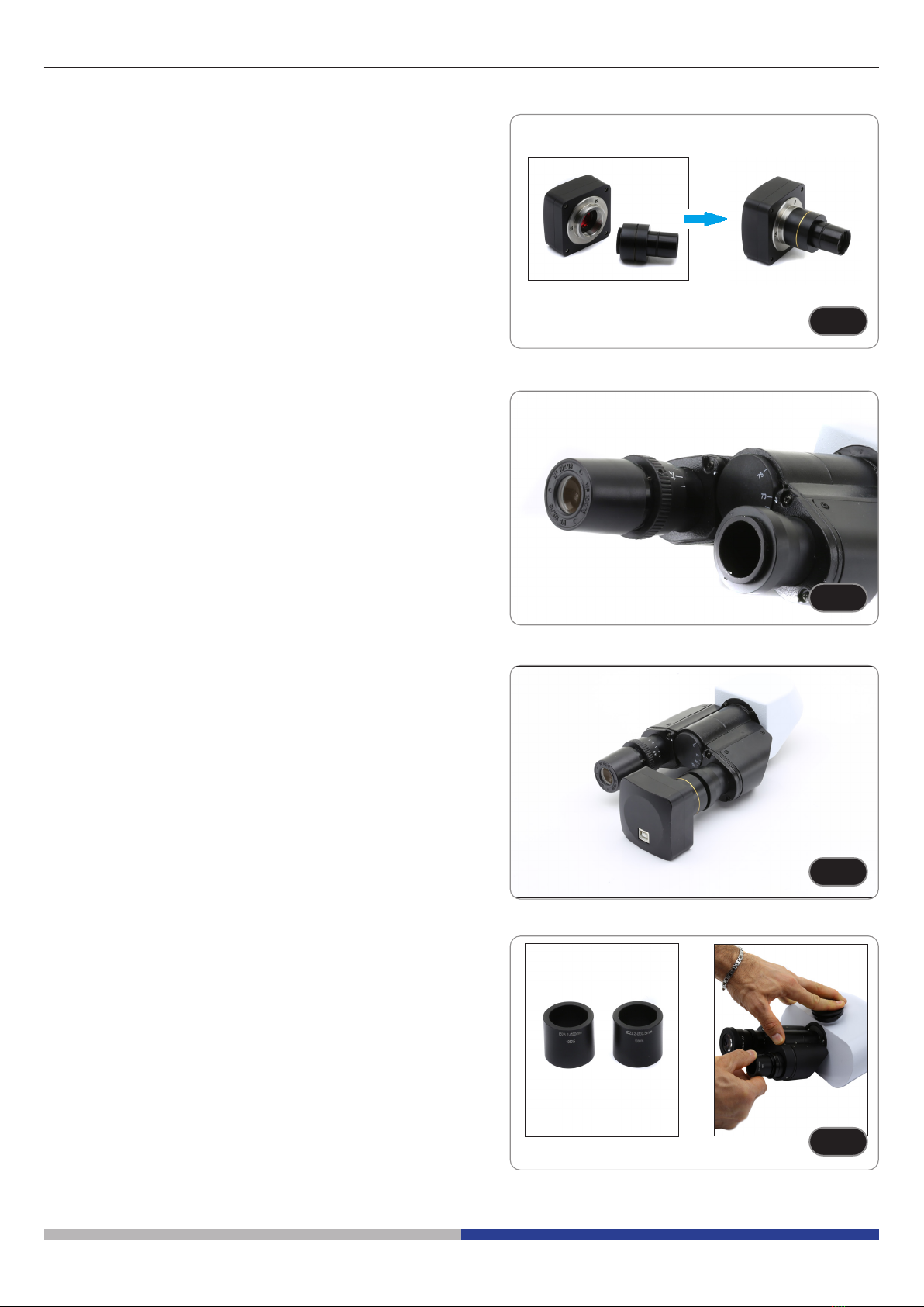
Page 10
The camera is driven by a software.
Two levels of software are available: PROVIEW and LITEVIEW.
Inside the package is enclosed a Function Table showing the several software functions.
It will be the user’s responsibility to decide which level of software best meets his needs.
The software can be downloaded from the site:
http://www.optikamicroscopes.com/optikamicroscopes/optika-support/download-drivers-softwares/
You will have to register to download the .zip le.
Once the le has been downloaded, you will have to run the setup.exe le.
At the end of the installation it is possible to start the software.
• NOTE: no driver installation is required for the cameras. The software setup procedure automatically
installs all the needed drivers for the correct operation of the camera.
The software’s User Manual is available in PDF format within the application itself and can be opened using the
“F1” function key.
You must have Acrobat Reader installed to view the manual.
The manual contains all the operating instructions for using the camera and for the various functions of the soft-
ware.
8.1 WiFi camera connection
The camera is powered via USB cable (C-WF version) or via USB cable and/or rechargeable batteries (C-WFR
version).
• The camera connects to PC only and exclusively with a WiFi connection.
• There is no possibility to connect the camera to PC with a USB cable.
1. Connect the USB power cable to the camera.
2. In the Settings of your PC select the WiFi device C-WFR-xxxxx.
• The password for connecting the camera is “12345678”.
3. Launch the PROVIEW or LITEVIEW software.
4. Manage the camera using the software to control all function.
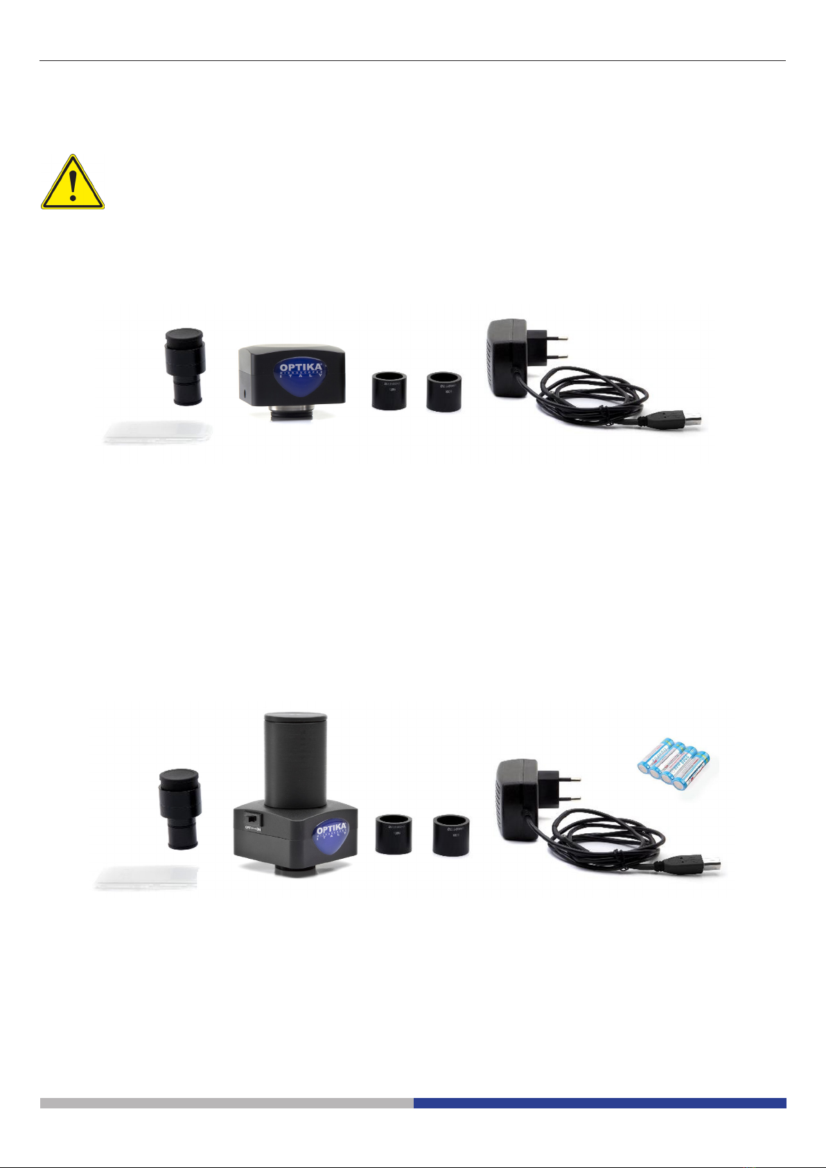
8.2 Use of the camera (C-WF)
1. Connect the USB power cable to the camera.
2. From “Camera List” panel in the software, click on the camera name (C-WFR).
3. Live image appears.
4. Manage the camera using the software to control all function.
8. Use of camera