Delmont imaging iCare User manual


delmont
imaging
D900 700 053 B Rev 09/2018 2
TABLE OF CONTENTS
1. General presentation............................................................4
2. Safety instructions...............................................................4
2.1. General instructions
2.3. Contraindication
3. Regulatory notice..................................................................5
3.1. Compliance
3.2. Electromagnetic interferences and electrostatic discharges
3.3. Materiovigilance
3.4. Life-end
4. Product description..............................................................6
4.1. Control unit description
4.2. Camera head description
5. Installation............................................................................8
6. Functioning...........................................................................9
6.1. Powering on
6.2. Connecting the endoscope to the camera head
6.3. White balance
6.4. Focus
6.5. Other camera head buttons features
6.6. Turning off the product
7. Disinfection procedure of the camera head........................10
8. Failure detection.................................................................10
8.1. The light indicator of the front panel does not illuminate when power is turned on
8.3. There is no more light but the fans continue to work
8.5. The image is blurred
9. After-sales service and maintenance.................................11
10. Technical data.....................................................................11
Instructions for use: iCare imaging station

delmont
imaging
D900 700 053 B Rev 09/2018 3
TABLE OF CONTENTS
11. Electromagnetic compatibility............................................12
11.1. Guidance and manufacturer’s declaration: electromagnetic emissions
11.2. Guidance and manufacturer’s declaration: electromagnetic immunity
11.3. Recommended distances between portable and mobile RF communication systems
for this product
12. Used symbols.....................................................................15
Instructions for use: iCare imaging station
delmont
imaging
D900 700 053 B Rev 09/2018 4
1. General presentation
gynecologists when performing surgical endoscopy or diagnostic procedures.
This equipment was delivered to you in a cardboard box. Keep it for possible transport.
It includes all the following elements:
- a control unit that contains a 64 Watts LED light source,
- an HD CMOS sensor with its 22mm C-mount focal length lens, 3 pre-programmed buttons, 2.99mm
cable, connector and sealing cap,
- an EU power cable,
- an HDMI cable,
- a USB key,
- two WiFi keys that allow our imagyn application to work properly (for more information on the imagyn
software, please refer to the corresponding instructions for use).
Phrases with the symbol correspond to points that require special attention.
Phrases with the symbol are information.
2. Safety instructions
2.1. General instructions
- Read the instructions for use.
- Comply with the conditions of use and storage.
- The unit may only be opened by a competent technician authorised by the manufacturer.
- Do not expose the unit to water splashes or in a place that is too humid.
- Use only accessories supplied with the unit or offered as an option by the manufacturer.
- Do not place heavy objects on the unit.
- This unit is not sterile.
- If the power cord is damaged, turn off the power immediately. It is dangerous to operate this unit with a
damaged cord. To disconnect the cord, pull it by the plug. Never pull on the cord itself.
- Unplug the unit from power if it is not going to be used for a few days or more.
Do not cover it and make sure that the feet of the unit are present.
On the camera side, it is a 1/4’’ colour CMOS mono HD camera with remote electronics. Its small ergonomic sen-
sor, automatic shutter, sensitivity, excellent resolution and exceptional colour rendering make it the ideal medical
tool.
On the light source side, this LED source is specially designed for use in various endoscopic diagnostic or surgical
applications. Its ease of use and powerful lighting make it the ideal multidisciplinary medical tool.
To get the most out of iCare while taking all the necessary precautions, it is essential that you read this manual.
Instructions for use: iCare imaging station

delmont
imaging
D900 700 053 B Rev 09/2018 5
- Do not use corrosive or abrasive products to clean the device but only the disinfectant liquids recom-
mended in the corresponding chapter.
- Before each use, make sure that the unit has no rough surfaces, sharp edges or protrusions that could
cause safety problems.
- The surface temperature can exceed 41°C (after a few minutes of use). Therefore, avoid contact with
skin.
- To avoid the risk of electric shock, this device may only be connected to a power supply equipped with
a protective ground.
- The devices connected to the inputs/outputs must comply with IEC 60950-1.
-
- Check with the manufacturer the compatibility of your endoscope before use.
- To prevent errors or delays in diagnosis, it is important to ensure that the monitor settings used are
optimized for the procedure being performed so that a clear, noise-free colour image is obtained.
- This device should be used on individuals (patients) suitable for endoscopic procedures.
- Applied parts of electro-medical devices that can be used in conjunction with iCare shall be of BF-type
or CF according to standard 60601-2-18. Check this compatibility before each operation for safe use.
It is recommended to have a second camera and light source available to use in case an absence or
degradation of performance is observed.
- Never look at the light output or the end of the light cable.
- Do not insert anything other than a light cable into the hole provided for this purpose, otherwise the
optical system may be damaged.
immunity of the unit.
-
- The light power at the light cable outlet can cause eye damage. Handle the light cable carefully when the
device is in use.
- Do not place the distal end of the light cable or endoscope directly on the patient or on any other flam-
This product is equipped with Group 1 LEDs according to IEC 62471. Do not look directly at the light
to avoid any ocular risk.
2.3. Contraindication
The use of iCare is contraindicated when endoscopic practice is contraindicated for the patient.
Instructions for use: iCare imaging station
3. Regulatory notice
3.1. Compliance
delmont
imaging
D900 700 053 B Rev 09/2018 6
3.2. Electromagnetic interferences and electrostatic discharges
Although this product complies with EMC standards, it is possible that under very special circumstances it may
cause interference to other devices, or may itself be affected by other devices or an adverse electromagnetic
environment.
To avoid these situations, it is recommended:
- to ensure the quality of the electrical network (especially the grounding of all equipment and trolleys),
- to keep the device away from electromagnetic sources (for example, a compressor, a motor, a transfor-
mer, an HF generator, etc.).
3.3. Materiovigilance
As any medical device, this device is subject to the provisions of the materiovigilance. Any serious malfunction
must therefore be reported to the competent authorities and the manufacturer as soon as possible and with the
greatest possible precision.
Manufacturer’s contact information: refer to the last page of this IFU.
3.4. Life-end
This device carries the recycling symbol in accordance with the European Directive 2002/96/EC on the Waste of
Electrical and Electronic Equipment (WEEE).
Proper disposal of this device will help prevent any harmful consequences for the environment and human health.
The symbol on the unit or accompanying documentation indicates that this product may under no circums-
tances be treated as household waste. It must therefore be handed over to a waste collection centrer responsible
for recycling electrical and electronic equipment.
For disposal, observe the waste disposal regulations in force in the country of installation.
For further details on the processing, recovery and recycling of this unit, please contact your nearest commercial
representative for instructions.
Instructions for use: iCare imaging station
It meets the requirements of European Directive 93/42/EEC on medical devices. It therefore complies with the
relevant electrical safety (IEC) and electromagnetic compatibility (EMC) standards.
4. Product description
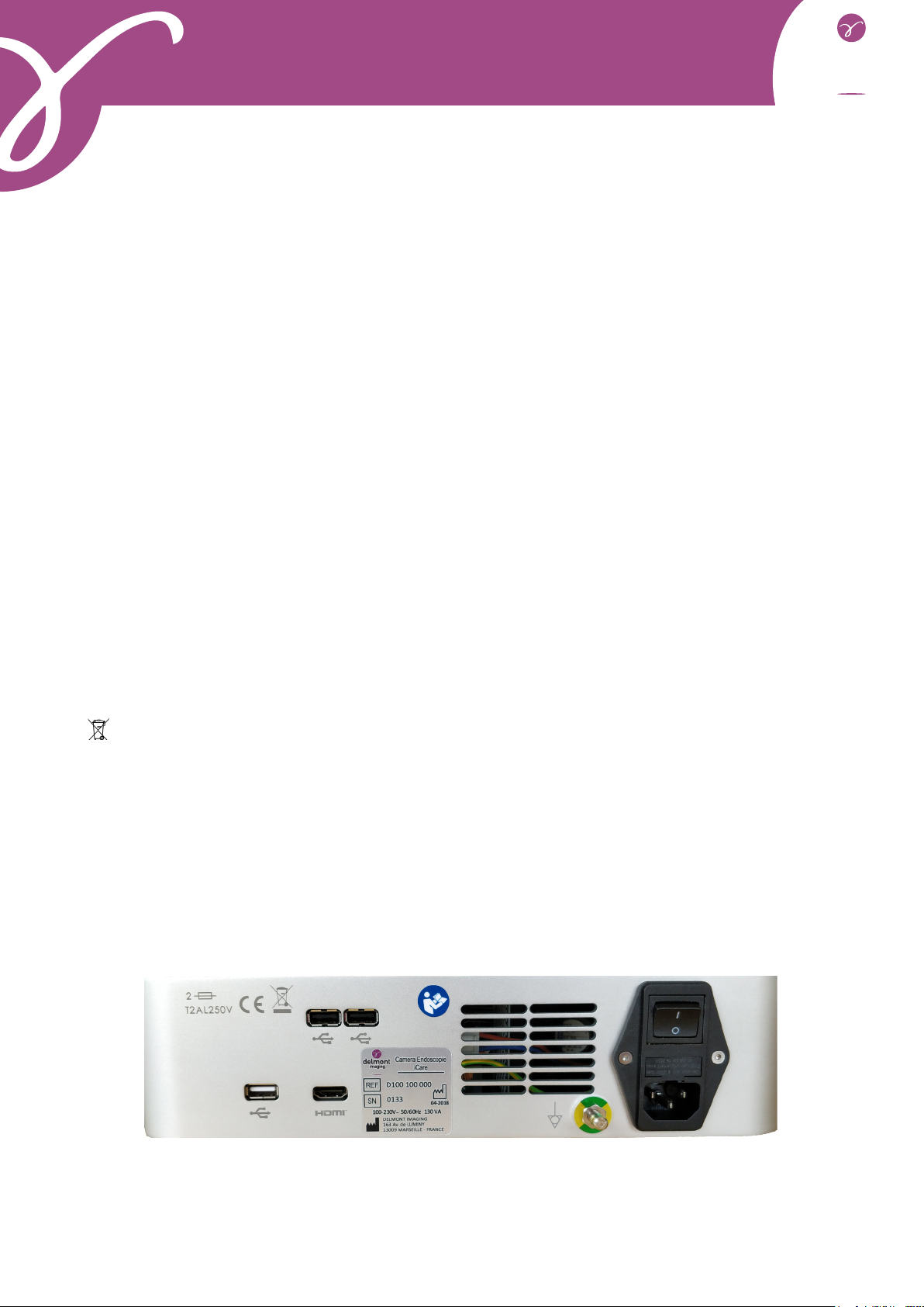
4.1. Control unit description
[1] [2]
[3]
[4]
[5][5]
[6]
[7]
[7]
[7]
[1] : The fans on the rear of the unit must not be blocked to prevent overheating. The light source is equipped with
an automatic safety feature that stops the lighting if the internal temperature becomes excessive.
Fans on the left side and under the unit are also present for better ventilation.
delmont
imaging
D900 700 053 B Rev 09/2018 7
[2] : Power is supplied through the rear panel power outlet, which must be connected to the power supply via the
cord provided with the product. This socket has a fuse cover and the main switch for powering on. When repla-
cing a fuse, it is imperative to disconnect the product from the network and use a fuse of the same type. The T
of «T2A» means «time-delay». Use only fuses marked UL/CSA.
[3] : The unit has an HDMI video output.
Devices that connect to the «VIDEO OUT» sockets must comply with IEC 60950.
[4] : The unit has a USB output for a storage key.
[5] : The unit has two USB outputs for two WiFi keys.
[6] : The unit has an equipotential plug, which can be connected to other electromedical devices in order to reduce
the formation of different electrical potentials.
[7]
standards IEC 60601-1, IEC 60601-2-18, IEC 60417 and EN 980 (see the corresponding section 12).
Instructions for use: iCare imaging station
[8]
[9]
[10]
[11]
[8] : The unit has a connector for plugging in the light cable.
[9] : The unit has a light indicator that alerts the user to the different phases of use of the product.
[10] : The unit has a connection for plugging in the iCare camera head.
[11] : The device has a stand-by button.

delmont
imaging
D900 700 053 B Rev 09/2018 8
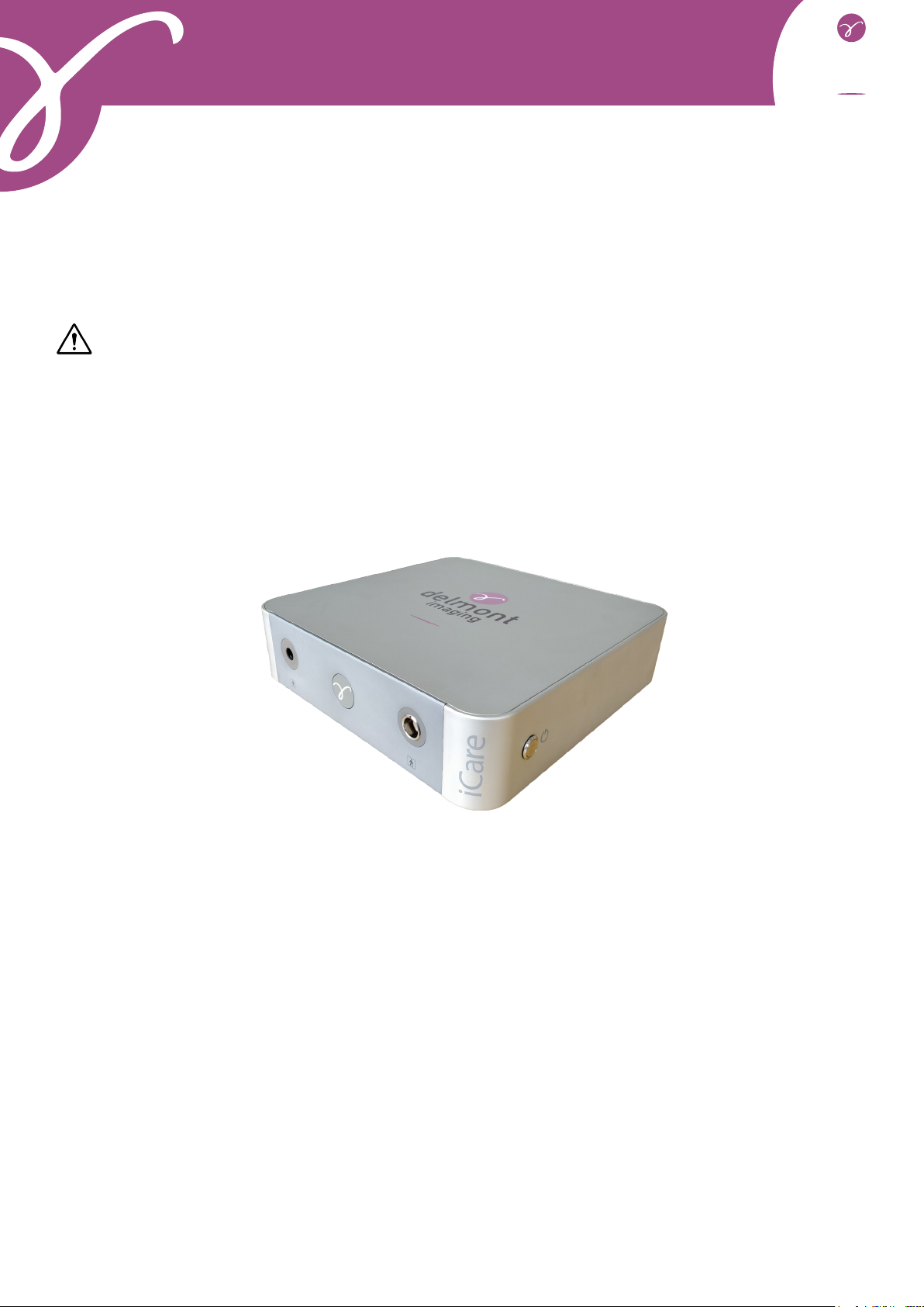
4.2. Camera head description
[12] : This button allows to launch a white balance by long press.
[13] : This button allows to switch from a minimum value of light power to a maxi-
mum value by short press, and vice versa. These values are 75% and 100% of the light
output respectively.
[14] : This button allows:
- by short press, to launch an image capture,
- by long press, to start or stop a video recording.
[12]
[14]
[13]
5. Installation
No special training is required to install this medical device. Please refer to the instructions in this section of the
user manual.
Once all iCare components have been unpacked from their original packaging, please perform the following ac-
tions:
- Place the control unit on a level and stable surface. If you place it in a compartment, make sure that it is
- Connect the power cord to the power outlet [2] on the rear of the unit.
- Connect the other end of the power cord to an electrical outlet.
- Connect the HDMI cable to the corresponding output [3] on the rear of the unit.
- Connect the second end of the HDMI cable to the corresponding input on the monitor.
- Place the two WiFi keys on the two USB outputs [5] on the rear panel.
- Place the USB key on the USB output [4] on the rear panel.
- Set the power supply button on the rear panel of the unit to position « I». The unit enters in a booting
sequence: the indicator light [9] on the front panel flashes rapidly. This booting sequence ends after approxima-
tely 1 minute 30. Then, the unit goes into standby mode: the front panel indicator light flashes slowly.
- Power on the monitor. A colour bar should appear on the screen.
- Plug the camera head connector to the front of the control unit [10]. A red coding is present on the
connector of the camera head and above the corresponding socket of the control unit. Align these two coded pins
to connect the camera head. A « click » lock must be heard.
- Insert the light cable into the hole provided for this purpose [8]. A « click » lock must be heard. Connect
the other end of the light cable to your endoscope.
- Place an endoscope in front of the camera lens.
- Press the stand-by button [11] on the right side of the control unit to start the device: the indicator light
Instructions for use: iCare imaging station
It is then possible to check the correct operation of the camera and especially to ensure that the settings of the
monitor give full satisfaction thanks to the colour bar which appears when disconnecting the sensor.

delmont
imaging
D900 700 053 B Rev 09/2018 9
6. Functioning
6.1. Powering on
The device is equipped with a switch placed on the rear of the device [2]. It is activated by changing this switch
to position « I». The light source is set to the last power used or to the value saved by the practitioner in imagyn.
6.2. Connecting the endoscope to the camera head
Bring the dewclaws on the ring of the lens closer together until the endoscope can be inserted. Once the endos-
cope is inserted, release the two dewclaws.
The endoscope can be unlocked by simply bringing the dewclaws closer together.
6.3. White balance
White balance is triggered by pressing and holding the [12] button on the camera head. The following procedure
should be followed:
- Start the white balance by pressing the corresponding button.
«Processing AWB...» is present on the screen.
- Keep the button pressed until the message «AWB OK» appears on the screen. This delay avoids unin-
tentional press.
6.4. Focus
Use the lens ring associated to the camera head to focus.
objects observed are sharp.
Instructions for use: iCare imaging station
focusing.
6.5. Other camera head buttons features
The [13] button on the camera head switches from a A value to a B value of the light source. These basic values
are set at 75% and 100% light output but can be adjusted via imagyn.
The [14] button allows:
- by short press, to launch an image capture,
- by long press, to start a video recording. Another long press stops the video recording.
6.6. Turning off the product
To turn off the device, put the switch on the rear panel of the control unit [2] to the « O» position.

delmont
imaging
D900 700 053 B Rev 09/2018 10
7. Disinfection procedure of the camera head
Pre-disinfection cleaning Disinfection Sterilization
Immersion in an enzymatic cleaning
solution (Aniosyme DD1, Hexanios
G+R or surface cleaning (Anios wipe))
Immersion in a glutaraldehyde so-
lution (Anios-Steranios Laboratory
2%)
Sterilization with ethylene oxide
Any other method of disinfection is prohibited. Damage caused by these other methods cannot be
borne by the manufacturer.
The use of a sterile protective cover on the camera is recommended regardless of the procedure fol-
lowed in stetilization service and for the duration of the surgical procedure.
The camera is not autoclavable.
The camera is not compatible with automatic washers/disinfectors.
Existing alkaline solutions for pre-disinfection of certain medical devices are FORBIDDEN for pre-di-
sinfection of our cameras.
Parts that have been in contact with the disinfectant must be rinsed thoroughly.
Use non-woven compresses to dry the optics so as not to scratch them.
The procedures described in this chapter are provided as advice, not in any way to replace ofcial
recommendations or guidelines.
Decontamination being linked to the selected products, methods and/or tools, remains the sole res-
ponsibility of the personnel concerned.
8. Failure detection
8.1. The light indicator of the front panel does not illuminate when power is turned on
- Check that the power cord is connected to the network and the device, and that the power switch on the
rear panel of the unit is in the « I» position.
- Check fuses for proper condition (use T2A - 250V - UL/CSA fuses only).
Instructions for use: iCare imaging station
Check if the light source power is set to Pmax (100% of the power) thanks to the button of the camera head. If
the problem remains even with Pmax value, check if the light cable is properly plugged. Check eventually the
condition of your light cable and optics.
8.3. There is no more light but the fans continue to work
Check that the light cable is properly connected. If this is the case, wait a few minutes: the light source is equip-
ped with a safety device that cuts off the power supply to the LED if the temperature inside the device is too high.
Once the temperature has dropped, the source can be used again.
nothing must obstruct the fan grids below, on the back and on the left side of the device.

delmont
imaging
D900 700 053 B Rev 09/2018 11
- Check that the camera head is connected to the control unit (otherwise a colour bar will be displayed).
- Check that the control unit is correctly connected to the monitor (cable in good condition and plugs
properly inserted).
- Check that the monitor is turned on, that the correct video input is selected and that the screen image
settings are not in the minimum position (colour, light and contrast).
- Check for the presence of light by inspecting the light source, light cable and endoscope.
8.5. The image is blurred
- Check that there is no fog on the camera lens or endoscope.
- Check the focus of the lens.
9. After-sales service and maintenance
Simply make sure that the aerators are not clogged with dust. If this is the case, unplug the unit and vacuum the
dust.
Errors of use are not covered by the warranty.
If a defect persists and it is necessary to return the device to the after-sales service, take care to ship it in its
original packaging. Also, the entire device (control unit, camera head, lens, cables, etc.) must be returned.
Please attach a small explanatory note to the shipment regarding the defect found.
-
ming them to the carrier by registered letter within 48 hours. In the case where a material dispatched by us would
suffer damage during its transport, the amount of the repair is charged either to the carrier if the reserves were
made within the time limits, or to the recipient in the contrary case.
In case of an incident, please contact our customer service or our nearest sales representative.
The equipment must be disinfected before returning for repair.
Instructions for use: iCare imaging station
If the defect persists and it is necessary to return the device to the after-sales service, take care to send it to us
in its original packaging after having disinfected it.
10. Technical data
Camera side:
- Waterproof HD CMOS sensor (IP67)
- 22 mm C-mount lens
- Resolution: 1920 x 1080
- Interlaced scanning
- Sensibility : 2 000 lux at F8
- Signal-to-noise ratio: 54 dB
- Automatic electronic shutter (1/50 to 1/10 000)

delmont
imaging
D900 700 053 B Rev 09/2018 12
Light source side:
- LED technology
- Nominal power: 64 W
- Colour temperature: 6 000°K
- LED typical lifetime: 50 000 hours
- Compatible light cable type: Storz
- Automatic thermal protection system
- Automatic detection of presence/absence of the light cable
General specications:
- Power supply: 100-230 V ~; 50/60 Hz
- Power consumption: 130 VA
- Two fuses T2A - 250V UL/CSA 5 x 20 mm
- Equipotential plug
- Dimensions of the control unit (w x h x d): 310 x 75 x 310 mm
- Weight of the control unit: 4 200 g
Environment :
- Working temperature: +10°C/+40°C
- Working humidity: 30 to 75%
- Transport and storage temperature: -10°C/+40°C
- Transport and storage humidity: 20 to 85%
- Working, transport and storage atmospheric pressure: 800 hPa to 1 060 hPa
- Not protected against water splashes (IPXO)
- Not suitable for use in the presence of flammable anesthetic mixture with air with oxygen or nitrous
oxide
Instructions for use: iCare imaging station
- Pre-programmed buttons on the camera head
- Colour bar
- White balance
- 1 HDMI output
- 2 USB outputs for WiFi connection
- 1 USB output for a storage key
Regulation :
- Electrical safety class 1, type BF
- Complies with European directive 93/42/EEC
- Complies with international norms: IEC 60601-1, IEC 60601-2-18, IEC 60601-1-2, IEC 60417 and
EN 80
- IEC 62471: group 1 risk
11. Electromagnetic compatibility
11.1. Guidance and manufacturer’s declaration: electromagnetic emissions
The « CMOS camera + LED source » reference equipment is intended for use in the electromagnetic environment
delmont
imaging
D900 700 053 B Rev 09/2018 13
Emissions test Compliance Electromagnetic environment: Guidance
RF emissions
CISPR 11 Group 1
This « CMOS camera + LED source » product only uses radio
power for its subsystems. It therefore emits very low RF ener-
gy and is not likely to interfere with nearby electronic devices.
RF emissions
CISPR 11 Class A This « CMOS camera + LED source » product must be used
in all installations, other than residential installations and
premises directly connected to the public low voltage power
distribution network intended to supply residential buildings.
Harmonic emissions
IEC 61000-3-2 Compliant
Voltage fluctuations/Flicker
IEC 61000-3-3 Compliant
11.2. Guidance and manufacturer’s declaration: electromagnetic immunity
The « CMOS camera + LED source » reference equipment has been designed for use in the electromagnetic envi-
Immunity test IEC 60601
Severity Level
Compliance
Level Electromagnetic environment: Guidance
Electrostatic
discharges
IEC 61000-4-2
± 6 kV via contact
± 8 kV via air
± 6 kV
± 8 kV
The floor must be made of wood, concrete
or tiles. If the floor is covered with a synthe-
tic material, the relative humidity must be at
least 30%.
Rapid transient
peaks
IEC 61000-4-4
± 2 kV power lines
± 1 kV input/output lines
± 2 kV
± 1 kV The quality of the main power supply must
be the one of a typical commercial or hospi-
tal environment.
Electric shocks
IEC 61000-4-5
Differential mode ± 1 kV
Common mode ± 2 kV
± 1 kV
± 2 kV
Power outages,
short power ou-
tages and voltage
fluctuations
IEC 61000-4-11
<5% U- for 10 ms
40% U- for 100 ms
70% U- for 500 ms
<5% U- for 5 ms
5% U10 ms
<40% U100 ms
<70% U500 ms
<5% U5 s
The quality of the main power supply must
be the one of a typical commercial or hospi-
tal environment. If the user of this product
must be able to continue working during
power outages, it is recommended that this
product be powered by UPS or battery.
mains frequency
(50/60 Hz)
IEC 61000-4-8
3 A/m 3 A/m
must be at a characteristic level of a loca-
tion (50/60 Hz) in a typical commercial or
hospital environment.
Instructions for use: iCare imaging station
Note: Uis the nominal value of the electrical voltage applied during the test.
Guidance and manufacturer’s declaration: electromagnetic emissions
The « CMOS camera + LED source » reference equipment is intended for use in the electromagnetic environment
delmont
imaging
D900 700 053 B Rev 09/2018 14
Safety test IEC 60601
Severity Level
Compliance
Level Electromagnetic environment: Guidance
Conducted RF
IEC 61000-4-6
Radiated RF
IEC 61000-4-3
3 Vrms
150 kHz to 80 MHz
3 V/m
80 MHz to 2.5 GHz
3V
3 V/m
Portable and mobile RF communication devices should
not be used at a distance, including cables, from this
product that is less than the recommended distance,
calculated by applying the formula that corresponds to
the transmitter frequency.
d = 1,16 P
d = 1,16 P 80 MHz to 800 MHz
d = 2,33 P 800 MHz to 2,5 GHz
Where P is the maximum output power of the trans-
mitter, in Watts (W), assigned by its manufacturer and
(d) is the recommended separation distance in metres
(m).
which must be established by in situ electromagnetic
measurement - must be below the compliance level in
each frequency band. Interference may occur with de-
vices marked with the following symbol:
Note 1: At 80 MHz and 800 MHz, the highest frequency band should be used.
Note 2: These recommendations may not be applicable in all situations. The propagation of electromagnetic
waves is altered by absorption and reflection from structures, objects and people. For transmitters whose maxi-
mum output power is not listed in the table above, the recommended separation distance d, in metres (m) can be
established using the equation applicable to the transmitter frequency, where P is the maximum output power of
the transmitter in Watts (W) assigned by the transmitter manufacturer.
Instructions for use: iCare imaging station
11.3. Recommended distances between portable and mobile RF communication systems
for this product
The « CMOS camera + LED source » reference equipment has been designed for use in the electromagnetic
environment in which the emitted RF interference is controlled. The user of this equipment can help avoid elec-
tromagnetic interference by maintaining a minimum distance between portable and mobile RF communication
systems (transmitters) and this equipment, as recommended below, as a function of the maximum output power
of the communication system.
Maximum assigned
transmitter output power
in W
Separation distance as a function of transmitter frequency (m)
150 kHz to 80 MHz 80 MHz to 800 MHz 800 MHz to 2.5 GHz
d = 1,16 P d = 1,16 P d = 2,33 P
0.01 0.116 0.116 0.233
0.1 0.366 0.366 0.736
1 1.16 1.16 2.33
10 3.66 3.66 7.36
100 11.6 11.6 23.3
delmont
imaging
D900 700 053 B Rev 09/2018 15
Note 1: At 80 MHz and 800 MHz, the separation distance given in the upper frequency band applies.
Note 2: These recommendations may not be applicable in all situations. The propagation of electromagnetic
waves is altered by absorption and reflection from structures, objects and people. For transmitters whose maxi-
mum output power is not listed in the table above, the recommended separation distance d, in metres (m) can be
established using the equation applicable to the transmitter frequency, where P is the maximum output power of
the transmitter in Watts (W) assigned by the transmitter manufacturer.
Instructions for use: iCare imaging station
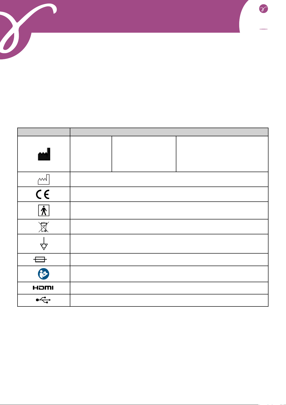
Symbol Description
Symbol for
«Manufacturer»
Legal Manufacturer:
DELMONT IMAGING
Zone Athélia V
390 Avenue du Mistral
13600 La Ciotat, FRANCE
Tel.: +33 (0) 9 51 51 30 30
Fax: +33 (0) 9 57 51 31 00
Email: [email protected]
Website: www.delmont-imaging.com
Symbol for «Manufacturing year»
Complies with European directive 93/42/EEC
Symbol for «Type BF device»
Electronic and electrical equipment put on the market after 13/08/2005. This symbol
indicates that this product should not be treated with household waste.
Symbol for «Equipotential plug»
TSymbol for «UL/CSA timed fuses»
Symbol for «Read the instructions for use»
Symbol for «HDMI video output»
Symbol for «USB output»
12. Used symbols

delmont
imaging
D900 700 053 B Rev 09/2018 16
SOMMAIRE
1. Présentation générale.........................................................18
2. Consignes de sécurité........................................................18
2.1. Consignes générales
2.2. Consignes propres à la source de lumière
2.3. Contre-indication
3. Avis réglementaire..............................................................20
3.1. Conformité
3.2. Interférences électromagnétiques et décharges électrostatiques
3.3. Matériovigilance
3.4. Fin de vie
4. Description du produit........................................................21
4.1. Description de la voie de commande
4.2. Description du capteur caméra
5. Installation..........................................................................22
6. Fonctionnement..................................................................23
6.1. Mise sous tension
6.2. Connexion de l’endoscope sur le capteur
6.3. Balance des blancs
6.4. Mise au point
6.5. Autres fonctionnalités des boutons du capteur
6.6. Arrêt du produit
7. Procédure de désinfection du capteur caméra..................24
8. Recherche de pannes.........................................................25
8.1. Le voyant en face avant ne s’éclaire pas à la mise sous tension
8.3. Il n’y a plus de lumière mais les ventilateurs continuent de fonctionner
8.5. L’image est floue
9. Servie après-vente et entretien..........................................25
10. Caractéristiquestechniques...............................................26
Manuel d’utilisation : station d’imagerie iCare

delmont
imaging
D900 700 053 B Rev 09/2018 17
SOMMAIRE
11. Compatibilité électromagnétique.......................................27
11.1. Guide et déclaration du fabricant : émissions électromagnétiques
11.2. Guide et déclaration du fabricant : immunité électromagnétique
11.3. Distances recommandées entre les systèmes de communication RF portables et
mobiles de ce produit
12. Symboles utilisés...............................................................30
Manuel d’utilisation : station d’imagerie iCare
delmont
imaging
D900 700 053 B Rev 09/2018 18
1. Présentation générale
iCare est un combiné unissant une caméra HD CMOS et une source de lumière à LED dans une unique voie de
chirurgicales ou de diagnostic.
Cet équipement vous a été livré dans un emballage cartonné. Le conserver pour un éventuel transport.
Il comprend l’ensemble des éléments suivants :
- une voie de commande qui contient une source de lumière 64 Watts à LED,
- un capteur HD CMOS avec son objectif monture C de focale 22mm, doté de 3 boutons préprogrammés,
d’un câble de 2,99mm, d’un connecteur et de son bouchon d’étanchéité,
- un câble secteur EU,
- un câble HDMI,
- une clé USB,
- deux clés WiFi qui permettent le bon fonctionnement de notre application imagyn (pour plus d’informa-
tions sur le logiciel imagyn, vous référez au manuel d’utilisation correspondant).
Les phrases comportant le symbole correspondent à des points nécessitant une attention particulière.
Les phrases comportant le symbole sont des informations.
2. Consignes de sécurité
2.1. Consignes générales
- Lire le manuel d’utilisation.
- Respecter les conditions d’utilisation et de stockage.
- L’ouverture de l’appareil doit être réalisée exclusivement par un technicien compétent habilité par le
fabriquant.
de court-circuit ou d’émission dangereuse.
- Cet appareil n’est pas stérile.
- N’utiliser que les accessoires fournis avec l’appareil ou proposés comme option par le fabricant.
- Ne pas poser d’objets lourds dessus.
- Si le cordon d’alimentation est endommagé, mettre immédiatement le dispositif hors tension. Il est
dangereux de faire fonctionner cet appareil avec un cordon endommagé. Pour débrancher le cordon, le tirer par
Côté caméra, il s’agit d’une caméra HD mono CMOS 1/4’’ couleur, à électronique déportée. Son capteur ergono-
mique de taille réduite, son shutter automatique, sa sensibilité, son excellente résolution ainsi que son exception-
nel rendu des couleurs en font l’outil médical idéal.
Côté source de lumière, cette source LED est spécialement conçue pour être utilisée dans des applications en-
doscopiques variées de diagnostic ou de chirurgie. Sa facilité d’utilisation, ainsi que sa puissance d’éclairage en
font l’outil médical pluridisciplinaire idéal.
-
sable que vous preniez connaissance du présent manuel.
Manuel d’utilisation : station d’imagerie iCare

delmont
imaging
D900 700 053 B Rev 09/2018 19
- Ne pas exposer l’appareil à des projections d’eau ou dans un endroit trop humide.
- Débrancher l’appareil du secteur s’il n’est pas prévu de l’utiliser pendant quelques jours ou plus.
moins 15cm tout autour de l’appareil. Ne pas le couvrir et veiller à ce que les pieds de l’appareil soient présents.
- Ne pas utiliser de produits corrosifs ou abrasifs pour nettoyer l’appareil mais uniquement les liquides
désinfectants recommandés au chapitre correspondant.
- Avant chaque utilisation, s’assurer que l’appareil ne présente aucune surface rugueuse, d’arête tran-
chante, ni de protubérances qui pourraient engendrer des problèmes de sécurité.
- La température de surface peut dépasser 41°C (après quelques minutes d’utilisation). Par conséquent,
éviter de maintenir cette zone en contact avec la peau.
- Pour éviter tout risque de choc électrique, cet appareil doit être raccordé uniquement à un réseau d’ali-
mentation équipé d’une terre de protection.
- Les appareils qui se connectent sur les entrées/sorties doivent être conformes à la norme IEC 60950-1.
sécurité.
- Pour prévenir toute erreur ou retard dans le diagnostic, il convient de s’assurer que les réglages du mo-
niteur utilisés soient optimisés pour l’intervention réalisée, de telle sorte qu’ils permettent d’obtenir une image
couleur nette, sans bruit.
- Cet appareil doit être utilisé sur des individus (patients) aptes à subir une procédure d’endoscopie.
- Les parties appliquées des appareils électro-médicaux pouvant être utilisés conjointement à iCare
intervention pour une utilisation sans risque.
Il est conseillé d’avoir à disposition une deuxième caméra et source de lumière an de l’utiliser au cas
où une absence ou une dégradation de performances est observée.
2.2. Consignes propres à la source de lumière
- Ne jamais regarder la sortie lumineuse, ni le bout du câble de lumière.
- Ne pas insérer autre chose qu’un câble de lumière dans le logement prévu à cet effet sous peine d’en-
dommager le système optique.
émissions ou diminuer l’immunité de l’appareil.
avec attention le câble de lumière lorsque le dispositif est en cours d’utilisation.
- Ne pas placer l’extrémité distale du câble de lumière ou de l’endoscope directement sur le patient ni sur
aucune autre matière inflammable (draps, gazes, champs opératoires, etc.) car celle-ci peut être très chaude et
Ce produit est équipé de LEDs de Groupe 1 selon la norme IEC 62471. Ne pas regarder directement la
lumière pour éviter tout risque oculaire.
Manuel d’utilisation : station d’imagerie iCare
2.3. Contre-indication
L’utilisation d’iCare est contre-indiquée dès lors que la pratique endoscopique est contre-indiquée pour le patient.

delmont
imaging
D900 700 053 B Rev 09/2018 20
3. Avis réglementaire
3.1. Conformité
la directive européenne 93/42/CEE, relative aux dispositifs médicaux. Par conséquent, il répond notamment aux
normes de sécurité électrique (IEC) et de Compatibilité Electromagnétique (CEM) ad hoc.
3.2. Interférences électromagnétiques et décharges électrostatiques
Bien que ce produit respecte les normes CEM, il est possible que dans des circonstances très particulières, il
perturbe d’autres dispositifs, ou bien qu’il soit lui-même perturbé par d’autres appareils ou un environnement
électromagnétique défavorable.
- de veiller à la qualité du réseau électrique (tout particulièrement de la mise à la terre de tous les appa-
reils et des chariots),
- d’éloigner l’appareil des sources électromagnétiques (par exemple, un compresseur, un moteur, un
transformateur, un générateur HF, etc.).
3.3. Matériovigilance
Comme tout dispositif médical, cet appareil est sujet aux dispositions de la matériovigilance, tout dysfonctionne-
ment grave doit donc faire l’objet d’un signalement aux autorités compétentes et au fabricant dans les plus brefs
délais et avec la plus grande précision possible.
Coordonnées du fabricant : se reporter à la dernière page du présent manuel.
3.4. Fin de vie
Cet appareil porte le symbole du recyclage conformément à la directive Européenne 2002/96/CE concernant les
Déchets d’Equipements Electriques et Electroniques (DEEE ou WEEE pour l’anglais Waste of Electrical and Elec-
tronic Equipment).
En procédant correctement à la mise au rebut de cet appareil, vous contribuez à empêcher toute conséquence
nuisible pour l’environnement et la santé de l’homme. Le symbole présent sur l’appareil ou sur la documen-
tation qui l’accompagne indique que ce produit ne peut en aucun cas être traité comme déchet ménager. Il doit
par conséquent être remis à un centre de collecte des déchets chargé du recyclage des équipements électriques
et électroniques.
Pour la mise au rebut, respectez les normes relatives à l’élimination des déchets en vigueur dans le pays d’ins-
tallation.
Pour obtenir de plus amples détails au sujet du traitement, de la récupération et du recyclage de cet appareil,
veuillez contacter votre représentant commercial le plus proche qui vous indiquera la marche à suivre.
Manuel d’utilisation : station d’imagerie iCare
Table of contents
Languages:
Popular Medical Equipment manuals by other brands


Invacare
Invacare BAR750 Owner's operating and maintenance manual

Mettler Electronics
Mettler Electronics Sonicator Plus 921 Operation manual

COOK Medical
COOK Medical ShortShot Quick reference guide

DENTSPLY
DENTSPLY Clamus DUAL manual

MGF
MGF DRAGER PP10 quick start guide

Synthes
Synthes Colibri II Instructions for use


