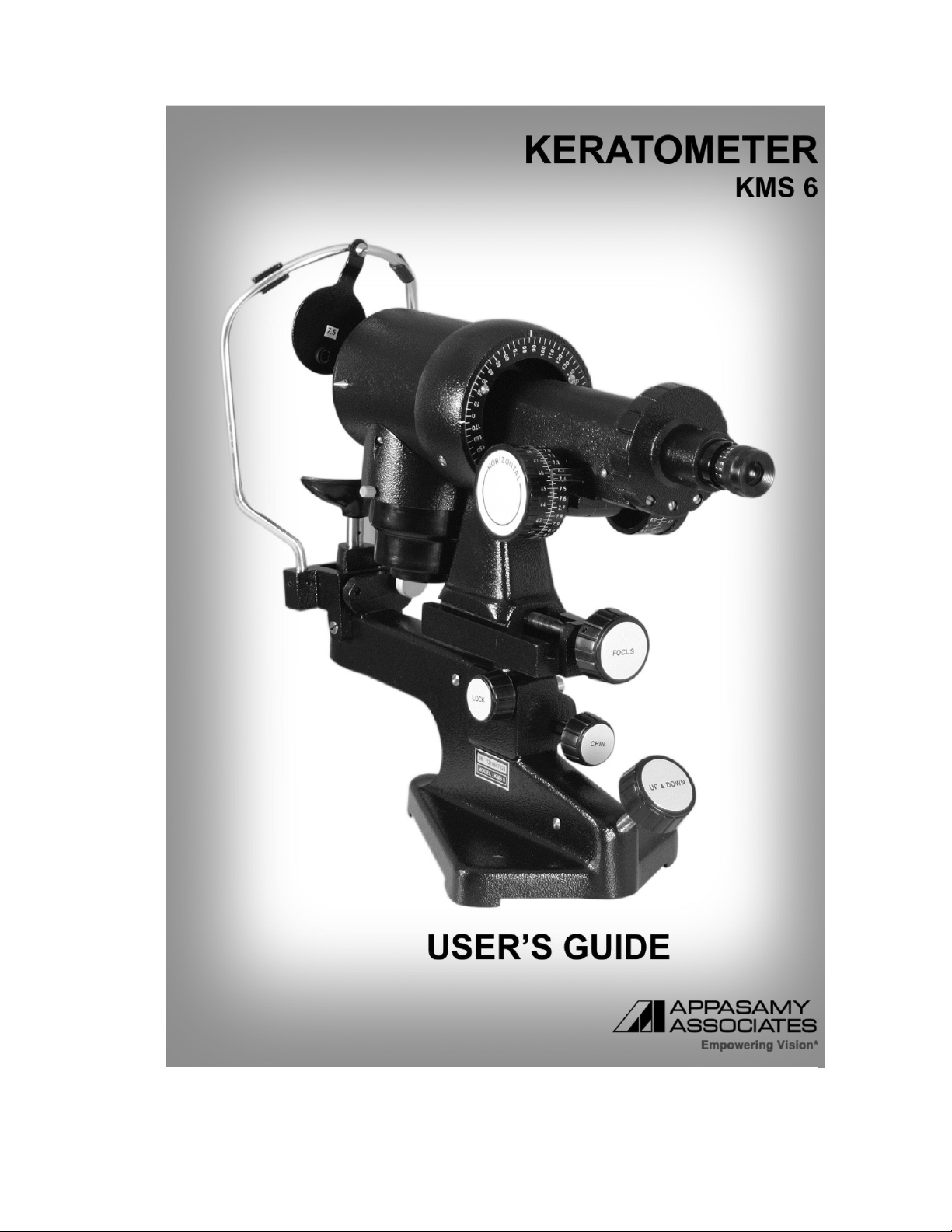
APPASAMY ASSOCIATES KMS 6 User manual


INTRODUCTION
Appasamy Associates proudly offers the finest and most precise Keratometer. All
adjustments on the instrument are manipulated conveniently from the practioner’s
position. All functional parts of the Instrument are manufactured and assembled to meet
customer’s requirements. High cared design of positioning, rotating, moving and
clamping assemblies is confirmed to meet the actual requirements efficiently.
DESCRIPTION OF KERATOMETER
A keratometer is a medical instrument that eye care professionals use to measure the
curvature and reflection of the anterior surface of the cornea.
2

TABLE OF CONTENTS
# Chapter Page No
1.
Introduction & Description of Keratometer 02
2.
Quality, Reliablity and Safety 04
3.
Warning 05
4.
Important Safety & Maintenance Instructions 06
5.
Safety Precautions 07
6.
Caution for use, General Specification 08
7.
Environmental Condition for use 09
8.
Technical Specification 10
9.
Cleaning and disinfection information 11
10.
Unpacking and Installation 12
11.
List of Parts 13
12.
Preliminary Adjustments 14
13.
Operating the Patient for Examination 15
14.
Instrument Orientation 15
15.
Patient Fixation & Way to focus the Instrument 16
16.
Locating the Cylinder Axis 18
17.
Measuring the Horizontal Principal Meridian 19
18.
Measuring the Vertical Principal Meridian 19
19.
Corneal Astigmatism & Axis of Astigmatism 20
20.
With and Against the Rule Astigmatism 20
21.
Schematic Diagram 21
22.
Trouble Shooting & Advisory Notice 22
23.
Warranty and Limitation of Liability 23
3

QUALITY, RELIABILITY AND SAFETY
Appasamy Associates is responsible for the safety, reliability and performance of the
equipment only if it is used in accordance with the instructions given in the Manual. The
Instrument is designed to perform as per the declared Intended for use with safer and
reliable.
Electrical installation of the room or building in which the equipment is to be used
must comply with regulations specified by the country in which the equipment to be
used as well the specified in the specification.
The repair / service will be taken care by a qualified technical person of Appasamy
Associates.
Read and follow the instructions, caution and warnings before installing the
Instrument
4

WARNING
Warnings are intended to alert you to importance of following the correct operating
procedures.
This is Class 1m equipment to avoid risk of electric shock. This equipment must
only be connected to supply mains with protective earth.
No modification of this equipment is allowed.
Use only the type of power source that indicated on label.
Connect the Equipment to properly grounded power outlets.
Unplug the Equipment before servicing / cleaning it.
Confirm the AC power cord meets the relevant local safety standards.
Don’t use damaged power cord.
Only trained personal can handle the equipment.
Remove the plug of Equipment from wall outlet before changing the Fuse.
Check the electrical connections periodically; any defects noticed, like loose
connections, damaged to insulation in the electrical wires etc., should be rectified
immediately.
Always place silica gel that packed in a packet near to optics e.g. Eyepiece and
lens base. It reduces the moisture contamination and fungus of optics.
Intake of silica is dangerous
Calibration process
If the customer needs any calibration please send the equipment for calibration
purpose.
TRAINING REQUIREMENT
The equipment is expected to operate by ophthalmic Doctors only. If at all it is used by
technician they should by trained well in usage of this equipment.
Appasamy associates well trained the personal if required.
5

IMPORTANT SAFETY & MAINTENANCE INSTRUCTIONS
Before using the Equipment, read instructions carefully.
Regular maintain and check the instrument and its parts.
Handle the Equipment carefully.
Use proper methods to clean optics and Equipment.
Before switch off, turnoff regulator.
Always cover the Equipment when not in use.
Don’t touch the exposed lenses, and keep them clean.
Don’t spill liquid into the Equipment.
Don’t use any hazardous solvents to clean the optics and parts.
Only use soft, dry cloth to clean the device.
When using the instrument after a prolonged period of inactivity, confirm normal and
safe operation beforehand
Unplug the Equipment and refer servicing to qualified service personnel under the
following condition
•If the product isn’t able to use after done all the troubleshooting in this
manual.
•If the liquid has been spilled into the optics / Equipment.
If the product has been exposed to rain or water.
•
If smudged target image formation occurs or the line damaged on
reticules
•If the Instrument is not able to use because of developed or exposed
to rain or water?
6

SAFETY PRECAUTIONS
Safety pointers for the manual and the device described therein are depicted according to
the following categories. Carefully read these sign codes & follow them when necessary.
The following code categories describe the degree of danger or damage likely to be
incurred in the event of user error made in ignorance of these codes.
This symbol is placed on bulb housing part to indicate that they may be hot.
Allow sufficient time to cool before touching the parts or changing the bulb.
In the event of user error, it is possible to injury.
WARNING: In the event of user error, death or serious injury is Possible
This icon denote cautions
This icon represents the productive earth
This icon represents Alternating current
This icon represents the power switch is turned OFF
This icon represents the power switch is turned ON
Type B applied part
LABELING INFORMATION
SN Number following this symbol indicate the serial number of the
Equipment
REF Character following the symbol indicate the Model number of the
Equipment
Classification CE-Regulation 93/42/EEC - Class 1m
Disposal
Dispose of the instrument according to local disposal and recycling laws.
7

CAUTION FOR USE
Cautions are intended to alert you to importance of following the correct operating
procedures where risk of injury to the patient or system.
Do not handle the plug with wet fingers ( To avoid electric shock)
The User are here by instructed not to touch the equipment and the patient simultaneously.
GENERAL SPECIFICATION
Equipment classification
Mode of operation : 5 minutes (Max)
Degree of mobility : Portable
Type of protection against
Electrical shocks - Class 1
Degree of protection against
Electrical shocks - Type ‘B’
Power Requirements
AC Input I/P: 110V AC /220V AC, 60Hz / 50Hz, 25VA
O/P: 6V DC, 2A
Fuses 1A Slow Blow (110V AC)
500mA Slow Blow (230V AC)
3A Slow Blow
IP Category 2X
Safety Rules:
•Leakage current to ground less than 0.5mA in normal condition.
•Leakage current to ground less than 1.0mA in single fault condition.
•Isolation voltage between ground and mains wires greater than 1500V.
•As per IEC 60601-1-2 standard, Emission and susceptibility requirements meets
class A.
8

ENVIRONMENTAL CONDITIONS FOR USE
Operating Temperature: 0°C ~ 50°C
Humidity : 35% ~ 95% (without dew condensation)
Air Pressure : 700hPa ~ 1060hPa
STORAGE, USAGE PERIOD AND OTHERS
1. Environmental conditions for installation (without package)
Temperature: -5°C ~ 70°C
Humidity : 35% ~ 95% (without dew condensation)
Air Pressure : 700hPa ~ 1060hPa
2. When storing the instrument, ensure that the following conditions are met.
a) The instrument should not be splashed with water.
b) Store the instrument where air pressure, temperature, humidity, ventilation,
sunlight, dust, salty/sulfurous air, etc. do not give any negative side effect.
c) Do not store or transport the instrument on a slope or uneven surface or in
an area where it is subject to vibrations or instability.
d) Do not store the instrument where chemicals are stored or gas is generated.
ENVIRONMENTAL CONDITIONS FOR PACKAGING IN
TRANSPORTATION
Temperature : -5°C ~ 70°C
Humidity : 35% ~ 95%
9

TECHNICAL SPECIFICATION
Key Features
Coincidence focusing system. Positive measurements to 0.02mm accuracy.
One-position instruments. Measure both meridians without change optical system.
15x Wide-angle piece.
Two-way adjustment to fit patient.
Accurate measurement of corneal curvatures
Allows subjective review of corneal surface
Measures concave surfaces for contact lens fitting
Easy elevation and lowering allows quick patient positioning
Engraved focusing scale on eyepiece
Dual eye level sighting system. Facilitates horizontal alignment.
Clear, uniform illuminated mires
Accurate of 0.012D from 36D to 52D
Measuring range : Radius of curvature, 9.4 ~ 6.4 mm
(In 0.05 mm steps),
: Diopter 36 ~ 52 (in 0.25 steps).
Instrument Dimensions : Height -16”, Width - 7”, Length - 19”
Weight of the Instrument : 7.0 Kgs approximately
Colour / Finish : Black
Mode of operation : Continuous
Electrical Input (~ AC) : (1) 230v, 50 Hz (2)110v, 60 Hz
Lamp : 230v-15w, 110v-25w, E12 Type
Standard Accessories : Dust Cover, Spare Bulbs (2 No’s)
Optional Accessories : Tripod Stand / Motorized Instrument
Table (Extra cost)
10
CLEANING AND DISINFECTION INFORMATION

Lens cleaning Method.
•Fold a piece of lens tissue four or five times to give a narrow triangular shape with a
finely pointed end.
•Moisten the pointed end of the lens tissue with a small amount of cleaner, keeping
fingers away from the solvent to avoid contamination by oils.
•Do not use excessive solvent such that the tissue is saturated.
•Place the tissue against the objective front lens, so wiping and rotating the lens in
one corkscrew motion from the centre of the lens to the outside edge to avoid going
back and forth over the same area.
•In this way the lens can be cleaned up to the edge of the cell. Further pressure will
cause the tissue to spread sufficiently to clean the whole surface. Remove any
obstinate marks by repeating the process. A new and clean piece of tissue should
be used each time.
Caution:
•Avoid cleaning lens with abrasive or lint cloths.
•Never dry wipe a lens as this may cause scratching. Begin by blowing off dust or
loose material with pressurized optical duster or bellows ball.
•Flooding or placing solvents directly on the lens is discouraged.
•Careful fogging of the lens with breath can assist in removing water-soluble residue
although caution should be exhibited in regard to excluding saliva.
•Keep lens upright allowing tissue to touch base surface so solvent does not run into
cements or other substances holding the lens in place.
•Disinfection instructions:
Clean the chinrest and Head stoppers with
Clean cloth after examine every patient.
11
UNPA
CKING & INSTALLATION

Unpack the Instruments and save all packing materials. They are specially designed to
protect the Instrument and will make repacking easy if you ever need to ship your
Keratometer.
After unpacking verifies the list of accessories us indicate below.
INSTALLATION
The initial installation will be done by Appasamy Associates Trained Engineers only.
After installation a performances check will be carried by Appasamy Associates
Engineers. Nobody is authorized to install the Medical Equipment except Appasamy
Associates Engineers.
Connect the Output of the power supply to the Keratometer.
Ensure the intensity control in minimum position.
Connect it to supply mains.
Do not position the medical equipment to make it difficult to operate the
disconnection devices. (appliance coupler)
Don’t close the equipment immediately with any cover wait until it cools and cover
with proper dust cover.
Always place silica gel that packed in a packet near to optics, e.g. eyepiece. It
reduces the moisture contamination and fungus of optics.
Intake of silica is dangerous. It is for external use only
Figure: 1
1. Keratometer 3. Spare Bulb
2. Dust Cover 4. User Manual
12
PARTS DESCRIPTION

13
PRELIMINARY ADJUSTMENTS

The operator must first adjust the instrument eyepiece for his eye.
1. Position a white background in front of occluder.
2. Turn the eyepiece cap counter clockwise as far as possible.
3. Switch on the instrument lamp and view through the eyepiece; a blurred cross will
be seen.
4. Continue viewing through the eyepiece and slowly turn the eye cap in a clockwise
direction to that position where the cross is in the sharpest focus. Do not turn eye
cap back and forth to focus the cross. This tends to stimulate accommodation and
may result in erroneous readings.
5. When the cross is at its sharpest focus, note the reading on the outer periphery of
the eye cap.
6. Repeat steps 3, 4 and 5 several times, if the results are approximately the same
each time the eyepiece is adjusted for your eye.
The calibrated scale on the outer periphery of the eye cap is for the operator’s
convenience. Once he has assured him self that the eyepiece is adjusted for his eye, he
should note and remember the setting. The next time he uses the instrument he merely
turns the eye cap to this setting. Occasionally, this setting should be verified.
14
ORIENTING THE PATIENT FOR EXAMINATION

Seat the patient comfortably before the instrument. The chin should fit snugly into the chin
rest. It is necessary that the patient hold his head firmly against the headrest during the
examination. The patients should grasp the horizontal arm of the instrument thereby
providing a better support while proper fixation is being maintained. For the patient with an
extremely receding forehead or with deep-set eyes and a protruding forehead, it may be
necessary to adjust the forehead or chin rest. The adjustment on the keratometer body is
sufficient to take care of most types of patients.
INSTRUMENT ORIENTATION
Grasp the “Axis Rotation Grip” and rotate the instrument body to allow the axial scale to
read exactly 90 degree and 180 degree at the axis marks. Release “Instrument position
looking knob” and swing the tube of the instrument to one side. Raise or lower the
instrument by means of “Elevation Knob” until the leveling sight pin on the side of the lamp
house and the white axis marker are aligned with the patient’s pupil or with his outlet
canthus.
For grater accuracy in axis specification, turn the instrument in the direction of the other
eye, and make the same adjustment. If, the patient's head is not held in the proper vertical
position, it will not be possible to line up both pupils and canthi at the same level. When the
correct position has been established, be sure the patient’s head remains in position while
the readings are being made. The corneal axis measurement is based on the 0-180 degree
imaginary line connecting the outer canthi or pupil centers of the two eyes.
Another method of adjustment is to elevate the instrument until the small, black, circular
shadow, caused by the aperture in the target, falls on the bridge of the nose at the level of
the inner canthi, and then swing the instrument to the right or left, depending upon which
eye is to be examined first. Cover the eye not under examination with the occluder.
15
Patient Fixation

After the patient has been properly oriented, turn the instrument to point directly at the eye
to be examined. Looking from the side, the practitioner will see a tiny, bright ring in the
centre of the cornea (the corneal image of the circular mire).
With the correct position established, the patient sees a red fixation light in the tube of the
instrument. The patient should be instructed to watch this target.
Way to focus the Instrument
* The Instrument is focus, horizontal & vertical
meridians are coincided by rotating horizontal and
vertical knobs.
* The Instrument is focus. To coincide the mires, it
is to be titled correctly at cylinder axis.
* The Instrument is focus and horizontal meridian is
coincided by rotating horizontal knob and vertical
meridian is to be coincided by rotating vertical
knob.
* The Instrument is slightly out of focus
.
16
Looking through the eyepiece, the operator will see the images of the target mire, perhaps

very blurred. These will be cleared with the focusing knob. By swiveling the instrument
slightly and by making fine adjustments of the elevation knob, the black cross is placed
near the centre of the doubled circle, (This doubled circle is called the focusing circle). This
accomplished, lock the instrument with “Instrument position locking knob,” and the corneal
surfaces are read for measure.
It is important that the cross in the eyepiece be near the centre of the focusing circle. At this
point, the optic axis of the instrument will coincide with the visual axis of the patient’s eye.
These conditions constitute the “triple alignment.” In the Appasamy keratometer’ the
focusing does not depend alone upon the apparent sharpness of the target image, but is
accomplished more precisely by coincidence of overlapped focusing circle appearing
single. When the instrument is out of focus, the central focusing circle and the plus and
minus signs are doubled. But when the exact focus is located, the focusing circle will
appear single and sharp.
It should he noted that with an astigmatic eye all of the central focusing circle will not
appear exactly in focus of the same time. Therefore for greater accuracy, direct attention to
the doubled plus sign and focus it sharply. The focus of the minus sign is disregarded until
later.
17
Locating the Cylindrical Axis

Two plus signs will be seen between the left-hand and central focusing circles in the
instrument. The axis of the cylinder can be found easily when the axis of these plus signs
just touch.
Turn the horizontal lines of the drum until the plus signs are barely separated.
If the horizontal lines of the plus sign appear to be continuous and unbroken, the
instrument is set at the position of the axis of the astigmatism.
If the horizontal lines axis appears discontinuous the keratometer is not at the cylinder axis.
Grasp the keratometer at the “axis rotation grip” and rotate the entire tube while looking in
to the eyepiece. Using a somewhat trial and error approach, a position is reached at which
the horizontal lines of the plus signs will appear continuous and unbroken.
A further check on the accurate location of the axis may be obtained by throwing the
instrument slightly out of focus. Then the plus sign of the central of using circle will be
doubled. If the axis is correct, the horizontal line in the plus sign of the left circle will be
exactly midway between the double horizontal lines of the other plus sign.
If it is not at the midpoint, a slight rotation of the instrument one way or the other will move
the line to the midpoint. The double plus should next be focused, became continuous. This
extra check on the corneal axis is particularly valuable in low astigmatic errors. When these
horizontal white fines of the plus, signs appear tip to tip and indicate the axis of
astigmatism.
18
Measuring the Horizontal principal meridian
After the axis has been established, turn the horizontal measuring drum and the left hand

plus sign will move to the (right or left). Move this plus sign until it is exactly superimposed
on the plus sign of the central focusing circle. This completes the setting for the near
horizontal meridian.
The scale of the left-hand or horizontal measuring drum indicates the actual dioptric power
of the cornea in the horizontal or near horizontal meridian. The drum may be left at this
position during the rest of the procedure.
Measuring the Vertical meridian
When the horizontal meridian is found with an astigmatic cornea, the minus signs above
and below the centre circle will be doubled. They must now be made single by focusing the
instrument. This brings the vertical meridian into proper focus.
To measure the curvature in the vertical meridian, it is necessary only to superimpose the
short minus sign and the split minus sing by turning the right-hand measuring drum. The
scale on the right-hand drum then indicates the actual dioptric power of the cornea
curvature in the vertical or neat – vertical meridian. Remember that if the cornea is
astigmatic, it is impossible to get both principal meridians in focus at one time.
Note: While measuring with either drum, the other hand should be constantly on the
focusing knob to keep the meridian being measured in sharp focus. This permits the
operator to keep the mire, whose images in the cornea he measures, a constant distance
form the eye in split of the small movements, which every eye constantly makes. Unless
the object distance is a fixed one, this measurement of the image size may be erroneous.
The operator should continuity take advantage of the critical focus capabilities provided by
the keratometer.
19
Corneal Astigmatism
On that left or horizontal measuring drum the power is established for the cornea in the

meridians nearest is established for the cornea in the meridians nearest to 0-180 degree.
On the right, or vertical measuring drum the power it established form the cornea in the
meridian nearest to 90 degree. The difference between these two readings is the amount of
corneal astigmatism in the eye under examination. If they are the same, there is no
measurable corneal astigmatism.
Axis of Astigmatism
The meridian indicators of the keratometer tube are white lines on the instrument body
facing the operator. The horizontal marks on each side indicate the meridian measured on
the vertical drum. The vertical mark on top indicates the meridian measured on the vertical
drum. If the astigmatism is to be corrected in a minus cylinder from, take for its axis the
mark representing the drum having the/lower dioptric reading. If the astigmatism is to be
corrected in a plus cylinder from, use the axis mark representing the drum having the
higher dioptric reading. The keratometer axis will usually corroborate that found with the
Retinoscope.
“With” and “Against” the Rule Astigmatism
The power as read at or 90 degree is usually greater than that at or near 180 degree. In
other words, the reading on the vertical measuring drum is usually greater than that on the
horizontal drum, if the power in the vertical is greater than that in the horizontal meridian,
the astigmatism is said to be “With the rule”. If the power at or near 30 degree is less than
that at or near 180 degree, the astigmatism is said to be “against the rule”.
20
Table of contents
Popular Measuring Instrument manuals by other brands

Furrion
Furrion FBM08A10C user manual

Aquametro Oil & Marine
Aquametro Oil & Marine CONTOIL DFM Series Mounting and operating instructions

TELEDYNE API
TELEDYNE API 465M user manual

Nexus
Nexus 1252 quick start guide

Honeywell
Honeywell enCore FC Series operating instructions

Thermo
Thermo 42i instruction manual