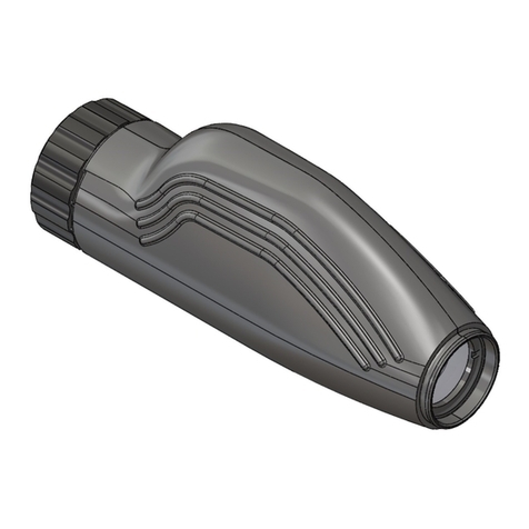
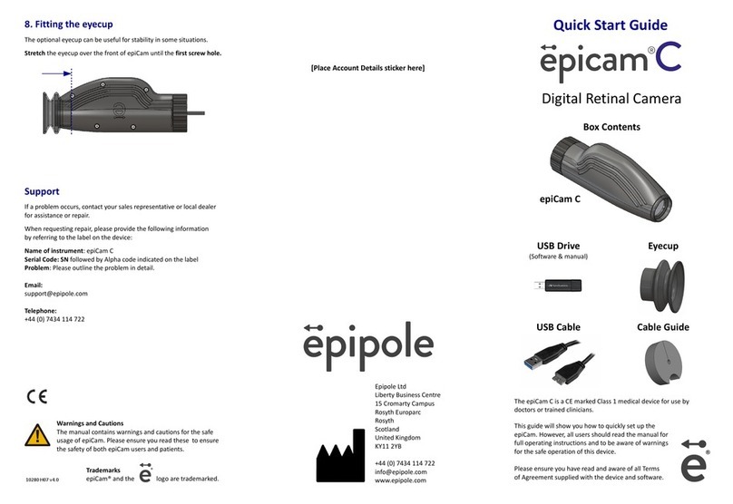
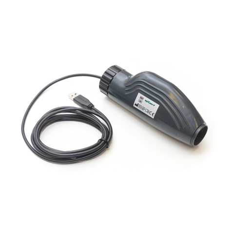
Epipole epiCam User manual

Digital Retinal Camera
epiCam
Please ensure you keep this manual in a safe place.
Read before operating the device.
epiCam H3 version 1.0

Table of Contents
1 Features......................................................................................................................................................
2 Components...............................................................................................................................................5
3 Instructions for use....................................................................................................................................7
3.1 Software installation...........................................................................................................................7
3.2 Connecting the epiCam......................................................................................................................9
3.3 Capturing images................................................................................................................................9
3. Reviewing patient images.................................................................................................................10
3.5 Synchronise with the epipole cloud service.....................................................................................10
3.6 After use...........................................................................................................................................10
3.7 Cleaning............................................................................................................................................10
Technical specifications and performance...............................................................................................11
.1 Intended use.....................................................................................................................................11
.2 Main specifications...........................................................................................................................11
.3 Environment of use..........................................................................................................................11
. Declaration of Conformity................................................................................................................12
.5 Software licenses..............................................................................................................................13
.6 Safety information ...........................................................................................................................1
5 Repair and return information.................................................................................................................16
6 Product failure..........................................................................................................................................16
7 Manual updates.......................................................................................................................................16
Page 2 of 17 epiCam H3 version 1.0

NOTE
1. Please contact your sales representative or local epiCam dealer if you require installation for your instrument.
2. Use a computer, monitor, and other equipment that conforms to the system standard IEC 60601-1/
UL60601-1 or IEC 60950-1/UL 60950-1 for the epiCam. Be sure that the entire system conforms to IEC 60601-1-1/UL
60601-1-1. Be sure to use an isolating transformer conforming to IEC60601-1/UL 60601-1 when a computer or
monitor conforming to IEC 60950-1/UL 60950-1 is used. For details, consult your sales representative or local epiCam
dealer.
3. To maintain compliance to IEC 60601-1-2, it is recommended that the computer, monitor, and other equipment
configured with the epiCam be evaluated to IEC 60601-1-2
. The user is responsible for the use and maintenance of the product. We suggest that a dedicated individual is
assigned responsibility for maintenance to ensure that the product is kept in good condition and can be used safely.
Medical products should only be used by a doctor or trained clinician.
5. It is unlikely but possible that this product may malfunction due to electromagnetic waves caused by mobile phones
or other radio wave generating devices. Be sure to avoid having objects such as these brought near to the product.
6. Disposal of this product in an unlawful manner may have a negative impact on human hea lth or on the environment.
When disposing of this product, therefore, be absolutely sure to follow the procedures which conform with the laws
and regulations applicable in your area.
7. Reading of images and storage of data must be performed in accordance with the law of the country where the
product is being used. Also, the user is responsible for maintaining the privacy of image data.
8. The cable supplied is designed to be used solely with this camera. Do not use it for any other product.
9. epipole Ltd reserves the right to change the specifications, configuration and appearance of the product without
prior notice. Updates may be found on the epipole website at http://www.epipole.com
European Union (and EEA*) only
This symbol indicates that this product is not to be disposed of with your household waste,
according to the WEEE Directive (2002/96/EC) and your national law. This product should be
handed over to a designated collection point, e.g., on an authorized one-for-one basis when
you buy a new similar product or to an authorized collection site for recycling waste electrical
and electronic equipment (EEE). Improper handling of this type of waste could have a possible
negative impact on the environment and human health due to potentially hazardous
substances that are generally associated with EEE. At the same time, your cooperation in the
correct disposal of this product will contribute to the effective usage of natural resources. For
more information about where you can drop off your waste equipment for recycling, please
contact your local city office, waste authority, approved WEEE scheme or your household
waste disposal service.
*EEA: Norway, Iceland and Liechtenstein
© EPIPOLE LTD 2015
Page 3 epiCam H3 version 1.0

1 Features
The epiCam is used to examine the retinas of patients’ eyes and take monoc romatic digital images of
them. The device has the following features:
Bot mydriatic and non-mydriatic use
epiCam may be used as a mydriatic or non-mydriatic camera without significant adjustment.
epiCam can take images through dilated or undilated pupils with a diameter of at least mm with a up to
35° field angle. We would normally recommend pupils over 5mm for wider fields.
The system may not be suitable for the following:
•neonate or premature babies.
•sufferers of albinism.
•those with severe degenerative retinal diseases.
•those of great age.
•those whose focal prescription is beyond +/- 6D.
•those with very high levels of retinal pigmentation.
Portable, and- eld, user-friendly design
Panning and tilting with the device is simple with such a compact, lightweight camera design.
The compact body makes it easier to work with patients and provides ease of operation, enabling the
clinician to significantly improve alignment.
Digital upload and processing
With the simple-to-use control software, images sessions photographed by epiCam can be browsed,
processed, stored locally or uploaded to epipole's proprietary Cloud storage.
EpiCam software
The epiCam Patient Manager provides a simple interface for managing patients and image captures. The
software is used to view the live image feed from the epiCam, adjust camera settings and save digital
images. The view can be adjusted to fit the window size and the software provides an indication of focus.
Page epiCam H3 version 1.0

2 Components
Contents in the box
Epicam
USB drive
Includes: the operation manual
epiCam Patient manager software
Carry case
For safe transportation of the device
Quick start guide
Includes details of the username and password for epipole
secure cloud storage account
USB cable
Type: USB 2.0 A to micro-B used for data and power (1.8m)
Page 5 epiCam H3 version 1.0

Components of epiCam:
Page 6 epiCam H3 version 1.0

3 Instructions for use
3.1 Software installation
System requirements
The epiCam Patient Manager requires:
•A personal computer (PC) with a 32-bit or 6 -bit x86 compatible processor
•Microsoft Windows Vista, Windows 7 or Windows 8/8.1
•At least 1 GB of RAM
•A free USB 2.0 (or 3.0) port
•As much free disk space as necessary for image storage and at least 8 MB for installation. Each
image can range in size from 500 to 1200kb and is store in png format.
•An active Internet connection to connect to epipole's cloud service
Limitations and restrictions
•The software can only operate a single epiCam. Do not attempt to use with more than one
device.
•The software product does not encrypt images or patient data stored on your PC system.
•You, the user, are responsible for safe and secure storage of all data.
•For best viewing it is recommended that the computer screen resolution is at least 1280 x 102
Be aware that the capture software may not display the feed from the camera at full-frame rate on
overloaded PC systems.
Installing t e software
•Insert the USB drive supplied with the epiCam into an USB port on your laptop or computer.
•The USB drive includes the installation file for the software (file ends in .exe). Run the installer by
double clicking on it.
•Accept the dialog to install the software.
•Read and agree to the license to use.
•Select where the software should be installed.
•A dialog will open asking for the username. This can be found on the card shipped with the
device. It is also printed on the silver label on the epiCam. The username is 6 characters long.
•When install is complete the Close button will become active:
•Once the software is installed, the following dialog will be display:
Page 7 epiCam H3 version 1.0

•The epiCam Patient Manager shortcut will be added to your Windows Start menu and an icon will
be added to your desktop.
•Launch the software. You will be prompted for a password which can be found on your Account
Card.
•The epiCam Patient Manager will open, this will enable you to add,edit and delete patient details
before taking images of their eyes. It is also used to synchronise (sync) images with epipole's
secure image storage service.
Add patient
Page 8 epiCam H3 version 1.0

The “Reference” field is optional for your own use. Each patient is given a unique Patient ID by the
software which is used to label the folder where that patient's images are stored in your computer.
Delete patient
This will delete the patient record from the Patient Viewer. It is not possible to undo this action or to
restore the patient record. Deleting a patient record will not remove images from your computer.
Edit patient
This enables you to change patient details. It does not make any changes to the folders where the
images are stored on your computer.
3.2 Connecting t e epiCam
To capture images, connect the epiCam to your computer using the USB cable. Do not touch the
objective lens whilst connecting or disconnecting the epiCam as any dirt, fingerprints, dust, or other
foreign objects on the objective lens could appear as artefacts in captured images.
Sudden changes in temperature may cause condensation to form on the objective lens or on optical
parts inside the instrument. In this case, wait until condensation disappears before using the device.
CAUTION Before connection or disconnecting the cables, be sure to hold the epiCam firmly to ensure safety.
Otherwise the main unit may fall over, causing possible injury.
NOTE To ensure epiCam can be detected by your laptop/ PC, you must use only use cables with type A to
micro-B connector plug supporting USB 2.0 Hi-Speed, maximum length of 3 meters.
•Insert the USB cable into the USB port on the epiCam.
•Insert the USB cable connector into an USB port on your laptop or desktop PC.
•The Status LED will begin to flash.
3.3 Capturing images
•In the epiCam Patient manager, add a new patient entry or select an existing entry and click on
the patient ID.
•Select Capture left eye / Capture rig t eye as appropriate
•The epiCam viewer will launch. The Status LED will flash faster as images begin to stream.
•Move the epiCam towards the patient's eye until retinal tissue becomes visible. The correct
working distance is 13mm from the corneal surface.
•Adjust the focus by turning the focus ring left or right
◦The epiCam Viewer has a Focus indicator to aide users. The automatic assessment shown by
this indicator is only approximate and so the user's own judgement should be used. Images
can be improved by manually altering the camera sensor parameters – in most cases altering
Exposure and Gain will be sufficient.
•Locate area to be imaged
◦The most obvious structure in the retina is the optic disk which appears as a large, bright
circular object in the retina. Navigating relative to this structure is generally the most
straightforward way of locating the required location.
◦Rotate the epiCam slowly from side to side and up-and-down to locate the area of interest.
◦Ask the patient to locate a distant object and gaze at this to prevent their eye from
Page 9 epiCam H3 version 1.0

wandering.
•Capture the image
◦Save image will save the image currently displayed on screen. You can also press your
computer spacebar
◦Close the patient viewer when you have captured all the images wanted for this eye. Return
to the epiCam Patient Manager software and repeat the steps above to capture images of
the other eye or for another patient.
3.4 Reviewing patient images
Patient images are stored in individual folders identified by epiCam Patient ID within the directory
selected at install. Patients' personal details are not used in the naming of folders to ensure that data
synced to the epipole servers is anonymous. The only way to identify the folder is to link the Patient ID
with the information in the Patient Manager – personal details entered here are only stored locally on
the laptop/PC.
Each folder contains subfolders for each eye.
3.5 Sync ronise wit t e epipole cloud service
epipole provide secure storage of images on our servers. To upload images select Sync from the epiCam
Patient Manager window. You will be asked to enter your password the first time you access the cloud
service. Syncing will securely transmit images to the server. The data is only identified using the epiCam
patient ID – we do not store patient name, birthdate or your reference on our servers.
3.6 After use
After imaging is finished and the software is closed, the device will enter standby.
It is recommended that the epiCam is disconnected from the PC/laptop and stored within the carry case
when a session is completed to protect both the device and computer from accidental damage. Do not
pull on the USB cable as this can cause damage – remove the cable by holding onto the USB plug.
3.7 Cleaning
•If you select to use a blower to clean the objective lens, do not let the tip of it touch the objective
lens.
•Do not wipe off or rub the objective lens when there is dust or other substances on it as this
could scratch the lens surface.
•Never wipe the objective lens with disinfecting ethanol, eyeglass lens cleaner, or cleaning paper
containing silicon as the lens surface could be damaged or the surface may not be completely
wiped off.
Page 10 epiCam H3 version 1.0

4 Tec nical specifications and performance
4.1 Intended use
The device is intended to be used for taking digital images of the retina of the human eye.
For European Union
Intended use:
This medical device is intended to observe and record images of retinal fundus through the pupil without
making contact with patient's eye. The purpose of these images is for differentiation between healthy
and unhealthy tissue and the identification of unexpected phenomena.
4.2 Main specifications
Field angle
Non-mydriatic Min 35° x 35°
Magnification view 1.2 x image size on the sensor
Diameter of pupil required
Non-mydriatic ø mm or more
Working distance 13mm
Focus adjustment range -15D to +15 D
Illuminant intensity 2000 mCd at source
Illuminant wavelength 589mm
Camera sensor Y800 format 1280 x 102
Moving range
Panning
Tilting
30° to the right and left
15° up and 10° down
Rated power supply USB 2/3 5V@500mA
Dimensions 63 × .5 × 152.7 mm
Weight 170g (not including cable)
NOTE The International System of Units (SI), the expression 1 D (diopter) = m-1
NOTE Technical specification is sub ect to modification without prior notice
4.3 Environment of use
Use, store, and transport the camera in an environment that satisfies the following requirements. Use
the supplied hard case to store or ship it.
In use Storage and transportation
Temperature 10°C to 35°C –30°C to 50°C
Humidity 30% to 90% RH (no condensation) 10% to 95% RH (no condensation)
Atmospheric pressure 700 hPa to 1060 hPa 700 hPa to 1060 hPa
Do not install or store the camera or leave it standing in an adverse environment where the temperature
or humidity levels are high. Doing so may cause mis-operation and/or malfunction.
Dust in the air may attach to the objective lens and to the optical parts inside the instrument. This will
affect the quality of images that can be taken. Keep the environment of use dust free.
Page 11 epiCam H3 version 1.0

4.4 Declaration of Conformity
Page 12 epiCam H3 version 1.0

4.5 Software licenses
The epiCam software:
•uses libpng licensed under the libpng license.
•uses zlib licensed under the zlib license.
•includes software developed by the OpenSSL Project for use in the OpenSSL Toolkit
(http://www.openssl.org/)
Page 13 epiCam H3 version 1.0

4.6 Safety information
Follow the safety instructions in this manual and all warnings and cautions printed on the warning labels.
Ignoring such cautions or warnings while handling the product may result in injury or accident. Be sure
to read and fully understand the manual before using this product. Keep this manual for future
reference.
Meaning of Caution Signs
To protect the safety of users and others and to prevent accidents, this operation manual utilizes the
symbols and text shown below in warnings and cautions. Read the meanings of these caution signs and
the safety precautions (see page 5 onwards), and follow the safety instructions.
WARNING This indicates a potentially hazardous situation which, if not heeded, could result in death or serious
injury to you or others.
CAUTION This indicates a hazardous situation which, if not heeded, may result in minor or moderate injury to
you or others, or may result in machine damage.
NOTE This is used to emphasize essential information.
Be sure to read this information to avoid incorrect operation.
Safety precautions
Be sure to follow the safety instructions below to ensure correct operation of the instrument.
Installation and Environment of Use
WARNING Do not use or store the instrument near any flammable chemicals such as alcohol, thinner, or benzine.
If chemicals are spilled or evaporate, it may result in fire or electric shock through contact with
electric parts inside the instruments.
Also, some disinfectants are flammable. Be sure to exercise caution when using them.
CAUTION Do not use or store the instrument in a location with the conditions listed below. Otherwise, it may
result in failure or malfunction, fall or cause fire or injury.
•Close to facilities where water is used.
•Where it will be exposed to direct sunlight.
•Close to air-conditioner or ventilation equipment.
•Close to heat source such as a heater.
•Surfaces or areas prone to vibration.
•Dusty environment.
•Saline or sulphurous environment.
•High temperature or humidity.
•Freezing or condensation.
CAUTION Do not cover the vent holes in the cover.
Otherwise, the temperature in the instrument may rise and cause fire.
Installation Operation
WARNING Do not connect the instrument except in the manner specified. Otherwise, fire or electric shock may
result.
Also, when the instrument is going to be connected to other equipment using the cable, be sure that
leakage current is within the tolerable value. For details, please contact your sales representative or
local epiCam dealer.
Page 1 epiCam H3 version 1.0

Power Supply
WARNING Do not cut or process the cable. Do not place heavy objects on, step on, pull or bend the cable.
Otherwise, the cable may be damaged, which may result in fire or electric shock.
Handling
WARNING Never disassemble or modify the product as it may result in fire or electric shock.
WARNING Do not place anything on top of the instrument.
If metal objects such as a needle are jammed into the instrument, or if liquid is spilled on it, it may
result in fire or electric shock.
WARNING When the instrument is going to be carried, be sure to remove the cable and pack
the instrument in its appropriate box, bag or cover (as appropriate).
Do not hold the device by the cable or eyepiece as it may detach resulting in damage.
WARNING When the instrument is going to be carried, be sure to remove the cable and pack
the instrument in its appropriate box, bag or cover (as appropriate).
Do not hold the device by the cable or eyepiece as it may detach resulting in damage.
CAUTION To prevent the risk of infection, wipe contact surfaces with disinfectant ethanol for each patient. For
details on how to disinfect, consult a specialist.
If a problem occurs
WARNING Should any of the following occur, immediately unplug the instrument and contact your sales
representative or local epiCam dealer.
•When there is smoke, an odd smell or abnormal sound.
•When liquid has been spilled into the instrument or a metal object has entered through an
opening.
•When the instrument has been dropped and it is damaged.
Maintenance and inspection
WARNING For safety reasons, be sure to unplug the device when the inspections indicated in this manual are
going to be performed. Otherwise, electric shock may result.
WARNING When the instrument is going to be cleaned, be sure to unplug the instrument.
Never use alcohol, benzine, thinner or any other flammable cleaning agents.
Otherwise, fire or electric shock may result.
WARNING Clean the plug of the cable periodically by unplugging it and removing dust or dirt from the plug, its
periphery and USB inlet with a dry cloth.
If the cable is kept plugged in for a long time in a dusty, humid or sooty place, dust
around the plug will attract moisture, and this could cause insulation failure which
could result in a fire.
WARNING The instrument must be repaired by a qualified engineer only.
If it is not repaired properly, it may cause fire, electric shock, or accident.
System use
WARNING Do not simultaneously touch a patient and non-medical electrical equipment.
Otherwise, electric shock may result.
CAUTION This instrument utilizes a CCD sensor, if any pixel becomes full activated [white] or fully deactivated
[black] at all times, please return for repair.
Page 15 epiCam H3 version 1.0

5 Repair and return information
Repair
If a problem occurs, contact your sales representative or local dealer for repair.
When requesting repair, please provide us with the following information by referring to the label on the
device:
Name of instrument: epiCam
Serial number on the instrument: Alpha code indicated on the label
Problem: Please outline the problem in detail.
Limit for supplying performance parts for repair
Parts (parts required to main the functioning of the product) of this product will be stocked for 5 years
after production ceases to allow for repair.
6 Product failure
Out of box failure
If your device is not functioning on receipt or fails within 3 days of arrival, please contact us either via
our website or using the contact information below.
Incorrect functioning
If your device is not functioning correctly on receipt or within 3 days of arrival, please contact us either
via our website or using the contact information below.
Contact support:
http://www.epipole.com/contact
Support:
+ (0) 7 3 11 622
7 Manual updates
Updates to this manual and epiCam software will be available to download on www.epipole.com. It is
recommended that users check to ensure they always have the latest updates.
Page 16 epiCam H3 version 1.0

epipole Ltd
Liberty House Business Centre
15 Cromarty Campus
Rosyth Europarc
Rosyth
Scotland
United Kingdom
KY11 2YB
Made in Scotland
Page 17 epiCam H3 version 1.0
Table of contents
Other Epipole Digital Camera manuals
