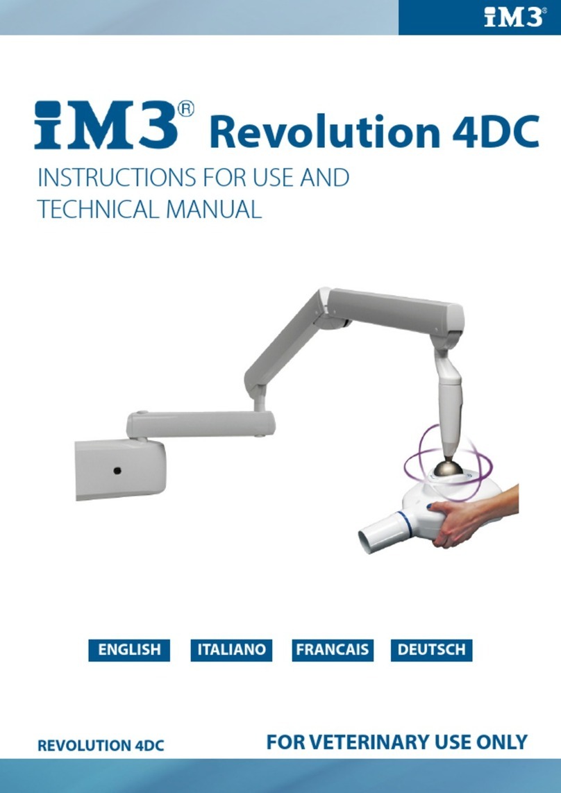
iM3 REVOLUTION 4DC User manual

Revolution 4DC
97050748
rev. 002
07/2014
EN
IT
FR
DE
中文


OPERATING INSTRUCTIONS
EN OPERATING INSTRUCTIONS
CONTENTS
1. General precautions .......................................................................................................................................................4
1.1. Description of the symbols used ................................................................................................................................4
1.2. Purpose and use of the equipment ............................................................................................................................5
1.2.1. Classication..................................................................................................................................................5
1.2.2. Environmental conditions ...................................................................................................................5
1.2.3. Guarantee...........................................................................................................................................5
2. Description of the system ........................................................................................................................................6
2.1. Description of the x-ray unit ............................................................................................................................6
2.2. Operating mode ..............................................................................................................................................6
3. Operation..................................................................................................................................................................7
3.1. Turning the x-ray unit on and off ...................................................................................................................7
3.1.1. Turning on the basic X-ray unit...........................................................................................................7
3.1.2. Turning on the handheld.....................................................................................................................7
3.1.3. Automatic handheld shut off ..............................................................................................................8
3.1.4. Handheld stand-by according to time .................................................................................................8
3.2. Handheld display functions .............................................................................................................................8
3.3. Control panel...................................................................................................................................................8
3.4. Checking the parameters................................................................................................................................9
3.5. Factory settings.............................................................................................................................................10
3.6. Batteries and charge level indication ...........................................................................................................10
3.7. X-ray generator indicator light.......................................................................................................................10
4. Using the x-ray unit ................................................................................................................................................11
4.1 Position of the patient ....................................................................................................................................11
4.2. Positioning the x-ray head .......................................................................................................................... 11
4.2.1. Hypersphere technology...................................................................................................................11
4.3. Setting the exposure mode and time ...........................................................................................................12
4.4. Setting the mode and exposure time in USER mode ...................................................................................12
4.5. Procedure to be followed when taking the x-ray..........................................................................................13
5. Advanced options ...................................................................................................................................................14
5.1. Setting the safety unlock mode ....................................................................................................................14
5.2. Setting the operating mode ..........................................................................................................................14
5.3. Restoring factory settings ............................................................................................................................15
6. Error messages ......................................................................................................................................................15
7. Routine maintenance..............................................................................................................................................16
8. Cleaning and disinfection .......................................................................................................................................16
9. Dismantling the equipment when no longer used...................................................................................................17
10. Technical specications........................................................................................................................................17
11. Dimensions ...........................................................................................................................................................18
12. Identication nameplates......................................................................................................................................20

4EN
4OPERATING INSTRUCTIONS
1. General precautions
These instructions explain how to properly use the x-ray unit. Please carefully read and become familiar with the content of this manual
before attempting to use the apparatus.
NOTE: This manual does not specify the rules and regulations for acquiring, possessing or using a source of ionizing radiation as
each country has its own laws. Only the most common ones shall be mentioned and this means that it is the user’s responsibility to
check local standards and observe the relevant laws.
No part of this manual is to be reproduced, stored in a retrieval system or transmitted in any form or by any means, i.e. electronic, mechanical, photocopying,
translation or otherwise, without the prior written permission of the manufacturer.
CEFLA SC - CEFLA DENTAL GROUP has a policy of ongoing development and is committed to improving its products. Therefore, some instructions,
specications and illustrations given in this manual may differ slightly from the product actually purchased. The manufacturer reserves the right to
make changes to this manual without giving prior notice.
The original version of this manual is written in Italian. Translation from the original in Italian
1.1. Description of the symbols used
The following symbols are used:
WARNING!
Indicates a situation where failure to observe the instructions could cause damage to the
equipment or injury to the user and/or patient.
NOTE:
Indicates important information for the user and/or technical support personnel.
Earth connection.
Alternating current.
On.
Off.
ionizing radiation.
237835
©
Equipment abides by the essential requirements established by USA and Canada.
FCC ID F.C.C. mark (Federal Communication Commission)

5
EN 5
OPERATING INSTRUCTIONS
1.2. Purpose and use of the equipment
This x-ray unit is designed for use in the dental veterinary surgery to make endo-oral x-rays for diagnostic purposes.
This equipment can be used to produce traditional x-rays developed using chemicals or, alternatively, it can be used with digital x-ray sensors.
ATTENTION!
This x-ray unit is FOR VETERINARY USE ONLY.
This x-ray unit is NOT FOR HUMAN USE.
1.2.1. Classication
RADIO EQUIPMENT AND TELECOMMUNICATIONS TERMINAL EQUIPMENT Classication
Equipment classication in accordance with directive 99/05/EEC art.12: Class I.
EMC classication
Classication of the equipment according to standard CEI EN 55011: Group 1 - Type B.
1.2.2. Environmental conditions
The equipment should be installed in an area where the following conditions are present:
• Temperature from +10° to +40° C;
• relative humidity from 25 to 75% without condensate;
• atmospheric pressure from 700 to 1060 hPa;
• The electrical wiring in the room where the equipment is installed must conform to the I.E.C. 60364-7-710;V2 specication (i.e. the regulations
concerning the electrical wiring to be used in surgeries) or equivalent standards in force in the country where the equipment is installed.
• ELECTRICAL CONNECTIONS : The electrical wiring must have an effective ground conductor as set forth by I.E.C. – US National Electrical Codes
and C.E.I. standards. In Italy, electrical wiring must comply to standards IEC 60364-7-710 which require that a ground fault circuit interrupter is
installed upstream. The ground fault circuit interrupter must be as specied below:
- contact capacity: 250 V 10A in compliance with standards IEC 60898-1 and IEC 60947-2.
- differential sensitivity: 0.03A.
- power supply: 3 x 2.5 sq.mm
The color of the 3 wires should be as specied in the standards (brown power, BLUE neutral, YELLOW/GREEN ground).
1.2.3. Guarantee
CEFLA SC - CEFLA DENTAL GROUP guarantees the safety of the operator as well as the reliability and high performance of the equipment. The
guarantee shall be voided if the following precautions are not taken:
• Observe the conditions specied in the guarantee certicate itself.
• The equipment must only be used by following the instructions contained herein.
• Equipment installation, expansion and technical support must be performed exclusively by personnel authorized by the manufacturer to carry out
these operations.
• Never open the equipment casing. Installation, repairs and, in general, any other operations requiring the casing to be opened are to be performed
exclusively by personnel authorized by the manufacturer to carry out these operations.
• The equipment must be installed in areas which comply with that stated above at point 1.2.2 “Ambient Conditions”.
• The area where the x-ray unit is installed must comply with ofcial regulations regarding protection against radiation in the country where the
equipment is used.
SAFETY PRECAUTIONS
• If any person who is not an authorized technician changes the product in any way by replacing parts or components with other ones not used by
the manufacturer they shall assume responsibility for the product.
• Do not forget to turn off the main switch on the equipment before leaving the surgery.
• This equipment is not water-proof (risk of electrocution).
• This equipment is not suitable for use in the presence of a mix of inammable anaesthetic gas with oxygen or nitrous oxide.
• This equipment must be stored properly so that it is kept in top working order at all times.
• Use of electric scapels or other electric apparatus that do not comply to standard I.E.C. 60601-1-2, in the doctor’s ofce or in the immediate vicinity
of it may cause electromagnetic interferences or interferences of other types, resulting in equipment malfunctions. In these cases shut off the
power supply to the equipment before hand.
• The manufacturer shall not be held responsible for misuse, carelessness or improper use of the equipment.
• This equipment is to be used exclusively by qualied personnel with the proper medical training.
• The user must be present at all times when the equipment is turned on or ready for start-up. In particular, never leave the equipment unattended
in the presence of children/the mentally disabled or other unauthorised personnel in general.
• If the x-ray equipment is damaged or oil leaks, do not use the equipment and contact customer service immediately.
Protection against radiation
X-rays are hazardous and adequate precautions must be taken when using them. Areas where it is possible to be exposed to the x-rays
shall be clearly indicated by using this symbol which should remind personnel to observe the safety rules laid down by the laws in force
in the country where the equipment is used.
• Control the emission of x-rays from the greatest distance possible (several metres) from the focal spot and the X-ray irradiation beam in the
opposite direction to where the rays are emitted.
• Only the authorized personnel and the patient can remain in the area when x-rays are being emitted.
• Always protect the patient’s thyroid and gonads under all circumstances.

6
EN
6OPERATING INSTRUCTIONS
2. Description of the system
2.1. Description of the x-ray unit
Description of the parts:
aX-ray generator.
Depending on the operating mode, the high frequency constant po-
tential x-ray generator works at 60KV 7ma (En60 mode, 63KV 6ma
(En63 mode) or 65KV 6ma (En65 mode).
The generator can turn on a horizontal plane endlessly; on the other
hand, as far as vertical movement is concerned, rotation is limited
upwards by a mechanical end-stop.
b Removable cone.
The generator can work with different types of collimators that are
automatically recognized:
• 8” ROUND COLLIMATOR (incorporated in the generator): mini-
mum skin/focus distance 20 cm and 60 mm output beam
• REMOVABLE 12” round CONE: minimum source/skin distance
30cm and diameter of x-ray beam exiting cone 55mm (with cone
attached).
cFocus spot.
d Double pantograph arm.
eExtension arm.
The extension is available in three lengths: 40 cm, 60 cm and 90 cm.
f Handheld.
The handheld can be placed either near the control unit or in a remote
position. As a result, the doctor can move conveniently around the
room and move out of the area where x-rays are emitted.
g Hand held holder.
h Control unit.
i Master switch.
2.2. Operating mode
The x-ray unit can function in different modes (MULTI-MODE technology):
1) En60 operating mode
X-ray emission at 60KV and 7ma. The X-ray unit automatically
calculates the best exposure time (from 0.01s to 1.00s) based on the
selected tooth and patient size.
2) En63 operating mode
X-ray emission at 63KV and 6ma. The X-ray unit automatically
calculates the best exposure time (from 0.01s to 1.00s) based on the
selected tooth and patient size.
3) En65 operating mode
X-ray emission at 65KV and 6ma. The x-ray unit automatically
calculates the best exposure time (from 0.01s to 1.00s) based on the
selected tooth and patient size.
4) AUTO operating mode
The x-ray unit automatically suggests the best operating mode (En60,
En63 o En65) and exposure time (from 0.01s to 1.00s) based on the
selected tooth and patient size.
The suggested exposure time can be corrected from the handheld.
In addition, the x-ray unit has a special USER mode that can be set
by the user. The user can select the best combination of load factors
(operating mode and exposure time) for each tooth and patient size.

7
EN 7
OPERATING INSTRUCTIONS
3. Operation
3.1. Turning the x-ray unit on and off
3.1.1. Turning on the basic X-ray unit
The control unit is turned on and shut off with switch (A).
The switch lights up to signal the control unit is energized.
NOTE: The technical specications of the switch are given
in paragraph 1.2.2.
The control unit is turned on and off with the main switch
(A), as illustrated in the gure below. The switch lights up
when the control unit is energized. Whenever turned on,
the equipment performs an operational test that takes a few
seconds. A beep is provided at the end of the test.
NOTE: The exposure time and the parameters displayed
when the unit is turned on are the last ones set before the
central control unit was turned off. If the central control
unit is left untouched for a few minutes it will go into stand-
by mode. Simply press any key on the control panel to
reactivate it.
3.1.2. Turning on the handheld
The handheld is turned on by pressing any key, except for the one for
x-ray emission. A buzzer rings to conrm the apparatus has been turned
on. The unit will be in the standard conguration described in detail in
paragraph 3.1.3 and then search for the base it works with.
If the base is off, the handheld will not indicate the eld nor status
“ready”. If the base is latter turned on, the handheld will detect it within
thirty seconds or by pressing any function key on the pushbutton panel.
NOTE:
To optimize the range of the handheld while it is being used,
keep it away from walls and metal instruments and above
all, do not cover its antenna found on top of the screen. In
addition, performance may be reduced if the handheld is
moved too quickly while x-rays are being taken. Error E 31
may be displayed if out of range.

8EN
8OPERATING INSTRUCTIONS
3.1.3. Automatic handheld shut off
Once the control unit has been turned off the handheld automatically shuts off after approximately one minute. The handheld also automatically shuts
off when it is further than the maximum allowable distance away from the control until.
3.1.4. Handheld stand-by according to time
The entire x-ray unit will switch over to stand-by (even if the base is on) and the handheld will automatically shut off after approximately ve minutes
of non-use to save battery power. The handheld turns back on displaying the last selection made by the user whenever any key, except for the X-ray
emission key, is pressed. To edit the standby time, refer to chapter 4 that deals with the handheld’s “Advanced options”.
3.2. Handheld display functions
3.3. Control panel
As illustrated in the gure below, the handheld has four function keys and a single x-ray emission key.

9
EN 9
OPERATING INSTRUCTIONS
The main functions of the keys on the handheld vary according to how they are pressed:
KEY BRIEFLY PRESSED (less than 3 sec.) PRESSED LONGER (more than 3 sec.)
Changes over from LARGE to SMALL and vice
versa (takes place when key is released).
Saves the selected setting (exposure time, sensitivity, etc…).
The memo icon ( ) lights up when the data item can be
saved.
Changes amongst the various types of teeth to se-
lect the area to be examined.
It displays the values corresponding to the tooth exposure ti-
mes in mGy and in mGy*cm2 if pressed again.
Increases the exposure times in steps, according
to the set scale.
Increases the scroll speed of the values in increasing order.
Increases the exposure times in steps, according
to the set scale. Increases the scroll speed of the values in decreasing order.
NO EFFECTS ARE OBTAINED IF THE KEY IS
PRESSED LESS THAN A SECOND.
STARTS X-RAY EXPOSURE (the button has to be held down
while the x-rays are being emitted, “dead man” function).
NOTE: “Dead man” function: the system that starts x-ray exposure with the dedicated key on the wireless handheld allows x-rays
to be emitted only when the user presses and holds down the exposure key. X-ray emission will stop if the key is released ahead
of time.
NOTE: The function related to pressing the key briey is performed by pressing the key which will activate the function assigned to
it. On the other hand, to perform the function carried out when the key is held down longer, press the key until the relative function
is started. The buzzer will ring shortly to signal the function has started.
NOTE: Warm-up: When the equipment has not been used for a prolonged period (more than 3 months) or when turned on for the
rst time, a number of emissions with short times (0.01-0.02 sec.) are recommended and then some pictures with 0.1 sec. intervals
to better stabilize operation of the x-ray tube before using it.
3.4. Checking the parameters
Before actually taking an exposure, make sure the exposure parameters for the examination in progress are correctly set.
• Checking the selected body build.
- “SMALL” selected: indicates the x-ray unit is set for patients with small builds.
- “LARGE” selected: Indicates the x-ray unit is set for patients with average-large builds.
NOTE: After the change has been made, the preset exposure times will automatically be modied.
Small build
selected
Average/large build
selected

10
EN
10 OPERATING INSTRUCTIONS
• Checking the selected type of intraoral exam.
Upper molars Lower incisors
Upper canines/bicuspids or rear ”bitewing” Lower canines/bicuspids
Upper incisors or front ”bitewing” Lower molars
3.5. Factory settings
The x-ray unit is supplied with the following factory settings:
• Operating mode: AUTO.
• Sensitivty: level 19.
• Handheld stand by: 5 minutes
• Exposure times as per standard R20: 0,010 - 0,011 - 0,012 - 0,014 - 0,016 - 0,018 - 0,020 - 0,022 - 0,025 - 0,028 - 0,032 - 0,036 - 0,040 - 0,045 -
0,050 - 0,056 - 0,063 - 0,071 - 0,080 - 0,090 - 0,100 - 0,110 - 0,125 - 0,140 - 0,160 - 0,180 - 0,200 - 0,220 - 0,250 - 0,280 - 0,320 - 0,360 - 0,400
- 0,500 - 0,560 - 0,630 - 0,710 - 0,800 - 0,900 - 1,000.
NOTE: These times comply with current standards I.E.C. 60601-2-7 (1999) and the ISO 497 series R’20 recommendations and
CANNOT BE MODIFIED.
3.6. Batteries and charge level indication
The handheld runs on two widely available AA alkaline batteries to assure sifcient stand-alone operation. The charge level of the batteries is given
on the screen as follows:
Battery fully charged (a symbol does not appear in the area that shows the battery charge level).
Battery half-charged.
Battery charge level low or almost dead (causing the handheld to automatically shut off).
NOTE: The batteries should be removed from the handheld if it is not going to be used for an extended period.
3.7. X-ray generator indicator light
The x-ray generator comes with an indicator light (B) that signals apparatus status:
• color (purple) > x-ray unit on (regular condition)
• Flashing color (purple) > stand-by (low consumption)
• Blue > x-ray one – head released
• Yellow > x-rays being emitted
• Red > fault

11
EN 11
OPERATING INSTRUCTIONS
4. Using the x-ray unit
4.1 Position of the patient
A positioner or alignment device specic for the selected image receiver should always be used to assure the x-rays are correctly aligned regardless
of the position the patient’s head is in.
4.2. Positioning the x-ray head
Position the x-ray head so that the cone is aligned with the image receiver.
4.2.1. Hypersphere technology
Hypersphere technology allows the x-ray head to turn endlessly on both the horizontal and vertical planes.
The x-ray head is initially blocked by an electromechanical brake.
The head can be tilted into the position required to take the x-ray by touching the unlock areas. To lock it again, release the unlock areas.
NOTE: Firmly hold the head with both hands when putting it in place.
It is possible to set a safety unlocking mode that allows the head to be turned only by pressing both unlock buttons. This prevents the head from
unlocking unexpectedly after one of the two unlock buttons has been accidentally pressed. To activate this mode, refer to “Advanced options” in
chapter 5.
Release area Release area

12 EN
12 OPERATING INSTRUCTIONS
4.3. Setting the exposure mode and time
The exposure parameters are set by following the directions given below:
1) select the tooth to be examined
2) select the patient size
The exposure time is automatically shown on the handheld screen.
NOTE: each tooth and patient size selected is displayed for approximately 1 second according to the operating mode (En60, En63
or En65) used.
The suggested exposure time can be changed with keys and . Exposure times ranging from 0.01s and 1.00s belonging to the R’20
scale can be set. Random exposure times different from the ones provided in the R’20 scale cannot be set.
When the exposure time displayed differs from the default setting, icon comes on.
To save the new setting, make sure icon is on and then press and hold down key for approximately 2 seconds. The handheld will beep
to conrm the setting has been saved. At this point, make sure icon is off.
NOTE: if the exposure time is not saved, the change made will be lost after a new entry or as soon as the handheld changes over
to stand-by.
Important: after customized settings have been made, the “Original exposure values charts” are no longer valid.
If icon is displayed while the exposure time is changed, it means the set time cannot be saved for the selected tooth-patient size combination.
In any case, the x-rays can be taken with the set time.
Important: when the suggested exposure time is changed, the sensitivity factor is also modied (by default set to F=19). Once
this change has been saved, it is applied to all the teeth and both patient sizes
The exposure time can also be modied by changing the sensitivity factor. Press keys and at the same time , the actual sensitivity
factor will be displayed.
Use keys and to change the value from 3 to 25. If the displayed value differs from the one previously saved, icon comes on. To
quit this mode, press key or . The change made to the sensitivity factor is applied to all the teeth and both patient sizes.
The selected operating mode is always used for each tooth and patient size combination in modes En60, En63 and En65.
In AUTO mode, each tooth and patient size combination is associated to the best mode from amongst the ones available. In this mode it is not
possible to assign a mode other than the default one to each combination. To set the mode, refer to paragraph 4.5 “Setting the mode and exposure
time in USER mode”.
To change the mode amongst En60, En63, En65 and AUTO refer to paragraph 5.2 “Setting the operating mode”.
4.4. Setting the mode and exposure time in USER mode
In USER mode, it is possible to assign an exposure time and a mode from amongst En60, En63 and En65 to each tooth-patient size combination.
The default setting corresponds to the AUTOmode settings with sensitivity factor F=19
To activate USER mode regardless of the mode currently being used, press keys and at the same time. Icon will come on to
signal USER mode is active.
To deactivate USER mode press keys and again (icon goes off).

13
EN 13
OPERATING INSTRUCTIONS
The exposure parameters are set as directed below:
1) select the tooth under examination
2) select the patient size.
The exposure time is automatically displayed on the handheld.
NOTE: It is not possible to access the sensitivity factor menu in USER mode. In addition, keys and are inoperative
in User mode.
The exposure times and mode assigned to the tooth – patient size combinations are custom set by following the directions given below:
1) press and hold down key about two seconds. Customized settings can be entered and icon comes on.
2) select the desired tooth-patient size combination
3) change the exposure time with keys and .
NOTE: it is possible to set exposure times ranging from 0.01s and 1.00s that are part of the R’20 scale.
4) press keys and simultaneously to open the menu used to select the operating mode
5) select the operating mode with keys and
6) quit the menu and press key to make the entry operative (if key is pressed, the menu will be quit without changing the previous
setting).
7) press and hold down key for approximately two seconds to conrm the entry and disable customized settings (icon goes out).
NOTE: it is possible to set the exposure parameters for several combinations. To do this, repeat steps 2 to 6 before going on to
step 7.
4.5. Procedure to be followed when taking the x-ray
• Pick up the handheld and go a safe distance away (at least 2 meters) maintaining visual contact with the patient and x-ray unit during the
exposure. Make sure “READY” is indicated.
• Tell the patient to stay still.
• Press and hold down the “Exposure” key on the handheld until the audible warning sound (beep) stops and the yellow light goes out.
“X-ray emission” key
Light on control panel illuminated during x-ray emission
NOTE: If the “EMIT X-RAY” key is released at any time, exposure will be interrupted and error code E01 will appear on the display.
• Once exposure has been completed, it is possible to proceed with the next exposure unless the x-ray unit has reached the maximum allowable
temperature. The percentage the cone exceeds the maximum allowable temperature is always shown on the screen (see icon below).

14 EN
14 OPERATING INSTRUCTIONS
• Once the temperature has been reached, wait the pause time for cooling signaled by symbol .
• At this point the exposure function will be disabled until the screen shows “READY” again.
• As soon as “READY” appears on the handheld, another exposure can be taken.
5. Advanced options
The handheld allows the user to view, edit and set some operating parameters by simply combining the keys provided. Follow the steps given below
to access:
Key combination Description of command
+
Press these two keys to adjust the sensitivity levels (determined based on the table given below and type of
sensor/receiver used), modifying the current value from the minimum to the maximum allowable one (on a
scale from 3 to 25), with keys "+" and "-". Press key "size" to conrm the desired level and go back to the
main screen.
This menu is not available in USER mode.
+
Hold down these two keys to go to the set up menu (from P 01 to P 07).
Press key “Build” to make the selection. Once within the individual congurations, they can be scrolled with keys
“+” and “-” and selected by pressing key “Build” again. Key "tooth" quits set up without saving the setting.
The congurations are given in detail below:
P 01: Sets the stand by time (from a minimum of 5 to a maximum of 30 minutes).
P 02: Assigns an identication tag to the x-ray unit’s base (from 1 to 5 or none).
P 03: Shows the list of software versions-
P 04: Handheld code display
P 05: Activates/deactivates the safety unlock mode (see section 5.1)
P 06: Selects the operating mode (En60, En63, En65 and AUTO).
P 07: Sets the type of removable cone used
+ Activating/deactivating the USER mode Icon comes on to signal USER mode is activated.
5.1. Setting the safety unlock mode
The x-ray unit has a safety unlock for the ball joint.
The default setting allows the ball joint to be disengaged by simply touching one of the keys present on the front of the head. To prevent accidental
contact with the keys from unexpectedly disengaging the ball joint (and therefore causing undesired movement of the head), the safety unlock mode
can be activated. In this mode, the ball joint is disengaged only if both keys are activated at the same time.
To set the safety unlock mode, press keys and to go to the set up menu. Scroll the parameters up to parameter P05 and press key
. Scroll the options to select “ON” and press key . Press key to quit the set up menu.
5.2. Setting the operating mode
The x-ray unit features the following operating modes:
• En60: all the x-rays are taken at 60KV and 7mA.
• En63: all the x-rays are taken at 63KV and 6mA.
• En65: all the x-rays are taken at 65KV and 6mA.
• AUTO: the system automatically selects the best setting from amongst En60, En63 and En65 for each tooth-patient size combination.
NOTE: the current setting is displayed on the handheld for approximately 1 second for each tooth-patient size selected before the
relative exposure time is shown.
To set the operating mode, press keys and to go to the set up menu. Scroll the parameters up to parameter P06 and then press key
. Scroll the options to nd the desired operating mode and then press key . Press key to quit the set up menu.

15
EN 15
OPERATING INSTRUCTIONS
5.3. Restoring factory settings
To restore the factory settings (see paragraph 3.5) press keys and to go to the set up menu. Press keys and
simultaneously. "rESS" will briey appear and the handheld will be rebooted.
6. Error messages
NOTE: In presence of strong wireless communication trafc, the connection between handheld and generator may break down.
To restore the connection run the "Restoring factory settings" procedure.
FAULT CAUSE SOLUTION
E01 X-RAY KEY RELEASED TOO EARLY Hold down the key until the image has been captured.
E02 SHOOTING SEQUENCE NOT COMPLETED
Handheld most likely lost the signal. Try to repeat
exposure. If the problem persists, contact technical
service.
E03 HANDHELD INTERNAL TEST ERROR
Take out the batteries and then put them back in after
waiting a few seconds. If the problem persists, contact
technical service.
E04
E05
E08
HANDHELD AUTO DIAGNOSIS TEST FAILED Contact technical service.
E06 GENERAL HANDHELD ERROR Try to repeat exposure. If the problem persists, contact
technical service.
E07 RF SIGNAL TOO LOW Handheld lost the signal. Try to repeat exposure. If the
problem persists, contact technical service.
E09 HANDHELD SERIAL NUMBER INCORRECT OR NOT
INITIALIZED Contact technical service.
E10
E12
E13
E16
X-RAY UNIT INTERNAL ERROR Contact technical service.

16 EN
16 OPERATING INSTRUCTIONS
E11 COLLIMATOR SELECTION NOT CONSISTENT After turning the rectangular collimator on or off, wait a few
seconds to allow the icon on the handheld to be updated.
E14
E15 GENERIC GENERATOR FAULT Contact technical service.
E17 DEVICE OVERHEATED
HEAD RELEASED
WAIT APPROXIMATELY 15 MINUTES FOR AUTOMATIC
SYSTEM RESET
E18
E19 SUPPLY VOLTAGE TOO HIGH/LOW
CHECK THE SUPPLY SYSTEM.
IF THE PROBLEM PERSISTS, CALL TECHNICAL
SUPPORT.
E30 INTERNAL ADJUSTMENT PROBLEM REPEAT THE X-RAY. IF THE PROBLEM PERSISTS,
CALL TECHNICAL SUPPORT.
E31
E32 REMOTE CONTROL ERROR
REDUCE THE DISTANCE BETWEEN THE REMOTE
CONTROL AND X-RAY HEAD AND THEN REPEAT THE
X-RAY. FOLLOW THE INFORMATION GIVEN ON HOW
TO PROPERLY USE THE HAND HELD’S ANTENNA.
E33 NO POWER SUPPLIED TO X-RAY GENERATOR X-ray generator or arm cord may be faulty. Contact
technical support service..
As regards the other error codes, CONTACT the technical service department.
7. Routine maintenance
ATTENTION!
Any technical maintenance work required must be carried out by qualied personnel or by a specialised technician authorised by the
manufacturer.
It is the user’s responsibility to check that routine maintenance is carried out by an authorised technician at least every 2 years.
The maintenance methods are specied in the Technical Service Manual possessed by the Authorised Technicians.
8. Cleaning and disinfection
The x-ray unit may be a source of infection passed from one patient to another if it is not cleaned properly. For this reason it should be disinfected
on the outside every day after use.
If digital X-ray sensors are used make sure they are always used with disposable hygienic covers. When disinfecting the x-ray unit use soft
disposable paper and avoid using corrosive substances or immersing the unit in liquids.
To avoid damaging the plastic materials use products containing:
• Ethanole 96%:
Concentration: maximum 30 g per 100 g of disinfectant.
• Propanole:
Concentration: maximum 20 g per 100 g of disinfectant.
• Combination of ethanole and propanole:
Concentration: the combination of the two should be maximum 40 g per 100 g of disinfectant.
Compatibility tests between plastics and the following products have been carried out with no negative consequences:
• Incidin Spezial (Henkel Ecolab).
• Omnizid (Omnident)
• Plastisept ( ALPRO ) (not tuberculocide as not an alcohol-based disinfectant)
• RelyOn Virkosept ( DuPont).
• Green & Clean SK ( Metasys ) (not tuberculocide as not an alcohol-based disinfectant)
ATTENTION!
• Do not use products containing isopropyl alcohol (2-propanol, iso-propanol).
• Do not use products that contain sodium hypochlorite (bleach).
• Do not use cleaners that contain phenol.
• Do not spray the selected products directly on the surfaces.
• Never combine products with each other or with liquids other than the products listed above.
• All products must be used as directed by the manufacturer.
Cleaning and disinfecting instructions.
Use soft disposable paper towels or sterile gauze to clean and disinfect.
Do not use sponges or in any case, any material that can be reused.
ATTENTION!
• Shut off the unit prior to clean and disinfecting the external parts.
• Never lubricate the pivot point of the x-ray cone as proper operation of the locking system may be compromised.
• All material used to clean and disinfect must be thrown away.

17
EN 17
OPERATING INSTRUCTIONS
9. Dismantling the equipment when no longer used
As set out in Directives 2002/95/ EC, 2002/96/ EC and 2003/108/ EC, on the restrictions of the use of certain hazardous substances in electrical and
electronic equipment along with collection, treatment, recycling and disposal of waste electrical and electronic equipment the latter must be treated
as municipal waste, therefore sorted and collected separately. When new equipment of equivalent type is purchased the waste equipment should be
returned to the distributor on a one-to-one basis for disposal.
As far as reuse, recycling and other forms of waste recovery mentioned above are concerned, the manufacturer is responsible for the actions specied
by individual local laws.
Efcient collection of sorted waste separately to recycle and treat waste electrical and electronic equipment aids in preventing negative environmental
impacts while protecting human health. In addition it facilitates recycling of the materials used to construct the equipment.
The crossed out wheeled bin placed on the equipment indicates that the waste equipment must be collected separately from other waste.
ATTENTION!
Illegal waste disposal carries heavy nes dened by local laws.
10. Technical specications
SPECIFICATIONS
• Rated voltage: 230Vac/115Vac (according to the model).
• Max. mains voltage uctuation: ±10%.
• Maximum current: 6A for the 230Vac version; 10A for the 115Vac version at
60KV 7mA
• Frequency: 50/60Hz.
• Maximum absorbed power: 1,4KVA.
• Apparent line resistance:: 0,5Ω(230Vac), 0,2Ω(115Vac).
• Fuses: 6.3A T for the 230Vac version; 10A T for the 115Vac version
• Generator: constant potential type.
• High nominal voltage: 60 / 63 / 65KV.
• Nominal current: 6 / 7mA.
• Power requirements at 0.1 sec: 420W (60KV 7mA), 378W (63KV 6mA),
390W (65KV 6mA).
• Current reference time: 0.7 mAs (7mA – 0.1s) / 0.6 mAs (6mA – 0.1s).
• Focal spot : 0,4mm
• Total ltration: 2.5mm Al @ 65KV.
• Half-value layer (HVL): >2mm Al @ 65KV.
• Leaked radiation: <0,25mGy / h at 1 metre from focusing point at 65KV 6mA,
duty cycle 1:60
• Ability to be reproduced: 0,05.
• Electrical classication: Class I - Type B, intermittent service.
• Set exposure time: from 0.010 to 1,000 seconds
• Accuracy of times indicated: ±10%.
• mGy display precision: ±30%.
WEIGHTS
• Weight of the unit with packaging: 38Kg max.
• Weight of the hand-held control panel: 0,3 Kg.
RADIOGENIC TUBE F.s.=0,4 mm
• Radiogenic tube: TOSHIBA D-041.
• Focal spot: 0,4 mm in compliance with IEC 336 / 1993.
• Tolerance for position of the focal spot along the reference axis: ± 2%.
• Nominal high voltage and maximum allowable current: (65KV, 7mA) ± 10%.
• Anode construction material: Tungsten (W).
• Anode inclination: 12,5°.
• Anode thermal load: 4,3 KJ (6 KHU).
• Maximum continous heat dissipation: 100 W.
• Operating cycle: 1:60 (1 second exposure - 60 seconds pause time).

18
1550 / 1750 / 2050
400 / 600 / 900
880
485
1145 200 95
950 / 1150 / 1450 350
245
60 1340
1085
(34.6)
(19.1)
(45.1) (7.9)(3.7)
(42.7)
(61.0 / 68.9 / 80.7)
(15.7/23.6/35.4)
(37.4 / 45.3 / 57.1)(13.8)
(9.6)
(2.4)(52.8)
90°
60°
EN
18 OPERATING INSTRUCTIONS
CONE TECHNICAL SPECIFICATIONS
• SSD=30cm (12”), X-ray beam less than or equal to Ø55mm.
•
A – REFERENCE AXIS
B – FOCAL SPOT IDENTIFICATION
HANDHELD BATTERIES
• Type 2 x AA Alkaline 1.5 V
TECHNICAL FACTOR MEASURE
The high voltage value is measured with a non-invasive instrument.
The anode current is controlled inside with measurement resistors and circuits to obtain very precise measurements. Operation of the circuits is
checked at the time of testing. Once assembled, the anode current can no longer be directly measured.
The exposure time should be evaluated by measuring the time that elapses from the moment in which high voltage exceeds 75% of the nominal va-
lue to the moment in which it drops below this value. Considering the high gradient of the rising and trailing edges of the anode voltage and squaring
due to inherent ltration, use of a threshold ranging from 25% to 75% may be considered non-inuential.
11. Dimensions
All dimensions are expressed in millimeters (inches).

19
725 / 925 / 1225
880
735
1145 200 95
950 / 1150 / 1450
350
245
915 1255
2185
1690
115
(34.6)
(28.9)
(45.1) (7.9)(3.7)
(36.0) (49.4)
(86.0)
(66.5)
(28.5 / 36.4 / 48.2)
(37.4 / 45.3 / 57.1)
(13.8)
(9.6)
(4.5)
2040 / 2240 / 2540
400 / 600 / 900
1550 / 1750 / 2050
(80.3 / 88.2 / 100.0)
(15.7/23.6/35.4)
(61.0 / 68.9 / 80.7)
165°
260°
EN 19
OPERATING INSTRUCTIONS

20 EN
20 OPERATING INSTRUCTIONS
12. Identication nameplates
WARNING!
Never remove the identication nameplates provided on the generator,
central control unit and collimator cone.
Central control unit (NAMEPLATE)
The nameplate is found beside the main switch.
Details shown on the nameplate:
• Name of the manufacturer
• Name of the equipment
• Rated voltage
• Type of current
• Rated frequency
• Maximum absorbed power
• Serial number
• Manufacturing date
X-ray unit head
The nameplate is found on the lower cover at the back of the radiogenic unit.
Details shown on the nameplate:
• Name of the manufacturer
• Name of the equipment
• Specications
• Model and serial number of x-ray tube
• Equipment serial number
• Date of manufacture
Collimator
The nameplate for the collimator is found outside it.
Details shown on the nameplate:
• Name of the manufacturer
• Type of cone
• Serial number
• Date of manufacture
Handheld
The nameplate for the handheld is found in the battery compartment.
Details shown on the nameplate:
• Name of the manufacturer
• Name of the equipment
• Rated voltage
• Number and type of batteries
• Serial number
Other manuals for REVOLUTION 4DC
2
Table of contents
Languages:
Other iM3 Medical Equipment manuals
