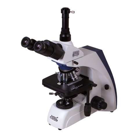
11
Microscope unpacking
Unpack the microscope carefully and install it on a smooth surface. Check the microscope package contents. Visually inspect all
elements included in the scope of supply, identify their purpose, make sure there are no damages and start assembling.
Microscope preparation for operation
Installation of modular units
• Install the decorative base on the microscope stand base (supply with the installed decorative base is possible).
• Install the halogen lamp ashlight (g. 1, 13) on the microscope stand base and x with a clamping knob (g. 4, 2).
• Install a uorescent illuminator (g. 2, 17) on the microscope stand ange (g. 1, 12). When installing the illuminator, rst
press the conical surface of the slip on ange to two supports arranged on the right in the stand seat, then clamp the ange
with a screw (g. 1, 28).
• Install the knob (g. 2, 1) in the position of beam of rays interception with a shutter, having extended it from the casing.
• Place the mercury-lled lamp ashlight (g. 1, 8) on a table, unscrew the cap clamping screw (g. 1, 9) and remove the cap
(g. 5, 1).
• Take the mercury-lled lamp from the microscope package, install it into bushings (g. 5, 2 and 5) on the cap (g. 5, 1) and
x with screws (g. 5, 3 and 6).
WARNING! DO NOT TOUCH THE MERCURY-FILLED LAMP BULB! AFTER LAMP INSTALLATION, DEGREASE THE BULB
SURFACE WITH ALCOHOL SOLUTION.
• Install the cap (g. 5, 1) into the mercury-lled lamp ashlight (g. 1, 8) and x with a screw (g. 1, 9).
• Install the mercury-lled lamp ashlight (g. 1, 8) on the uorescent illuminator (g. 2, 15), using the xator and the
adjusting ring (g. 5, 7 and 8), x with a bayonet ring located on the illuminator.
• Connect the cable from the ashlight to the slot on the rear surface of the mercury-lled lamp power supply unit (g. 6).
• Connect the power cord to the power socket on the rear surface of the mercury-lled lamp power supply unit. Make sure
that the switch is in “O” position (g. 1, 18).
• Install the trinocular head (g. 1, 1) on the uorescent illuminator ange (g. 2, 15), x with a lock knob (g. 1, 3). Install
the luminous ux switch (splitter) (g. 1, 2) into "Observation only" position.
• Install eyepieces (g. 2, 5) into eyepiece tubes.
• Install the beam splitting units switching ring (g, 1, 27) to position No.1.
• Put the stage (g. 2, 10) down by rotation of the coarse focusing mechanism knob (g. 2, 13) until stop.
• Install objective lenses (g. 2, 7) into revolving nosepiece seats in the ascending order of their magnications.
• Turn the lamp lament adjustment knob (g. 1, 17) in the direction of brightness reduction until stop.
• The switch (g. 1, 18) should be installed into "O" position.
• Connect the power cord to the power socket on the rear surface of the stand base (g. 1, 12).
• Install the UV protection screen (g. 1, 25) and x it with screws (g. 1, 26).
Microscope use
Safety precautions
The microscope may be handled by persons with special medical education. The source of hazard in microscope operation is
electric current. Microscope design prevents accidental contact with energized current-conducting parts.
WARNING! REPLACE LAMPS IN FLASHLIGHTS WHEN THE MICROSCOPE AND THE MERCURY-FILLED LAMP POWER
SUPPLY UNIT ARE DISCONNECTED FROM THE GRID. TO AVOID HAND SKIN BURN FROM THE LAMP BULB, REPLACE
THE LAMP 15—20 MINUTES AFTER DISCONNECTION.
When safety fuses are replaced, it is necessary to install new safety fuses with the same ratings as before.
After the end of operation, the microscope and the mercury-lled lamp power supply unit must be disconnected from the grid.
It is not recommended to leave the appliances connected to the grid unattended.
Perform repair and preventive maintenance only after disconnection of the appliances from the grid.
Observation of objects in transmitted light
Halogen lamp activation and illumination setup
Connect the microscope power cord to AC grid.
Activate the halogen lamp, having set the switch (g. 1, 18) in "I" position.
Adjust lamp brightness by rotation of the lament adjustment knob (g. 1, 17).
Image quality in the microscope to a large extent depends on illumination, therefore, illumination setup is an important
preparatory operation, which must be performed as follows:
• put the object on the stage (g. 2, 10) of the microscope;
• activate the objective lens with 4x or 10x magnication into the path of rays (it is recommended to start the process of
focusing from low or medium magnication objective lenses with suciently large elds and operating distances);
• focus the microscope by rotation of knobs (g. 1, 14 and 15);
• cover the eld diaphragm with a ring (g. 1, 23), and the aperture diaphragm of the condenser — with a knob (g. 3, 1);
• observing the object image, focus the condenser, moving it along the height with the knob (g. 3, 5) for sharp image of
the iris eld diaphragm;
• if the eld diaphragm image is displaced, bring the image into the eld center with condenser alignment screws (g. 3, 2);
