Nexcope NSZ818 User manual

NSZ818
ZOOM STEREO
MICROSCOPES
NSZ818
TRINOCULAR ZOOM STEREO MICROSCOPE
INSTRUCTION MANUALINSTRUCTION MANUAL

2 3
INTRODUCTION
MANY THANKS TO YOU
for the purchase of our professional trinocular zoom stereo
microscope NSZ818. It has been carefully manufactured with
materials of lasting value, and careful attention has been paid
to function and reliability. This microscope is designed for a
wide range of applications such as life sciences, research and
development. It has a 18:1 extra-class zoom ratio that delivers
images with outstanding sharpness. The integrated OIC (oblique
coherent contrast) illumination in the stand base enhances the
contrast of transparent specimen structures.
NEXCOPE microscopes are very robust, but need a certain
amount of attention and care to guarantee their longevity. We
therefore recommend that you read this user manual carefully
and keep it within reach for your reference while working with
the stereo zoom microscope. It contains all relevant information
on operation, user safety and care. If you follow the guidelines,
your microscope will function reliably and smoothly even after
years of intensive use.
We wish you much success in your work with your new zoom
stereo microscope!
The operating instructions can be downloaded
from the following web address:
www.bresser.de/PNX60818
i i
INTRODUCTION WARRANTY TERMS
GARANTIE · WARRANTY · GARANTÍA · GARANZIA
Besuchen Sie unsere Website über den folgenden QR Code oder Weblink um weitere Informationen
zu diesem Produkt oder die verfügbaren Übersetzungen dieser Anleitung zu nden.
Visit our website via the following QR Code or web link to nd further information on this product or
the available translations of these instructions.
Si vous souhaitez obtenir plus d’informations concernant ce produit ou rechercher ce mode d’emploi
en d’autres langues, rendez-vous sur notre site Internet en utilisant le code QR ou le lien correspon-
dant.
Bezoek onze internetpagina via de volgende QR-code of weblink, voor meer informatie over dit pro-
duct of de beschikbare vertalingen van deze gebruiksaanwijzing.
Desearía recibir unas instrucciones de uso completas sobre este producto en un idioma determin-
ado? Entonces visite nuestra página web utilizando el siguiente enlace (código QR) para ver las
versiones disponibles.
Desidera ricevere informazioni esaustive su questo prodotto in una lingua specica? Venga a visitare
il nostro sito Web al seguente link (codice QR Code) per conoscere le versioni disponibili.
DE
EN
FR
NL
ES
IT
www.bresser.de/warranty_terms
https://www.bresser.de/Nach-Hersteller/Nexcope/

4 5
i i
SERVICE SERVICE
DE
AT
CH
BE
Bei Fragen zum Produkt und eventuellen Reklamatio-
nen nehmen Sie bitte zunächst mit dem Service-Cen-
ter Kontakt auf, vorzugsweise per E-Mail.
E-Mail: [email protected]
Telefon*: +49 28 72 80 74 350
BRESSER GmbH
Kundenservice
Gutenbergstr. 2
46414 Rhede
Deutschland
*Lokale Rufnummer in Deutschland (Die Höhe der Gebühren je
Telefonat ist abhängig vom Tarif Ihres Telefonanbieters); Anrufe aus
dem Ausland sind mit höheren Kosten verbunden.
GB
IE
Please contact the service centre rst for any
questions regarding the product or claims,
preferably by e-mail.
Telephone*: +44 1342 837 098
BRESSER UK Ltd.
Suite G3, Eden House
Enterprise Way
Edenbridge, Kent TN8 6HF
United Kingdom
*Number charged at local rates in the UK (the amount you will
be charged per phone call will depend on the tariff of your phone
provider); calls from abroad will involve higher costs.
FR
BE
Si vous avez des questions concernant ce
produit ou en cas de réclamations, veuillez
prendre contact avec notre centre de services
(de préférence via e-mail).
Téléphone*: 00 800 6343 7000
BRESSER France SARL
Pôle d’Activités de Nicopolis
314 Avenue des Chênes Verts
83170 Brignoles
France
*Prix d'un appel local depuis la France ou Belgique
NL
BE
Als u met betrekking tot het product vragen
of eventuele klachten heeft kunt u contact
opnemen met het service centrum (bij voorkeur
per e-mail).
Telefoon*: +31 528 23 24 76
BRESSER Benelux
Smirnoffstraat 8
7903 AX Hoogeveen
The Netherlands
*Het telefoonnummer wordt in het Nederland tegen lokaal tarief
in rekening gebracht. Het bedrag dat u per gesprek in rekening
gebracht zal worden, is afhankelijk van het tarief van uw telefoon
provider; gesprekken vanuit het buitenland zullen hogere kosten
met zich meebrengen.
ES
IT
PT
Si desea formular alguna pregunta sobre el
producto o alguna eventual reclamación, le
rogamos que se ponga en contacto con el
centro de servicio técnico (de preferencia por
e-mail).
Teléfono*: +34 91 67972 69
BRESSER Iberia SLU
c/Valdemorillo,1 Nave B
P.I. Ventorro del Cano
28925 Alcorcón Madrid
España
*Número local de España (el importe de cada llamada telefónica
dependen de las tarifas de los distribuidores); Las llamadas des del
extranjero están ligadas a costes suplementarios..
You can obtain additional information from your customer service: We recommend that you study this manual thoroughly before operating the microscope for the rst
time. It will help you become fully familiar with the equipment and achieve optimum performance.
Keep this manual in an easily accessible place near the work area for future reference.
Changes in the interest of the technical development are reserved. These instruction manual is not
subject to the revision service. The reproduction of this document as well as the use and communi-
cation of its contents are not permitted unless expressly permitted. Contraventions obligate to com-
pensation for damages. All rights in the event of a patent being granted or utility model registration
reserved.

6 7
1 1
TABLE OF CONTENTS TABLE OF CONTENTS
Service
1. Important notes
1.1. Imprint/ validity information
1.2. About this instruction manual/intended use
1.3. General safety instructions
1.4. Safety symbols used in the manual
1.5. Sustainability
1.6. Check scope of delivery and handling
1.7. Site location
1.8. Cleaning/maintenance/transport
1.9. Protection and storage
1.10. Disposal
2. Introduction
What you can expect
3. Design of Trino Zoom Stereo Microscope
3.1. Product image NSZ818 – zoom stereo microscope
3.2. Assembly of the NSZ818 zoom stereo microscope
3.2.1. Detailed assembly procedure
1 Mounting the stand column
2 Mounting the zoom unit
3 Attaching the trinocular viewing head
4 Inserting the eyepieces
5 Attaching the plan Apo 1x objective
6 Placing the glass plate
7 OPTIONAL – Capturing images
4. Adjust microscope
4.1. Quick Guide
4.2. Detailed description of the Quick Guide steps
1 Setting up the power supply and start-up
2 Placing the specimen on the glass plate
3 Switching on the lighting and brightness control
4 Direct light to the eyepieces or to the camera
5 Settings on the trinocular viewing head
6 Settings on the zoom unit
7 Further adjustment options on the stand base
8 AFTER OBSERVATION: Switch off the microscope
5. Optical design: Parallel light (zoom type)
6. Scope of delivery
7. Technical specications
8. Trouble shooting guide
9. Dimensions
10. Warranty
11. Notes/comments
1. IMPORTANT NOTES
This chapter informs the user about the general instructions for the microscope and
the important safety symbols.
1.1. Imprint/validity information
Imprint
Bresser GmbH
Gutenbergstraße 2
46414 Rhede Deutschland
http://www.bresser.de
For information regarding liability claims or service requests, please refer to
chapters "Warranty" and "Service" in this documentation. Errors reserved -
technical specications subject to change.
Copyright: 2022 Bresser GmbH
All rights reserved.
It is not allowed to reproduce this documentation or parts of it in any kind (e.g.
photocopying, printing, etc.) without written a permission of the manufacturer or
the distributor, Bresser GmbH. It is also prohibited to use or transmit this documen-
tation with electronic systems (e.g. image le, website, etc.). The descriptions and
brand names of the corresponding rms used in this documentation are generally
protected under trademark, brand and/or patent law for Germany, for the European
Union and/or other countries.
Validity information
This documentation is valid for the products with the article numbers listed below:
NSZ818
Manual version: v122021a
Manual description: Manual_ NX60818_NSZ818_en_NEXCOPE_v122021a
For any inquiries, please state these information.
1.2. About this instruction manual/intended use
About this instruction manual
These operating instructions are to be considered a component of the
device.
PLEASE READ THE SAFETY INSTRUCTIONS AND THE OPERATING INST-
RUCTIONS CAREFULLY BEFORE USE. Keep these instructions available for
further reference when using the microscope. When the device is sold or given to
someone else, the instruction manual must be provided to the new owner/user of
the product.
Intended use
• This product is intended for private and corporate use.
• It was developed for the magnied display of things in nature.
• The device is intended only for indoor use.
This device is not intended for use by individuals (including children) with limited physical,
sensory or mental capabilities or those lacking in experience and/or knowledge, unless they are
supervised by an individual responsible for their safety or have received instructions from them
regarding the use of the device.
Table of contents NSZ818

8 9
1 1
IMPORTANT NOTES IMPORTANT NOTES
1.3. General safety instructions
Danger of an electronic shock!
This device contains electronic components that operate via a power source (power
supply and/or batteries). In case of any improper use of this device, there is a risk of
an electric shock. An electric shock can cause severe injury or even death. Therefore
please read the safety instructions below to avoid an explosion.
• Disconnect the device from the power supply by pulling the power plug when it
is not used or in case of longer interruption of operation and before starting any
work on maintenance and cleaning.
• Position your device so that it can be disconnected from the power supply at
any time. The wall socket you use should be located near the device and easily
accessible, since the plug on the power cable serves as a disconnecting device
for the power supply.
• Always pull on the plug to seperate the device from the power supply. Never pull
on the cable.
• Before operating, check the device, cables and connections for dammage.
• Never use a damaged unit or a unit with damaged power cables. Damaged parts
must be exchanged immediately by an authorised service centre.
• Only use the device in complete dry environment and do not touch it with wet or
moist parts of your body.
• The microscope is equipped with a power supply unit which allows the use of
mains voltage values of 12 V; 2 A.
• To avoid electric shock, connect the supplied power cord to a properly grounded
power outlet on. These mains cables are tted with three-pin plugs to ensure
proper earthing.
Danger of choking!
In case of any improper use of this device, there is a risk of choking, especially for
children. Therefore please read the safety instructions below.
• Keep packaging material, like plastic bags and rubber bans, out of the reach for
children, as these materials pose a choking hazard!
• This product contains small parts that can be swallowed by children! There is a
risk of choking!
• If small parts are swallowed, consult a doctor immediately!
Danger of explosion!
In case of any improper use of this device, there is a risk of an explosion. Therefore
please read the safety instructions below to avoid and explosion.
• Do not expose the device to high temperatures. Use only the supplied power
adapter. Do not short-circuit the device or throw them into a re. Excessive heat
or improper handling could trigger a short-circuit, a re or an explosion.
• Do not use the microscopes and the accessories supplied with them in potential-
ly explosive atmospheres, in the presence of ammable solvents such as alcohol,
petrol or volatile anaesthetics, etc..
CAUTION: Danger of injury!
This device contains components and/or accessories that can cause minor to severe
injuries in case of any proper use. Therefore please read the safety instructions be-
low to avoid any bodily injury.
• Tools with sharp edges and points are often used when working with this device.
Because there is a risk of injury from such tools, store this device and all the tools
and accessories in a location that is out of the reach of children.
• Children must not have access to the included chemicals and liquids. Do not
drink the chemicals. Wash hands thoroughly with running water after using the
chemicals. In the event that the chemicals come into contact with your eyes or
mouth, rinse thoroughly with water. If you are in pain after exposure, contact a
doctor immediately and take the substances with which you came into contact
with you.
CAUTION: Fire hazard!
In case of any improper use of this device, there is a risk of re. Therefore please read
the safety instructions below to avoid the initiation of burning.
NOTICE: Risk of property damage!
In case of any improper use of this device and/or its accessories, there is a risk of
property damage. Therefore only use the device according to the safety instructions
below.
• Do not disassemble the device. In the event of a defect, please contact your dea-
ler. The dealer will contact the Service Centre and can send the device in to be
repaired, if necessary.
• Do not expose this device to higher temperatures and protect it from water and
high humidity.
• Protect the device from severe shocks!
• For this device only use accessories and spare parts that comply with the techni-
cal information.
• Always use the power cord supplied by Nexcope. If an unsuitable power cord is
used, Nexcope can no longer guarantee the electrical functionality and safety of
the microscope.
• Use these microscope and the original accessories only for the applications de-
scribed in this manual.
• The manufacturer does not accept any liability for any other application, possibly
also for individual assemblies or individual parts. This also applies to all repair
and service work that is not carried out by authorised service personnel. There-
fore all guarantee / warranty claims expire.
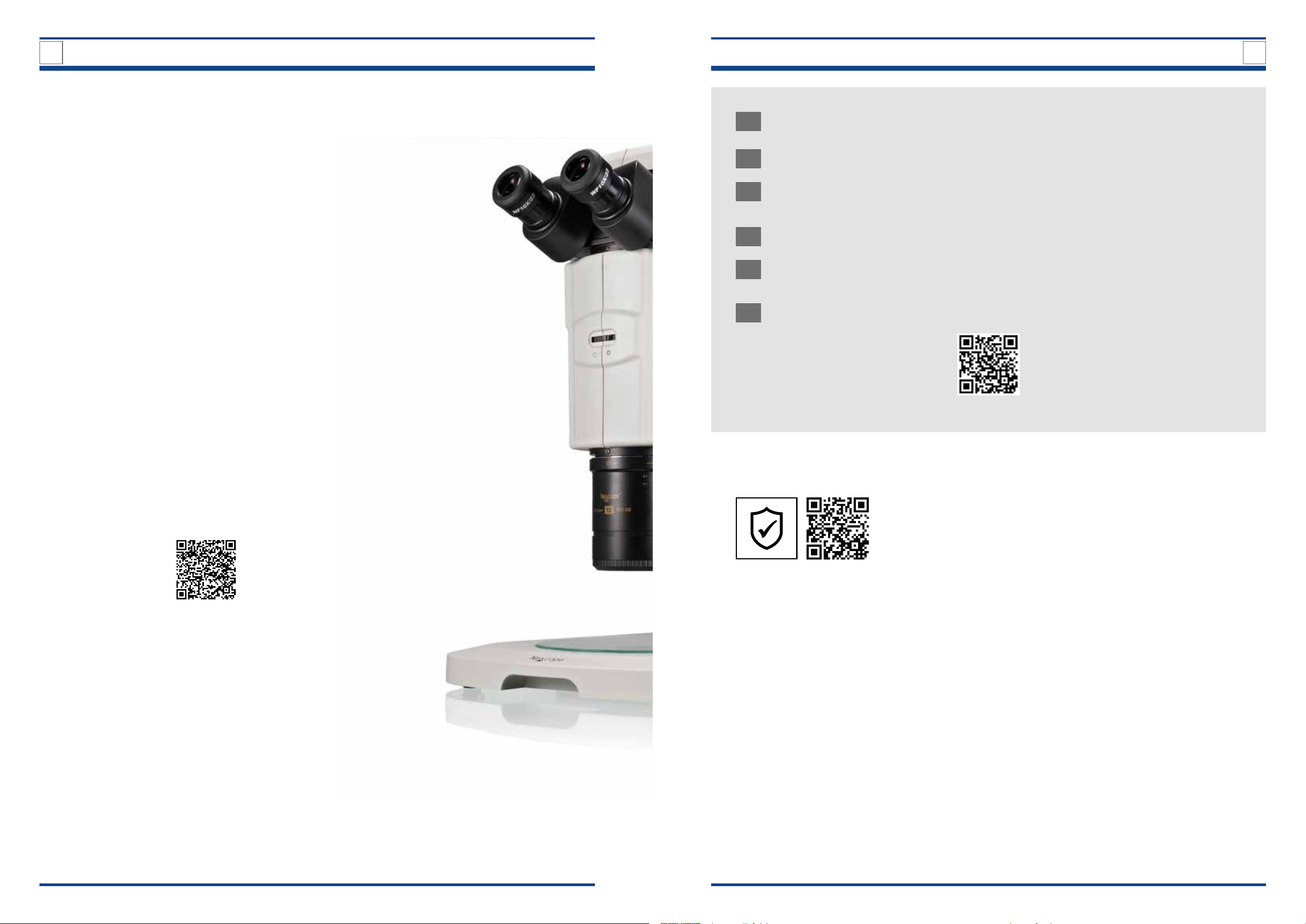
• Zoom stereo microscope NSZ818 is not equipped with any special device to
protect against corrosive, toxic, potentially infectious or radioactive samples or
other samples that are harmful to health equipped. All legal requirements, in
particular national regulations for accident prevention, must be observed when
handling such samples.
Note!
If you have any complaints or queries please contact your national service centre by
telephone. The address is included in these instructions.

10 11
IMPORTANT NOTES IMPORTANT NOTES
1
UKCA DECLARATION OF CONFORMITY
A “Declaration of conformity” in accordance with the applicable
directives and corresponding standards has been prepared by
Bresser GmbH. This can be viewed at any time upon request at
www.bresser.de/download/NX60818/UKCA/NX60818_UKCA.pdf
BRESSER UK Ltd. • Suite 3G, Eden House
Enterprise Way, Edenbridge, Kent TN8 6HF, GB
Caution LED Radiation
The LED module of the incident light uorescence lighting system
emits class 3B LED light. Direct exposure to the light and direct in-
cidence of light on the skin must therefore be avoided at all costs.
When microscopes are used, the protective devices provided with
the microscope must always be used. Never - neither with nor
without optical instruments - look into the light beam, not even
to simply look at the sample. Failure to observe this warning may
result in eye damage!
1.6. Sustainability
The protection of our environment is close to our heart.
That is why this manual is printed on 100% recycled paper.
1.7. Check scope of delivery and handling
• Open the packaging with care. Avoid ngerprints and sweat on the objective lens
and the glass plate. Prevent the objective and the glass plate from falling and
being damaged.
• Remove all components from the packaging and check for completeness accor-
ding to the delivery note. The microscope could be packaged in more than one
carton depending on the model or accessories.
• Handle the microscope with care and always protect it against violent impact and
vibrations.
• Keep the original packaging for possible longer storage or return of the device to
the supplier in case a repair or revision is needed.
Never lift the microscope by the trinocular viewing head or coarse/ne fo-
cusing knobs. This can damage the microscope!
• Always carry the microscope with both hands.
• It is recommended to carry the microscope by the carrying handle, stand
base as well as by the stand column.
1
1.4. Safety symbols used in the manual
Danger symbols
Hazard symbols are recognisable symbols intended to warn of hazardous ma-
terials, objects or places, including electric currents, radioactivity and poi-
sons.
Warning of a danger point
This symbol indicates information that must be read and obser-
ved.
Non-observance can lead to:
o Risk of injury
o Malfunctions or equipment damage
Warning of dangerous electrical voltage
This symbol precedes information that must be read and obser-
ved.
Non-observance can lead to:
o Risk of injury
o Malfunctions or equipment damage
Warning symbol
Warning symbols indicates a possible source of danger.
General warning
This symbol indicates information that must be read and obser-
ved.
Failure to follow the warnings may result in injury to the user
and/or damage to the microscope (including nearby objects).
Note symbol
The hint symbol provides you with important additional information and tips
about the microscope.
Important additional hint
Accompanying instructions serve to simplify operation and main-
tenance.
1.5. Symbols on the microscope
The following symbols are located on the microscope/accessories and should
always be observed:
Dispose of the packaging materials by type. Contact your local
waste-disposal service or environmental authority for informa-
tion on the proper disposal.
Do not dispose of electronic devices in the household garbage!
According to the European Directive 2002/96/EU on Waste Elect-
rical and Electronic Equipment and its transposition into national
law, used electrical equipment must be collected separately and
recycled in an environmentally sound manner.
EC DECLARATION OF CONFORMITY
A “Declaration of conformity” in accordance with the applicable
directives and corresponding standards has been prepared by
Bresser GmbH. This can be viewed at any time upon request at
www.bresser.de/download/NX60818/CE/NX60818_CE.pdf

12 13
IMPORTANT NOTES IMPORTANT NOTES
1.8. Site location
BEFORE SETTING UP THE MICROSCOPE: select a suitable location.
Place the microscope on a resistant surface away from strong sunlight, heat sources,
high humidity, high dust exposure as well as strong vibration.
• The ambient temperature range of the microscope is 5°C to 40°C (41°F to
109°F)!
• If the device is exposed to temperatures outside this range, this will cause
irreparable damage to optical and/or mechanical parts which are not
covered by warranty.
• Place the microscope on a surface that meets the following criteria:
› Flat
› Vibration-free
› Level
› Hard surface and non-ammable
› Stable (the unit weighs approximately 10.5 kg)
• Chemical and mechanical resistant
• Place the microscope so that:
› There is at least 10 cm clearance around the device or to other devices or the
wall.
› You can disconnect it from the mains at any time.
• The mains cable must be freely accessible at all times, as the mains cable is inten-
ded as a disconnecting device from the mains.
• Make sure the residual moisture is fully eliminated before use.
• For use in warm and humid climates, all optical components of the microscope
are already equipped with protection against fungus infestation.
1.9. Cleaning/maintenance/transport
IN THE EVENT OF A LONG OPERATING INTERRUPTION and especially
maintenance and cleaning work, disconnect the power supply by pulling out
the mains plug.
BEFORE EVER CLEANING: Dismantle optical elements (e.g. objective, eye-
piece, etc.).
BEFORE USING THE MICROSCOPE: The residual moisture should be com-
pletely removed.
DO NOT USE ORGANIC SOLVENTS (e.g. alcohol, ether, acetone, xylol or
other dilutions) to clean lacquered parts or plastic parts!
ALCOHOL IS HIGHLY FLAMMABLE!
AVOID DAMAGE TO THE ELECTRONICS: by not using cleaning uid.
1 1
IF YOU USE PREPARATIONS that pose a potential risk of infection, all parts
that have come into contact with the preparation must be thoroughly cleaned.
The PLAN APO 1x objective, the zooming body, the coarse/ne focusing
mechanism and the aperture diaphragm are precisely designed and matched
to each other. Please avoid disassembling the components. This will affect the
performance of the microscope.
TRANSPORT: Please remove all loose parts (e.g. glass plate, eyepieces)
and the object to be observed from the glass plate. Only use the original
packaging for transport.
Cleaning microscope body / optical elements
• Only use a dry cloth to clean the exterior of the device.
• BEFORE CLEANING: Disassemble optical elements (e.g. objective, eyepiece, etc.).
• Blow away loose dust from the lens surfaces rst.
• Use a soft disposable paper cloth soaked in 30%-70% pure alcohol (available at
the pharmacy or drugstore). Do not apply the cleaning solution directly to the
optical parts. Gently wipe the lens surface by cleaning the surface with circular
movements from the centre to the edge of the optic. Apply light pressure to the
optics while doing this.
1.10. Protection and storage
• PROTECT THE DEVICE against dust and moisture.
• AVOID PUTTING FINGERPRINTS and equal contaminations on any optical surfa-
ces.
• PULL A DUST PROTECTION COVER OVER THE MICROSCOPE. Before covering the
microscope, always check that the microscope is also switched off.
• IT IS RECOMMENDED to store this microscope in a protected place where there
are no acid gases, alkalis, organic solvents and other harmful substances in the
vicinity.
• STORE THE MICROSCOPE in a closed container at a dry and mould-free place.
• STORE THE MICROSCOPE AND THE ACCESSORIES in the relevant containers
when they are not used for a longer time.
• IT IS RECOMMENDED to store also the objective and the eyepieces in closed con-
tainers with desiccant.
A dust protection cover is included in delivery.
REMEMBER: A well maintained microscope will keep its optical quality for
years and thus maintain its value.

14 15
1
IMPORTANT NOTES
2
INTRODUCTION
1.11. Disposal
Dispose of the packaging materials properly, according to their type, such as
paper or cardboard. Contact your local waste-disposal service or environmental
authority for information on the proper disposal.
Please take the current legal regulations into account when disposing of your de-
vice. You can get more information on the proper disposal from your local waste-
disposal service or environmental authority.
DO NOT DISPOSE of electronic devices in the household garbage!
As per Directive 2002/96/EC of the European Parliament on waste electrical
and electronic equipment and its adaptation into German law, used electron-
ic devices must be collected separately and recycled in an environmentally
friendly manner.
2. INTRODUCTION
2.1. What you can expect
OUTSTANDING OPTICAL ZOOM PERFORMANCE
• The stereo microscope offers a zoom ratio of 18:1
• High resolution and 18x zoom ratio, with 7.5x to 135x zoom range
under 10x eyepiece
• PLAN APO 1x objective with numerical aperture (NA) up to 0.15 pro-
vides excellent images with lifelike colours
LARGE RADIUS OF OPERATION
• The 1x objective offers a working distance (W.D.) of 60 mm to facili-
tate sample placement and movement.
• * The large eld of view (FOV) allows you to cover a larger and more
comprehensive sample area.
EXTRA WIDE STAND BASE WITH EASY TO USE OIC ILLU-
MINATION
• The extra wide stand base provides stable support for the micro-
scope.
• LED stand base supports OIC illumination (illumination method
using oblique light to enhance contrast of colourless, transparent
specimen structures).
• Three-dimensional LED illumination base has a long life and gener-
ates low amounts of heat.
COMFORTABLE IN LONG-TERM OBSERVATION
Trinocular viewing head
• By quickly attaching a camera, you can create a sharp image of the
sample to be observed at any time
• Wide-eld 10x eyepieces with 23 mm eld of view (FOV)
• Adjustable diopter and interpupillary distance on both eyepieces
› Minimize user fatigue through long-term observation
WIDE RANGE OF APPLICATION
• Life Sciences
• Research
• Development
Zoom ratio 18:1
Zoom range 0.75x-13.5x
Total magnication 7.5x-135x
Working distance (W.D.) 60 mm
Field of view (FOV)
Objective
Eyepiece
31.4 mm
23 mm
NA
0.15
W.D.
60 mm

16 17
3. NSZ818
3.1. Product image NSZ818 – Zoom stereo microscope
Illustration 002: Product image: NSZ818 back/side view.
A
B
NSZ818 Front/Side view
NSZ818 Back/Side View
3
DESIGN OF TRINO ZOOM STEREO MICROSCOPE
3
DESIGN OF TRINO ZOOM STEREO MICROSCOPE
Equipment
1. 1x C-mount photo adapter
2. Trinocular viewing head
3. Eyepiece
4. Fine focusing knob
5. Coarse focusing knob
6. Aperture diaphragm handwheel
7. 1x Objective (Plan Apo)
8. Glass plate
9. Carrying handle
10. Zoom unit
11. Stand base
12. OIC adjustment slider
13. Rod without function (in development)
14. Stand column
15. Holder for zoom unit
16. Zooming knob (range: 0.75x-13.5x)
17. Switching lever for visual observation/photo
b
g
Illustration 001: Product image: NSZ818 front/side view.
Equipment
18. AC adapter input terminal
19. Connection for uorescent controller
20. LED brightness control selection switch
21. Main power switch ON/OFF
22. Brightness control knob (Transmitted light)
23. Subplate
24. Type plate with important information
Power Supply:
DC-12V 2A via power adapter

18 19
3.2. Assembly of the NSZ818 zoom stereo microscope
The diagram below shows the sequence of assembly of the various modules.
The numbers indicate the order of assembly.
When assembling the microscope, make
sure that all parts are free of dust and dirt and
avoid scratching any parts or touching glass
surfaces.
Please use the three hexagonal screwdriv-
ers for assembling and replacing components
3.2.1.Detailed assembly procedure
1Mounting the stand column
Illustration 004: Mounting the stand column.
V
II
IV
III
I
BEFORE YOU START: Select a lo-
cation free of vibration.
Put the stand base (I) at on the
workbench.
Align the connecting plate (II) of the
stand column (III) with the mounting
plate (IV) of the stand base (I).
Use the four supplied hexagon socket
head screws (V) to connect the stand
base and the stand column rmly.
USE the supplied hexagonal
screwdriver for assembling.
Check input voltage: The input voltage and
supply voltage indicated on the subplate of the
microscope must be consistent, otherwise the
microscope will be seriously damaged.
3
DESIGN OF TRINO ZOOM STEREO MICROSCOPE
3
DESIGN OF TRINO ZOOM STEREO MICROSCOPE
7
3
4
5
2
1
6
Illustration 003: Assembly of zoom stereo microscope NSZ818. Three hexagonal screwdivers.

20 21
3
DESIGN OF TRINO ZOOM STEREO MICROSCOPE
3
DESIGN OF TRINO ZOOM STEREO MICROSCOPE
3Attaching the trinocular viewing head
IIllustration 006: Attaching the trinocular viewing head.
III
II
IV
I I
Completely loosen the viewing head hol-
ding screw (I).
USE the supplied hexagonal
screwdriver.
If present, pull off the black protective
cap (II) from the tube lens at the bottom
of the trinocular head (III).
MAKE SURE not to touch any
optical lenses.
Place the trinocular viewing head (III)
into the round dovetail of the zoom unit
(IV) so that the eyepieces are in front.
ENSURE CORRECT ALIGNMENT
along the microscope body (blue
lines).
Fasten the viewing head by tightening
the viewing head holding screw.
2Mounting the zoom unit
IIllustration 005: Attaching the zoom unit.
III
IV
IV
II
I
V
Turn the coarse focusing knob (I) to pla-
ce the holder for the zoom unit (II) to the
upper range of the stand column (III).
Loosen the two set screws (IV) on the
right side of the zoom unit (V).
USE the supplied hexagonal
screwdriver.
Place the dovetail groove of the zoom
unit onto the dovetail of the holder for
the zoom unit.
Firmly tighten the two set screws.

22 23
3
DESIGN OF TRINO ZOOM STEREO MICROSCOPE
3
DESIGN OF TRINO ZOOM STEREO MICROSCOPE
4Inserting the eyepieces
IIllustration 007: Inserting the eyepieces.
II
I
III
Remove the black protective caps (I)
from the eyepiece sleeves (II). Press eyepieces (III) gently into the eye-
piece sleeves.
5Attaching the plan Apo 1x objective
HANDLE THE 1X OBJECTIVE WITH CARE
The objective is very heavy. Therefore, make sure that you hold it with both
hands when you tighten or loosen the objective holding screw.
BEFORE EACH USE, check the front lens of the objective for dirt. A contamina-
tion will degree the microscope images. Therefore clean the objective regularly.
Illustration A: Loosen the objective holding screw on the zoom
unit.
If present, remove the protective cap (I)
from the 1x objective lens (II).
Completely loosen the objective holding
screw (III) on the lower front of the zoom
unit (IV).
USE the supplied hexagonal
screwdriver.
B
II
Start position
a
End position
b
Illustration B: Aligning the objective with the zoom unit.
Insert the 1x objective (II) loosely into the
xture without pushing it upwards.
Align the mark on the top of the objective
according to the starting position a.
Rotate the objective in the direction of
the arrow until the mark on top of the
objective aligns exactly according to the
end position b.
C
III
Illustration xx C: Fixing the objective and the zoom unit.
After the 1x objective is properly seated
and aligned with the zoom unit, tighten
the objective holding screw (III).
B
I
III
III
IV
IV
II
II
I
III
III
IV
IV
II
II

24 25
6Placing the glass plate
III
Illustration A: Placing the glass plate.
BE CAREFUL WITH THE GLASS
PLATE. First assemble all other
components before placing the
glass plate on the stand base to
avoid glass breakage.
Use the recess (I) in the rear part of the
stand base (II) to place the glass plate
(III) securely.
NOTE: The glass plate only rests
loosely on the stand base. It is not
additionally fastened.
3
DESIGN OF TRINO ZOOM STEREO MICROSCOPE
4
QUICK GUIDE
7OPTIONAL – Capturing images
IV
III
I
II
Attaching a camera.
A 1x C-mount photo adapter (I)
is already attached to the micro-
scope imaging interface (II) of the
trinocular viewing head (III).
You can put a suitable C-mount camera
(IV) on the photo adapter to take photo-
micrographs or performing videomicro-
graphy.
When installing the camera,
please always make sure that you
HOLD IT FIRMLY IN PLACE to pro-
tect it from damage due to drop-
ping.
NOTE: A camera is not included
in the scope of delivery.
REMEMBER: Cameras usually
show a smaller image section than
eyepieces. This is partly due to the
sensor size and cannot be changed.
4. ADJUSTING THE MICROSCOPE
This chapter describes the individual microscopy steps.
4.1. Quick Guide
1Setting up power supply and start-up
2Placing the specimen on the glass plate
3Switching on the lighting and brightness control
4Direct light to the eyepieces or to the camera
5Settings on the trinocular viewing head
a) Adjusting the interpupillary distance
b) Diopter adjustment
6
Settings on the zoom unit
a) Adjust the focus using the focusing knobs
b) Set desired magnication on the zooming knob with scale
c) Contrast adjustment by means of aperture diaphragm (conden-
ser diaphragm)
7Further adjustment options on the stand base
a) OIC (oblique coherent contrast) illumination
b) Rod without function (in development)
8AFTER OBSERVATION: Switch off the microscope
II I

26 27
4
QUICK GUIDE
4
QUICK GUIDE
2Placing the specimen on the glass plate
Illustration xx: Placing the specimen on the glass plate.
1
Place the sample vessel with the sample
to be examined (1) on the glass plate (2)
of the stand base (3).
3Switching on the lighting and brightness control
Illustration xx: Adjusting the brightness.
1
This microscope has an illumi-
nation unit with a permanently in-
stalled LED light source.
To adjust the brightness, proceed as fol-
lows:
Turn the brightness control knob (1) in
the direction of the arrow the light in-
tensity increases and vice versa.
1Setting up the power supply and start-up
Illustration xx: Setting up power supply.
A
3
14
2
Setting up the power supply
Connect the AC adapter (1) of the power
supply to the AC adapter input terminal
(2) on the back of the microscope.
Insert the main power plug (3) into the
power socket (4).
MAKE SURE the supplied voltage
matches the instrument specica-
tions: DC 12 V; 2 A.
4.2. Detailed description of the Quick Guide steps
B
Illustration xx: Starting up the microscope.
5
Start-up
Move the main power switch (5) to
position I (ON) to turn on the device.
MAKE SURE that the main power
switch ON/OFF is set to O (OFF) be-
fore connecting the power plug.
Cables and cords are vulner-
able when bent or twisted. Never
subject them to excessive force.
The unit may only be operated
with the mains adapter supplied.
32

28 29
4
QUICK GUIDE
4
QUICK GUIDE
4Direct light to the eyepieces or to the camera
A
Illustration xx A/B: Light path of the trinocular viewing head.
2
1
3
You have the option of directing the light
either to the eyepieces of the binocu-
lar part (1) or to the camera (2) via the
switching lever for visual observation/
photo (3). To microscope the specimen
set the lever to 100 % to the eyepieces of
the binocular part.
B
Icon Proportion on the
light for the binocular
part
Propor-
tion of
the light
for the
camera
left right
¡100% 100% 0%
,100% 0% 100%
CONSIDER: The light amount
of the left eyepiece is always 100%.
The light amount of the right
eyepiece is diverged to the photo
adapter. Only the images of the
right eyepiece optical path are
recorded by camera. To do this,
ip the switching lever for visual
observation/photo to the symbol.
5Settings on the trinocular viewing head
Trinocular viewing head with 20° xed inclined eyepieces.
A
Illustration xx: Adjusting the interpupillary distance.
two elds of view merge
Adjusting the interpupillary distance
(Distance between the eyepieces)
Look through the eyepieces (1) and ad-
just the interpupillary distance (2) so that
the left and right elds of view are com-
pletely aligned.
NOTE: Proper interpupillary dis-
tance is crucial to the comfort of the
user.
B
Illustration xx: Diopter adjustment.
1
3
Diopter adjustment
Look through one of the eyepiece (1) and
focus the image (eyepiece freely selecta-
ble). Then look through the other eye-
piece with the other eye and turn only
the diopter adjustment ring (3) on this
eyepiece to focus the sample.
THE DIOPTER COMPENSATION
is an adjustment possibility on both
eyepieces and serves to compen-
sate for near - or farsightedness
(max. +/-8 dpt. difference between
both eyes can be compensated). If
the difference is higher, or if you
suffer from astigmatism etc. we
recommend observing with your
prescribed glasses on.
C
Illustration xx: Eyepieces with rubber cups.
4
DIOPTER COMPENSATION is
possible on both eyepieces but is
only performed on one eye if the
difference is +/- 4 dpt. or lower.
ADDITIONAL FEATURE:
Protection through rubber cups
Both eyepieces are suitable for spectacle
wearers. They are protected with rubber
cups (4) so that the user's eyeglasses are
not damaged when accidentally coming
in contact with the eyepiece.
1
2

30 31
4
QUICK GUIDE
4
QUICK GUIDE
6Settings on the zoom unit
A
Illustration xx A: Focusing controls.
2a 2b
1b1a
Focusing controls
To adjust the focus, rotate the focusing
knobs (1,2) on the right and left sides of
the stereo microscope.
Coarse focusing knobs (1a, 1b):
Pre-focusing, large distance between
specimen and objective.
Fine focusing knobs (2a, 2b):
The ne focus is used for image sharp-
ness. Fine and precise regulation of the
distance between the specimen and the
objective.
B
Illustration xx B: Relationship between focusing knobs and zoom
unit.
2
3
1
Relationship between focusing knobs
and zoom unit
NOTE: Familiarize yourself with
the correlation between the di-
rection of rotation of the focusing
knobs and the vertical movement
of the zoom unit.
Turning the focusing knobs (1,2)
forward
Zoom unit (3) moves downwards
Turning the focusing knobs (1,2)
backward
Zoom unit (3) moves upwards
a) Adjust the focus using the focusing knobs
A
Illustration xx/ A: Zooming knobs.
Zooming knobs
Turn the zooming knobs (1a, 1b) on the
right and left of the zooming body (2) to
change the magnication.
Turning the knob forward
Magnication decrease
Turning the knob backward
Magnication increase
The magnication values are marked on
the right-hand zooming knob (1b).
B
Illustration xx/B: Magnication details.
Zoom ratio 18:1
Zoom range 0.75x – 13.5x
Total magnication
(1x objective, 10x eyepiece)
7.5 – 135x
3
Magnication values
Turning the zooming knobs cause the
magnication to change in the range
from 0.75x to 13.5x (18:1 zoom ratio).
The following magnication steps (3) are
possible:
0.75x, 1x, 2x, 3x, 4x, 5x, 6x, 7x, 8x, 9x, 10x,
11x, 12x, 13x and 13.5x
NOTE: At the following magni-
cation steps, the zooming knobs
engages with an audible “click”:
0.75x (starting position), 1x, 2x, 3x,
6x, 10x, 13.5x (nal position).
b) Set desired magnication on the zooming knob with scale
1a 1b
2

32 33
4
QUICK GUIDE
Illustration xx A/B: Adjust the aperture diaphragm.
A
Aperture diaphragm handwheel
The zoom unit contains an aperture dia-
phragm. The aperture diaphragm can be
adjusted with the help of the aperture
diaphragm handwheel (1).
NOTE: Adjust the aperture dia-
phragm opening according to the
characteristics of the specimen.
B
Aperture diaphragm open/closed
OPEN CLOSED
Focus depth Shallow Deep
Brightness Bright Dark
Resolution High Low
Position of the aperture diaphragm
Opening and closing the aperture dia-
phragm causes a change in focus depth,
brightness and resolution.
c) Contrast adjustment by means of aperture diaphragm
(condenser diaphragm)
4
QUICK GUIDE
7Further adjustment options on the stand base
A
3
2
1
Illustration xx/A: OIC illumination.
OIC adjustment slider
The stand base (1) supports OIC illumina-
tion.
OIC means oblique coherent contrast il-
lumination. An illumination method in
which contrast gain is achieved by par-
tial or complete coverage of the normal
illumination beam path. For this purpose,
a black OIC plate (2) is brought into the
beam path that can be moved via the OIC
adjustment slider (3) on the right-hand
side of the stand base (1). The specimen
is then radiated through at an angle, re-
sulting in a shadow cast on the ne struc-
tures of the preparation. This enables
the observation of colorless, transparent
samples structures with relief and high
contrast.
a) OIC (oblique coherent contrast) illumination
B
42
Illustration xx/B: OIC illumination settings.
3
OIC illumination settings
Move the OIC adjustment slider (3) along
the scale (4). You can observe how the
black OIC plate (2) moves into the beam
path.
Adjust until the optimum observation
condition is reached in combination with
the selected magnication and the sam-
ple to be examined.
• OIC plate completely out of the
beam path
(OIC adjustment slider either at the
furthest or closest position on the
scale)
No contrast and relief enhancement
visible
• OIC plate partially in the beam path
Contrast and relief enhancement of
colourless and transparent sample struc-
tures visible
• OIC plate completely in the beam
path
(OIC adjustment slider in the middle
position of the scale)
Completely darkened beam path
Helpful: Thanks to the gradua-
tion on the scale, you can save and
reproduce the desired lighting set-
ting.
1

34 35
4
QUICK GUIDE
5
OPTICAL DESIGN
Illustration xx: Rod currently without function.
2
1
Rod without function
At present, the rod (1) on the right-
hand side of the stand base (2) is wit-
hout function.
It is intended for future accessories and
is currently still under development.
NOTE: As soon as the mod-
el is equipped with an additional
function, you will nd information
about it on our website: https://
www.bresser.de.
b) Rod without function (in development)
8AFTER OBSERVATION: Switch off the microscope
A
Illustration xx A: Switch off the microscope.
Switch off the microscope
Switch the microscope off using the
main power switch ON/OFF (1). To do
this, select position O (OFF).
MAKE SURE the supplied voltage
matches the instruments
specications: DC 12 V; 2 A
5. OPTICAL DESIGN: PARALLEL LIGHT (ZOOM TYPE)
NSZ818 is a parallel microscope, using plan apochromatic optical system and can ob-
tain perfect high three-dimensional and visual softer image. Large zoom range and
eld of view may be applied to imaging and observation of the image from macro to
micro. It has an 18:1 high zoom ratio, providing superior optical system, high resolu-
tion and ergonomic design.
NA
0.15
W.D.
60 mm
Zoom ratio 18:1
Zoom range 0.75x-13.5x
Total magnication 7.5x-135x
Working distance (W.D.) 60 mm
Field of view (FOV)
Objective
Eyepiece
31.4 mm
23 mm
Long working distance (W.D.) and large eld of view (FOV)
Plan Apo 1x objective to provide a working distance (W.D.) of 60 mm. Up 0.15 nume-
rical aperture (NA) offers you a clarity to study in-depth. The objective`s wide eld of
view (FOV) of 31.4 mm enables to see a wider and more comprehensive sample area.
Plan apochromatic 1x objective
The newly developed plan apochromatic 1x objective with a high numerical aperture
of 0.15 is used as the optical system. This ensures a uniform brightness of the eld
of view and a clear and reliable image at high magnication in bright eld mode as
well as at lowest magnication.
Disconnect main power supply
Disconnect the main power plug (3) from
the power socket (4).
Disconnect the AC adapter (1) of the
power supply from the AC adapter input
terminal (2) on the back of the microsco-
pe.
Cables and cords are vulner-
able when bent or twisted. Never
subject them to excessive force.
MAKE SURE that the LED power
ON/OFF switch is set to O (OFF) be-
fore disconnecting the power plug.
2
43
5

36 37
6
SCOPE OF DELIVERY
7
TECHNICAL SPECIFICATIONS
6. SCOPE OF DELIVERY
• 1x Trinocular zoom stereo-microscope (stand base, stand column, zoom unit, trino-
cular viewing head) (total magnication: 7.5x-135x)
• 1x Plan APO 1x objective
• 2x Eyepieces WF 10×/23
• 1x Glass plate
• 1x C-mount photo adapter
• 1x Nexcope power cord
• 3x screwdrivers
• 4x Hexagon socket head screws for the attachment of the stand column
• 1x Dust cover
• 1x Manual instruction
7. TECHNICAL SPECIFICATIONS
Device Technical Specications
Zoom
Optical system Parallel light (zoom type) apochromatic optical system
Manual Zoom
Zoom ratio 18:1
Zoom range 0.75-13.5x
Objective lens NA, WD PLAN APO 1x 0.15, 60 mm
Total magnication
(1x large object,
10x eyepiece)
7.5-135x
Eyepiece (FOV mm) 10x (23)
Lens tube (eye-piece/port) Trinocular 20° xed inclination lens barrel (100/0, 0/100)
Focusing device (stroke) 60 + 99 mm
Adapter DC 12V, 2A
Base LED three-dimensional lighting base (OIC built-in illuminator)
Observation method Bright eld, oblique illumination
Observation methods with optional
accessories
Fluorescence, simple polarized light, dark eld
Weight (approx.) 10.5 kg
Power consumption (approx.) 10 W
Operating environment • Indoor use
• Altitude: up to 2000 meters
• Ambient temperature: 5°C~40°C (41°F~109°F)
• Maximum relative humidity: relative humidity at a tempera-
ture of 31°C (88°F)
• 80%, then linearly decrease
• The relative humidity is 70% when the temperature is 34°C
(93°F)
• The relative humidity is 60% when the temperature is 37°C
(99°F)
• The relative humidity is 50% when the temperature is 40° C
(104° F)
• Pollution degree: Level 2
• Atmospheric pressure: 80kPa~106kPa
• Overvoltage category: Class II

38 39
8. TROUBLE SHOOTING GUIDE
Under certain conditions performance of the unit may be adversely affected by factors
other than defects. If problems occur, please review the following list and take remedial
action as needed. If you cannot solve the problem after checking the entire list, please
contact Bresser GmbH for assistance.
TROUBLE CAUSE SOLUTION
The eld of view is still dark The power supply unit is not
connected to the socket or the
input connector AC adapter
Check the connection between
the main power plug and the
power socket as well as the con-
nection between the AC adapter
and the AC adapter input terminal
The light intensity is adjusted too
low
Adjust the light intensity accor-
dingly via the brightness control
knob
There is dirt in the eld of view Dirt/dust on the sample Please use clean samples
Dirt/dust on the eyepiece Wipe the eyepiece
The two images do not match Improper interpupillary distance
adjustment
Correct interpupillary distance
Improper diopter adjustment Readjust
The left and right eyepieces have
different magnications
Change to the same eyepieces
The image is out of focus Incorrect focus Adjust the focus via the coarse
focus-ing knob and ne focusing
knob so that the sample can be
clearly seen
8
TROUBLESHOOTING
9
DIMENSIONS
9. DIMENSIONS
10. WARRANTY
The regular guarantee period is 2 years and begins on the day of purchase. To benet
from an extended voluntary guarantee period as stated on the gift box, registration on
our website is required. You can consult the full guarantee terms as well as information
on extending the guarantee period and details of our services at www.bresser.de/warran-
ty_terms.
10
WARRANTY
Table of contents
Other Nexcope Microscope manuals