SamanTree Medical Histolog Scanner v2 User manual

Instructions for use
v2
Histolog® Scanner v2 003
Histolog® Dish 004
1223 version 1.11_EN (2022-05-26)

SamanTree Medical SA
Avenue de Provence 12
1007 Lausanne
Switzerland
Tel: +41 21 625 0940
Customer service:
Tel: +41 21 623 0073
SamanTree Medical France SASU
c/o A.A.C.I.
2 avenue des Allobroges
74200 Thonon-les-Bains
France
UK Responsible Person
UKMDR Ltd.
71-75 Shelton Street, Covent Garden
London WC2H 9JQ
United Kingdom
Copyright © 2015–2022
This publication is protected by copyright and all rights are reserved. No part of this document may be
reproduced or transmitted in any form, or by any means, electronic or mechanical, including photocopying and
recording for any purpose other than the purchaser’s personal use without permission of SamanTree Medical.
Histolog is a registered trademark of SamanTree Medical. All other trade names and trademarks are the
property of their respective holders.
The Histolog® Scanner and the Histolog® Dish are patent protected products.
See www.samantree.com/ip for more information on intellectual property.
1223 version 1.11_EN (2022-05-26) Page 2 | 30

1. Revision history
Version
Date
Change description
1.0
2018-01-28
Initial Version.
1.1
2018-04 - 23
Update for release of software application version 3.0.0.
Changes requested by IEC 61010-1, IEC 61010-2-101 safety tests.
1.2
2018-05-29
Changes requested by IEC 60825-161010-1, IEC 61010-2-101 and IEC
608251-1 safety tests.
1.3
2018-07-05
Add information on the environmental pollution degree, required by IEC
61010-1:2010 + AMD1:2016 §5.4.2.
Add recommended material for specimen staining.
Add additional approved cleaning agents.
1.4
2019-05-17
Update with new product catalog codes.
Add a patent protection statement.
Update for release of software application version 3.1.0.
Update for release of Histolog Dip product.
1.5
2019-06-21
Removed §11.
Improve image quality for print.
Add description of symbol for “quantity in package”.
1.6
2020-08-31
Update SamanTree Medical’s address.
1.7
2020-11-16
Update list of compatible cleaning agents.
Change contact information.
Update patent protection statement with latest issued patents.
1.8
2021-09-15
Update for European Directive EU 2015/863.
1.9
2022-01-13
Update SamanTree Medical’s address.
Update copyright and patent protection statements.
1.10
2022-02-10
Update information for safety in line with risk analysis.
1.11
2022-05-26
Restructure the document and move the detailed instructions of Histolog®
Scanner Software Application to a separate, dedicated document.
Reformulate the intended purpose.
Update content of §6, §7, §8, §9, §14.
Other changes to get in conformity with Regulation (EU) 2017/746 (“IVDR”)
and with the Medical Devices Regulations 2002 (UK).
1223 version 1.11_EN (2022-05-26) Page 3 | 30

2. Table of contents
1. Revision history 3
2. Table of contents 4
3. Description of symbols 6
4. Introduction 7
5. Safety instructions 8
5.1. Warnings 8
5.2. Cautions 8
5.3. Laser safety 9
6. Intended purpose 10
6.1. Intended use 10
7. Product information 11
7.1. Overview 11
7.2. Accessories 12
7.2.1. Histolog® Dish 12
7.2.2. Histolog® Dip 12
7.2.3. Other accessories 12
7.3. Operating principle 12
8. Use of the Histolog® Scanner 13
8.1. Before use 13
8.1.1. Installing in use location 13
8.1.2. Switching ON 13
8.1.3. Mounting Histolog® Dish 13
8.2. After use 14
8.2.1. Switching OFF 14
8.2.2. Prepare for storage 14
8.3. Imaging a tissue specimen 15
8.3.1. Staining the tissue specimen 15
8.3.2. Positioning the tissue specimen 15
9. Cleaning 16
10. Moving 17
11. Storage 18
12. Maintenance 19
12.1. Update of Histolog® Scanner Software Application 19
1223 version 1.11_EN (2022-05-26) Page 4 | 30

12.2. Fuse replacement 19
13. Disposal 20
13.1. Histolog® Dish 20
13.2. Histolog® Scanner 20
13.3. Histolog® Dip 20
14. Troubleshooting 21
14.1. Test procedure 21
14.2. Troubleshooting 22
14.3. Export logs 24
15. Product catalog codes 25
16. Device specifications/technical information 26
16.1. Technical specifications 26
16.1.1. Histolog® Scanner 26
16.1.2. Histolog® Dish 27
16.2. Electromagnetic immunity 28
16.3. Electromagnetic compatibility 30
1223 version 1.11_EN (2022-05-26) Page 5 | 30

3. Description of symbols
Batch Code
Temperature limit
Catalog Number
Atmospheric Pressure Limitation
Serial Number
Humidity Limitation
In vitro diagnostic medical device
Fragile; handle with care
Manufacturer
This way up
Manufacturing date
Keep dry
Consult instructions for use
Do not reuse
Standby or power indicator
Use-by (expiry) date
Orientation mark
Non-sterile
Non-ionizing radiation
Quantity in package
Authorized EC Representative
Importer
Unique Device Identification
Class 1 laser product, as per IEC 60825-1:2014-05.
Follow the local regulations for separate collection of Electrical and Electronic Equipment.
CE marking of conformity with Directive 98/79/EC and Regulation (EU) 2017/746.
UKCA marking of conformity with the Medical Devices Regulations 2002 (UK).
● When followed by “ WARNING ” this symbol means “Warning! Failure to observe could result in
injury or death.”
● When followed by “ CAUTION ”, this symbol means “Caution! Failure to observe could result in
damaged equipment, forfeited time or effort, or the need to abort use of the device.”
● When found on equipment, this symbol means: “Attention: consult accompanying instructions
for use”.
NOTE
A NOTE sign draws attention to useful information regarding a function or procedure.
1223 version 1.11_EN (2022-05-26) Page 6 | 30

4. Introduction
These instructions for use describe the set-up, use and maintenance of SamanTree Medical’s Histolog®
Scanner device, including its Histolog® Dish accessory.
These instructions for use contain important information for the safe use of the device.
◼ Read these instructions for use before setting up and using the device the first time.
◼ Keep the instructions for use accessible for future reference.
Refer to the Histolog® Dip instructions for use for information on the Histolog® Dip fluorescent histological stain.
Refer to the Histolog® Scanner Software Application instructions for use for detailed instructions on how to
operate the Histolog® Scanner Software Application.
1223 version 1.11_EN (2022-05-26) Page 7 | 30

5. Safety instructions
Use only for the purpose described in these instructions for use. Do not use the device if it is not working
properly, or if it has suffered any damage. Misuse can cause electrocution, burns, fire and other hazards.
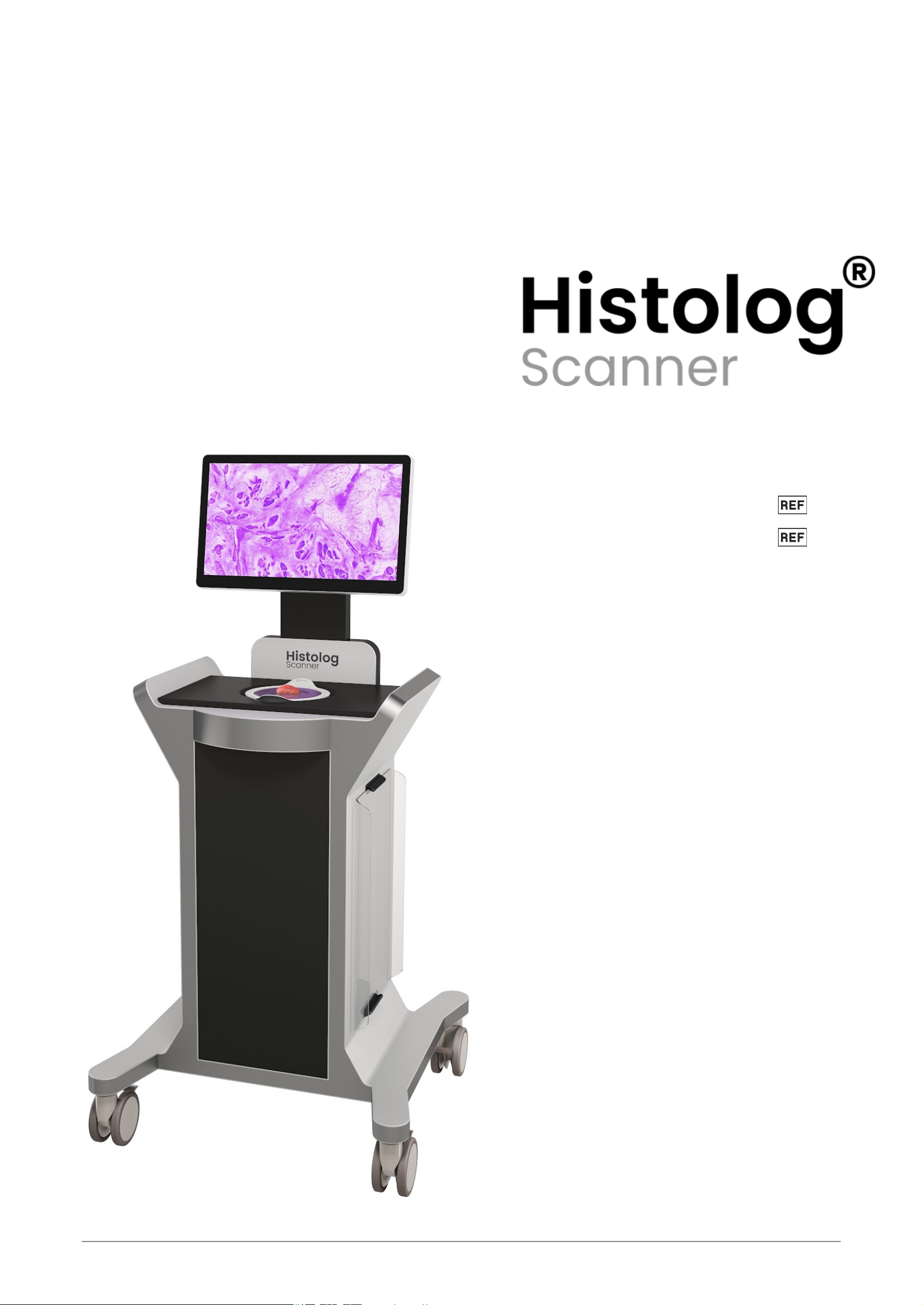
Prior to first use, the Histolog® Scanner device is to be put in service by SamanTree Medical authorized service
personnel.
The components located in the inner cavity of the device are not intended to be manipulated by the user. The
interior of the Histolog® Scanner may be serviced only by SamanTree Medical authorized service personnel.
Report any serious incident related to the Histolog® Scanner and its accessory devices to SamanTree Medical
SA and to your national competent authority.
5.1. Warnings
WARNING
■ The Histolog® Scanner and its accessory devices are intended to be used in vitro for the examination of
specimens. They are not intended for use on patients.
■ The Histolog® Scanner and the Histolog® Dish are non sterile devices. For use in an intraoperative
setting, the Histolog® Scanner and its accessory products shall be located outside the sterile surgical area.
■ When handling potentially infectious tissue specimens, avoid skin contact by wearing gloves or other
protective equipment.
■ This device is not compatible with magnetic resonance (MR) or explosive environments.
■ Handles [H] are not provided for lifting the device, but exclusively have a function for pushing/pulling.
■ The Histolog® Scanner imaging window [G] is fragile. Avoid impact with foreign objects. Do not load with
mass above 1 kg.
■ Do not operate the Histolog® Scanner in case of damage to the imaging window [G].
■ The protective earthing connection of the Histolog® Scanner is performed through the power cord [P]
connection. If this cable is damaged, the device must not be connected to the mains power.
■ Only use a power cord [P] having adequate ratings for the Histolog® Scanner.
■ Comply with the product safety instructions described in the Histolog® Dip instructions for use.
■ Cleaning when the device is connected to the mains power may be hazardous to the operator and/or
destructive to the device.
5.2. Cautions
CAUTION
■ Only use compatible accessories and parts supplied by SamanTree Medical or by an authorized
distributor.
■ Do not re-use a Histolog® Dish. A new Histolog® Dish shall be used for every new surgical procedure.
■ Do not use a Histolog® Dish, if it is damaged or if its optical interface is visibly dirty.
■ If the information about the tissue specimen orientation is critical, the tissue specimen should be marked
for orientation before starting the imaging procedure with the Histolog® Scanner.
■ Do not apply tissue marking dyes on the tissue specimen before imaging it with the Histolog® Scanner.
Tissue marking dyes prevent proper imaging with the Histolog® Scanner.
■ Do not use disinfecting products that contain glycolic acid. These disinfecting products may damage the
Histolog® Scanner.
■ The Histolog® Scanner is intended for use in the electromagnetic environment specified in §16.2 & §16.3 .
■ Do not use this device in close proximity to sources of strong electromagnetic radiation (e.g. unshielded
intentional radio frequency (RF) sources), as these can interfere with proper operation.
1223 version 1.11_EN (2022-05-26) Page 8 | 30

5.3. Laser safety
The Histolog® Scanner is a Class 1 laser product, as per IEC 60825-1:2014-05.
Laser radiation in the visible wavelength range is emitted from the imaging window [G] during imaging.
The Histolog® Scanner embeds a Class IIIB laser component with the following characteristics:
Wavelength: 488 ± 5 nm
Max output power: 200 mW (CW)
Beam divergence: < 0.7 mrad
1223 version 1.11_EN (2022-05-26) Page 9 | 30

6. Intended purpose
6.1. Intended use
The Histolog® Scanner is a confocal microscopy device intended for imaging the surface of excised human
tissue specimens to visualize morphological microstructures.
The Histolog® Scanner is intended to be used in combination with the following accessory devices:
■ The Histolog® Dish, a single-use protection preventing direct contact of the tissue specimen with the
Histolog® Scanner during imaging.
■ The Histolog® Dip fluorescent histological stain.
The Histolog® Scanner and its accessory devices are intended to be used in vitro for the examination of
specimens. They are in-vitro diagnostic medical devices.
The Histolog® Scanner and its accessory devices are intended to be operated by trained healthcare
professionals.
1223 version 1.11_EN (2022-05-26) Page 10 | 30

7. Product information
7.1. Overview
The Histolog® Scanner consists of the following components:
[A] Touchscreen monitor
[B] Histolog® Scanner Software Application
[C] ON/OFF power switch
[D] USB port
[E] Ethernet port
(only for maintenance purposes, under guidance of SamanTree Medical authorized service personnel)
[F] Orientation mark
[G] Imaging window
[H] Handles
[I] Lid
[J] Swivel caster wheels with brakes
[K] Appliance power inlet with fuse compartment
[L] Document holder
[M] Lid storage supports
[N] Histolog® Dish
[O] Cable winder
[P] Power cord
[Q] Ventilation openings
1223 version 1.11_EN (2022-05-26) Page 11 | 30

7.2. Accessories
7.2.1. Histolog® Dish
The Histolog® Dish is a single-use protection preventing direct contact of the tissue specimen with the Histolog®
Scanner during imaging. The Histolog® Dish is provided separately (see §15 for catalog codes).
The Histolog® Dish is placed over the imaging window [G] of the Histolog® Scanner. It is composed of:
1. Two handles protruding from the rigid base for manipulation;
2. An optical interface on which the tissue specimen is placed and through which it is imaged.
It is not intended to be used as a receptacle for transporting or storing tissue specimens.
The focal plane of the Histolog® Scanner is configured in-factory to lie just above the top surface of the
Histolog® Dish optical interface, so that the surface of a specimen in contact with the optical interface is imaged.
7.2.2. Histolog® Dip
The Histolog® Dip is a fluorescent histological stain for imaging tissue specimens with the Histolog® Scanner.
The Histolog® Dip is provided separately (see §15 for catalog codes).
7.2.3. Other accessories
A USB storage device is needed to export images and technical data (see §14.3 ) from the Histolog® Scanner
device. USB storage device is not provided by SamanTree Medical. It is recommended to use a USB 3.0 (or
better) storage medium with read/write speeds of at least 300 MB/s and with storage capacity of at least 256 GB.
NOTE USB storage devices with security lock features (such as PIN protection) are not compatible with the
Histolog® Scanner.
Refer to the Histolog® Scanner Software Application instructions for use for detailed instructions on how to
export images and for information on supported export formats.
7.3. Operating principle
The Histolog® Scanner is a digital microscopy scanner for use on excised human tissue. Its operating principle
is based on confocal fluorescence imaging modality and uses non-ionizing, low-power optical radiation.
In a conventional wide-field fluorescence microscope, the entire specimen is flooded in light from a light source
and the detected fluorescence includes a large part of out-of-focus signal. In contrast, a confocal microscope
uses a point illumination conjugated with a pinhole in front of the detector to eliminate out-of-focus signals so that
only the fluorescence produced very close to the focal plane in the specimen can be detected. This thin optical
sectioning makes the Histolog® Scanner particularly well suited for imaging the superficial cell layer of thick
tissue specimens: excised tissue can be immediately imaged, slide-free, without the need for freezing or fixing.
The Histolog® Scanner acquires digital images with high, micrometer-range resolution and enables the
visualization of tissue microstructures down to the cellular level.
The Histolog® Scanner is based on a massively parallel signal acquisition and processing technology providing
fast digital imaging over large areas.
1223 version 1.11_EN (2022-05-26) Page 12 | 30

8. Use of the Histolog® Scanner
Prior to first use, the Histolog® Scanner device must be put in service by SamanTree Medical authorized service
personnel.
8.1. Before use
8.1.1. Installing in use location
1. Move the Histolog® Scanner to the desired location with level ground.
WARNING The Histolog® Scanner and the Histolog® Dish are non sterile devices. For use in an
intraoperative setting, the Histolog® Scanner and its accessory products shall be located outside the
sterile surgical area.
NOTE Leave at least 15 cm of free space around the ventilation openings [Q].
2. When in place, lock the four wheel brakes [J] in downward position to prevent unintended movement of the
Histolog® Scanner.
3. Plug the power cord [P] to the mains power outlet.
WARNING
■ The protective earthing connection of the Histolog® Scanner is performed through the power cord [P]
connection. If this cable is damaged, the device must not be connected to the mains power.
■ Only use a power cord [P] having adequate ratings for the Histolog® Scanner. Suitable power cord
accessories are listed in §15 .
8.1.2. Switching ON
1. Switch ON the power switch [C] and wait for the Histolog® Scanner Software Application to initialize.
NOTE When switched ON, the power switch [C] is pressed and lighted in white.
8.1.3. Mounting Histolog® Dish
1. Remove the Histolog® Scanner lid [I], and store it in its lid storage supports [M].
2. Unpack a Histolog® Dish and mount it over the imaging window [G]. When properly mounted, only the
optical interface of the Histolog® Dish is in contact with the Histolog® Scanner.
CAUTION
■ Do not re-use a Histolog® Dish. A new Histolog® Dish shall be used for every new surgical procedure.
■ Do not use a Histolog® Dish, if it is damaged or if its optical interface is visibly dirty.
1223 version 1.11_EN (2022-05-26) Page 13 | 30

8.2. After use
8.2.1. Switching OFF
Quit the Histolog® Scanner Software Application:
1. Tap the button.
If User Authentication mode is used in the Histolog® Scanner Software Application, then select the “Turn
off” option in the graphical popup control.
2. Confirm the action by tapping “Turn off” to shutdown the device.
Or tap on “Cancel” if you do not want to shutdown the Histolog® Scanner.
Power OFF the Histolog® Scanner device:
3. Wait 15 seconds or until the ‘No Signal’ message is displayed on the monitor, and then switch OFF the
power switch [C].
8.2.2. Prepare for storage
1. Dispose of the Histolog® Dish according to §13.1 .
2. Unplug the power cord [P] from the mains power outlet and wind it on the cable winder [O].
3. Clean the Histolog® Scanner according to §9 .
4. Place the lid [I] back on the Histolog® Scanner to protect the imaging window [G].
1223 version 1.11_EN (2022-05-26) Page 14 | 30

8.3. Imaging a tissue specimen
WARNING When handling potentially infectious tissue specimens, avoid skin contact by wearing gloves or
other protective equipment.
CAUTION
■ If the information about the tissue specimen orientation is critical, the tissue specimen should be marked
for orientation before starting the imaging procedure with the Histolog® Scanner.
■ Do not apply tissue marking dyes on the tissue specimen before imaging it with the Histolog® Scanner.
Tissue marking dyes prevent proper imaging with the Histolog® Scanner.
8.3.1. Staining the tissue specimen
Stain the tissue specimen with the Histolog® Dip fluorescent histological stain. Refer to the Histolog® Dip
instructions for use for tissue specimen staining procedures.
WARNING Comply with the product safety instructions described in the Histolog® Dip instructions for use.
8.3.2. Positioning the tissue specimen
1. Place the tissue specimen on the optical interface of the Histolog® Dish, with the surface of interest
pointing downwards.
NOTE Specimen size should not exceed that of the Histolog® Dish (see §16.1 ). Furthermore, the tissue
specimen should be positioned in a way to only contact the optical interface of the Histolog® Dish, but
never its rigid base or handles.
2. Make sure that the area of interest of the tissue specimen surface is positioned over the image area and
that it is in contact with the optical interface of the Histolog® Dish to guarantee its complete imaging.
3. When the stained tissue specimen is properly positioned for imaging, refer to the instructions for use of the
Histolog® Scanner Software Application to perform the imaging and other associated procedures.
NOTE Image quality is negatively impacted by:
■ excess of fluid (especially blood) on the tissue specimen,
■ any treatment (such as cauterization) that alters the tissue specimen surface,
■ any residues or stains on the optical interface of the Histolog® Dish or on the tissue specimen
surface,
■ motion of the tissue specimen during image acquisition.
1223 version 1.11_EN (2022-05-26) Page 15 | 30

9. Cleaning
WARNING
■ Cleaning when the device is connected to the mains power may be hazardous to the operator and/or
destructive to the device.
The device may be cleaned with conventional alcohol-based disinfecting agents for medical devices. Read and
follow the instructions supplied with the cleaning product.
Make sure that the disinfecting agent does not enter into the device's inner cavity.
The following products are compatible with the Histolog® Scanner:
Active
compound
Tested with
Alcohol
Ethanol, 71%
Propanol, 50%
Quaternary
ammonia
compounds
DDAC - Didecyldimethylammonium chloride - (CAS 7173-51-5), 0.075%
Benzalkonium Chloride - benzyl-C12-16-alkyldimethyl, chlorides (CAS 68424-85-1), 0.095%
CAUTION
■ Do not use disinfecting products that contain glycolic acid. These disinfecting products may damage the
Histolog® Scanner.
NOTE
■ Take care not to scratch the surface of the touchscreen monitor [A] or imaging window [G]. Avoid contact
with hard or abrasive materials.
■ The Histolog® Scanner device must be dry before subsequent use.
1223 version 1.11_EN (2022-05-26) Page 16 | 30

10. Moving
Before moving the Histolog® Scanner, please ensure that:
1. The power cord [P] is disconnected from the mains outlet and secured on the cable winder [O].
2. The brakes of all four of the caster wheels [J] are released (in upwards position).
Use only the handles [H], and not other parts of the device, for pushing/pulling.
Move the device no faster than walking speed. Move the device over a flat surface. Avoid surfaces and
obstacles that could hamper the normal rolling of the wheels [J]. Beware of physical collisions of the device with
possible obstacles during the moving.
WARNING Handles [H] are not provided for lifting the device, but exclusively have a function for
pushing/pulling.
1223 version 1.11_EN (2022-05-26) Page 17 | 30

11. Storage
After use of the device, follow the instructions of §8.2 . The device can then be moved to the storage area. When
in place, lock the four wheel brakes [J] in downward position to prevent unintended movement of the Histolog®
Scanner.
See §16.1 for specified environmental storage conditions.
1223 version 1.11_EN (2022-05-26) Page 18 | 30

12. Maintenance
SamanTree Medical recommends an annual maintenance for every Histolog® Scanner device. Such an annual
maintenance may be performed only by SamanTree Medical authorized service personnel.
The components located in the inner cavity of the device are not intended to be manipulated by the user. The
interior of the Histolog® Scanner may be serviced only by SamanTree Medical authorized service personnel.
12.1. Update of Histolog® Scanner Software Application
Update of Histolog® Scanner Software Application may be installed only by SamanTree Medical authorized
service personnel.
12.2. Fuse replacement
Fuses are located in a compartment above the appliance power inlet [K] located at the back of the device.
To replace fuses:
1. With the device switched off, disconnect the power cord [P] from the appliance power inlet [K].
2. Using a flat screwdriver, open the fuse drawer on the back of the device.
3. Replace the blown fuses with new ones meeting the specifications of §16.1 .
4. Close the fuse drawer.
5. Reconnect the power cord [P] to the appliance power inlet [K].
1223 version 1.11_EN (2022-05-26) Page 19 | 30

13. Disposal
13.1. Histolog® Dish
Dispose of the Histolog® Dish according to local procedure for handling biomedical waste.
13.2. Histolog® Scanner
Follow the local regulations for separate collection of electrical and electronic equipment.
13.3. Histolog® Dip
Dispose of the Histolog® Dip in accordance with the information contained in its instructions for use.
1223 version 1.11_EN (2022-05-26) Page 20 | 30
This manual suits for next models
3
Table of contents
Other SamanTree Medical Medical Equipment manuals
Popular Medical Equipment manuals by other brands

Aqua Creek
Aqua Creek Tidal Wave manual

Acutus Medical
Acutus Medical Qubic Force Technical manual

Chattanooga Group
Chattanooga Group INTELECT LEGEND Series user manual

MIETHKE
MIETHKE SENSOR RESERVOIR Instructions for use and Technical description

Hologic
Hologic SecurView DX Quick reference guide

OrthoSensor
OrthoSensor Knee Balancer user guide