Soredex CRANEX 3D User manual

ENGLISH
Digital Panoramic X-Ray System
User Manual
206718 rev. 9
CRANEX™ 3D


CRANEX™ 3D
Copyright Code: 206718 rev 9Date: 2018-11-28
Copyright © 2018by SOREDEX.
All rights reserved.
SOREDEX™, SCANORA™, CRANEX™, ClearTouch™,
SMAR™, Wise Dose Scaler™, EasyScout™ and
Minidose™ are either registered trademarks or trademarks
of SOREDEX, Palodex Group Oy.
All other trademarks are property of their respective
owners.
U.S. patents US6731717, US6829326 and USRE41197.
Finnish patents 114383.
Documentation, trademark and the software are
copyrighted with all rights reserved. Under the copyright
laws the documentation may not be copied, photocopied,
reproduced, translated, or reduced to any electronic
medium or machine readable form in whole or part, without
the prior written permission of SOREDEX, PaloDEx Group
Oy.
The original language of this manual is English.
SOREDEX, PaloDEx Group Oy reserves the right to make
changes in specification and features shown herein, or
discontinue the product described at any time without
notice or obligation. Contact your SOREDEX, PaloDEx
Group Oy representative for the most current information.
Manufacturer SOREDEX, PaloDEx Group Oy
Nahkelantie 160
FI-04300 Tuusula
FINLAND
Tel. +358 10 270 2000
www.soredex.com
For service, contact your local distributor.

CRANEX™ 3D

rev i
Table of Contents
1 Introduction..................................................................................................................1
1.1 CRANEX™ 3D ...................................................................................................... 1
1.2 Intended use ......................................................................................................... 2
1.3 Intended user profile ............................................................................................. 2
1.4 Associated documentation .................................................................................... 2
1.5 References............................................................................................................ 2
1.6 Abbreviations used in this manual ........................................................................ 3
1.7 Warnings and precautions .................................................................................... 4
1.7.1 Warnings to be observed during use ......................................................... 4
1.7.2 Warnings for cross infection....................................................................... 5
1.7.3 General warnings ....................................................................................... 6
1.8 Disclaimer ............................................................................................................. 8
1.9 Disposal ................................................................................................................ 8
2 Unit description ........................................................................................................... 9
2.1 Main parts and controls......................................................................................... 9
2.2 Patient positioning lights ..................................................................................... 11
2.3 Patient positioning panel ..................................................................................... 14
2.3.1 Cephalometric unit patient positioning panel (optional) ........................... 14
2.4 Accessories......................................................................................................... 15
2.5 Other detachable partsEmergency stop switch................................................... 16
2.6 Patient contacting parts....................................................................................... 18
3 Imaging programs ..................................................................................................... 19
3.1 Panoramic programs........................................................................................... 19
3.2 Sectional imaging................................................................................................ 22
3.3 Default exposure settings................................................................................... 22
3.4 User Configurable Default Program .................................................................... 22
3.4.1 SMAR™, SOREDEX™ Metal Artifact Reduction..................................... 23
3.5 Cephalometric programs..................................................................................... 24
3.6 3D programs ....................................................................................................... 26
3.7 Exposure settings for 3D imaging ....................................................................... 27
3.8.1 AES values for Panoramic imaging.......................................................... 28
3.8.2 AES values for Cephalometric imaging.................................................... 29
4 Controlling the unit.................................................................................................... 31
4.1 ClearTouch™ control panel ................................................................................ 31
4.2 Modality section .................................................................................................. 32
4.3 Exposure indicators and settings ........................................................................ 32
4.4 Status section...................................................................................................... 32
4.5 Other sections ..................................................................................................... 33
5 Using the unit............................................................................................................. 35
5.1 Attaching and removing the sensor..................................................................... 35
5.1.1 Attaching the sensor ................................................................................ 35
5.1.2 Removing the sensor ............................................................................... 36
5.2 Preparing the system .......................................................................................... 37
5.3 Panoramic exposures ......................................................................................... 37

ii rev
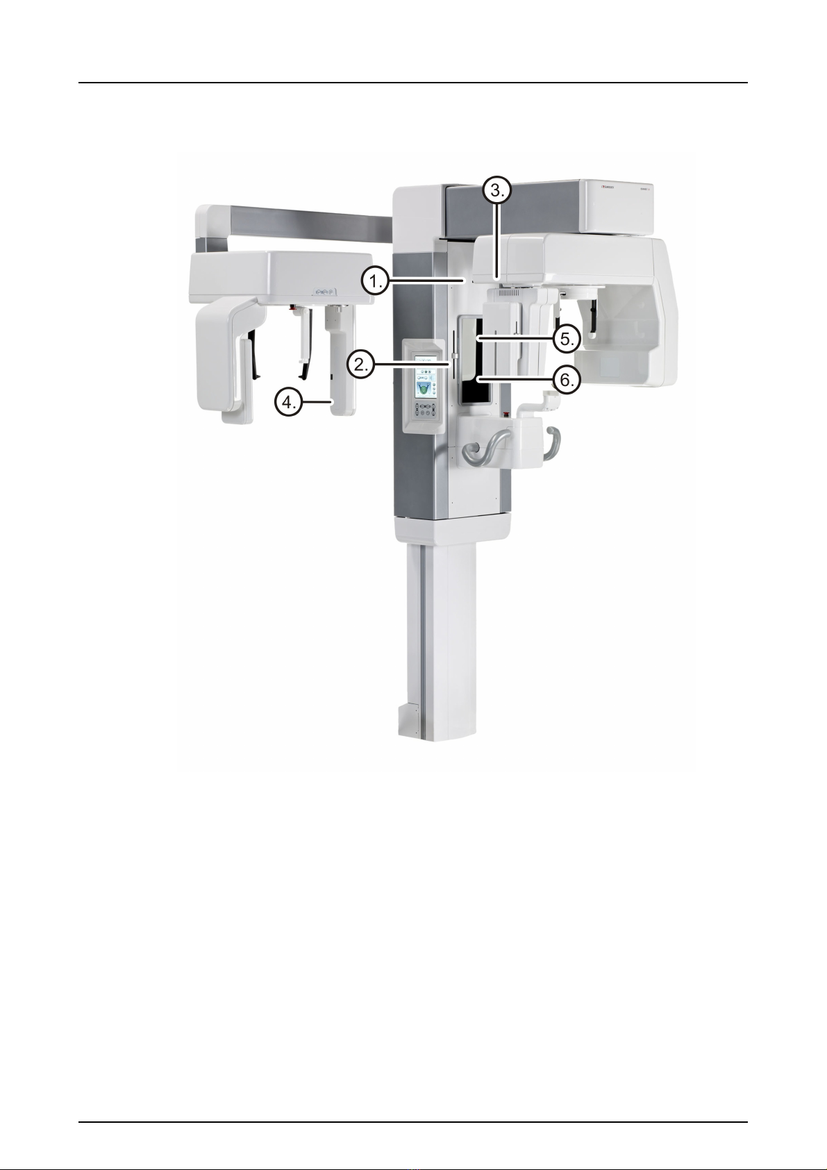
5.3.1 Positioning devices .................................................................................. 37
5.3.2 General instructions ................................................................................. 38
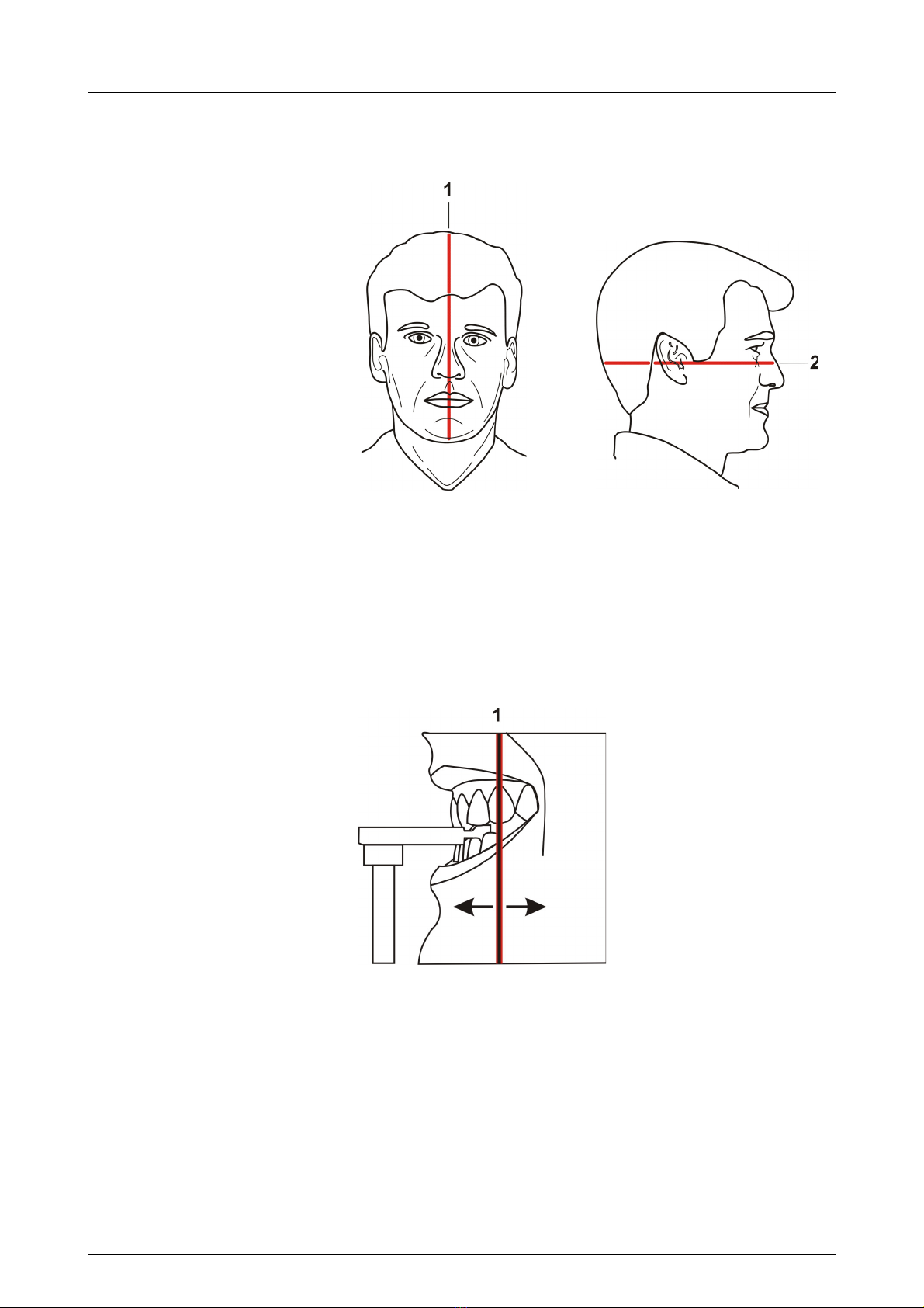
5.3.3 Patient positioning.................................................................................... 39
5.3.3.1 Panoramic exposure.................................................................. 39
5.3.3.2 TMJ exposure............................................................................ 43
5.3.3.3 Maxillary Sinus exposure........................................................... 45
5.3.3.4 Taking a panoramic exposure ................................................... 47
5.4 Cephalometric exposures ................................................................................... 48
5.4.1 General instructions ................................................................................. 48
5.4.2 Patient positioning.................................................................................... 50
5.4.2.1 Full width and reduced width projection..................................... 50
5.4.2.2 PA projection ............................................................................. 52
5.4.2.3 Reverse towne projection .......................................................... 54
5.4.2.4 Waters view ............................................................................... 55
5.4.2.5 Carpus program (optional)
(Not available in USA and Canada)........................................... 57
5.4.2.6 Taking a cephalometric exposure.............................................. 58
5.5 3D exposures ...................................................................................................... 60
5.5.1 Positioning devices .................................................................................. 60
5.5.2 General instructions ................................................................................. 60
5.5.3 Taking a Scout image .............................................................................. 63
5.5.4 Taking a 3D image................................................................................... 65
5.5.5 Stone model and radiographic guide scan............................................... 66
5.6 Warnings and error messages ............................................................................ 68
5.6.1 Acknowledging errors............................................................................... 68
5.6.2 Image transfer errors................................................................................ 68
6 Troubleshooting ........................................................................................................ 69
6.1 Patient positioning............................................................................................... 69
6.2 Image appearance .............................................................................................. 72
6.3 Artefacts .............................................................................................................. 73
6.4 Unit operation...................................................................................................... 75
7 Maintenance............................................................................................................... 77
7.1 Maintenance procedure ..................................................................................... 77
7.1.1 Annual maintenance ................................................................................ 77
7.1.2 Calibration intervals.................................................................................. 77
7.2 Changing the fuses ............................................................................................. 78
7.3 Cleaning and decontaminating the unit............................................................... 78
8 Calibration and adjustment ...................................................................................... 81
8.1 Introduction ......................................................................................................... 81
8.2 Preparing for calibration ...................................................................................... 82
8.3 Panoramic calibration.......................................................................................... 83
8.3.1 Panoramic geometry calibration............................................................... 83
8.3.2 Panoramic pixel calibration ...................................................................... 84
8.3.3 Panoramic Quality Check (optional)......................................................... 85
8.4 3D calibration ...................................................................................................... 87
8.4.1 3D geometry calibration ........................................................................... 87
8.4.2 3D pixel calibration................................................................................... 88
8.4.3 3D Quality Check program....................................................................... 89

rev iii
8.5 Cephalometric calibration.................................................................................... 91
8.5.1 Ceph pixel calibration............................................................................... 91
8.5.2 This calibration should always be a pass.Ceph Quality check program
(Optional) ................................................................................................. 92
9 Technical data............................................................................................................ 95
9.1 Technical specifications ...................................................................................... 95
9.2 Unit dimensions................................................................................................. 105
9.3 Symbols that may appear in the unit................................................................. 106
9.4 Labels on the unit.............................................................................................. 107
9.5 Electromagnetic Compatibility (EMC) tables..................................................... 108
9.6 X-ray tube assemblies....................................................................................... 113
10 PC requirements ...................................................................................................... 115
10.1 Minimum PC requirements................................................................................ 115
10.2 The dental imaging software ............................................................................. 118

iv rev

206718 rev 9SOREDEX 1
1 Introduction
1.1 CRANEX™ 3D
CRANEX 3D is a dental X-ray system for producing high
quality digital images of dentition, TM-joints and skull. In
order to take images with CRANEX 3D, you need a
suitable PC hardware connected to the CRANEX 3D unit
and SCANORA™ software to capture and manage
images.
CRANEX 3D performs the following procedures:
Panoramic
• Adult panoramic
• Sectional panoramic
• Child panoramic (Reduced width and height)
• Bitewing
• TMJ, PA projection
• TMJ, lateral projection (axially corrected)
• Maxillary sinus
Cephalometric (optional)
• Cephalometric Full size Lateral
• Cephalometric Reduced size lateral
• Cephalometric PA/AP projection
• Reverse Towne projection
• Waters view
• Carpus program (optional)
(Not available in USA and Canada)
3D H x W (optional)
• XS 61x41 mm Field of View
• Midi 61x78 mm Field of View
NOTICE! The FOV heights are maximum values measured
at the center of the FOV, the measured height at the edges
of the FOV are smaller.

1 Introduction
2 SOREDEX 206718 rev 9
1.2 Intended use
The unit is intended for dental radiographic examination of
teeth, jaw and TMJ areas by producing conventional 2D x-
ray images as well as x-ray projection images of an
examined volume for the reconstruction of a 3D view.
The unit is also intended for carpus imaging in assisting
cephalometric analyses (Not in USA).
CAUTION! USA only: Federal law restricts this device to
sale by or on the order of a dentist or other qualified
professional.
1.3 Intended user profile
Only for professionally qualified dental / medical personnel.
Typical user is a dental assistant with specific training for
using dental x-ray units.
1.4 Associated documentation
• CRANEX 3D user manual
• CRANEX 3D installation manual
• SCANORA software user manual
• SCANORA software installation manual
• The user manual supplied with the 3D imaging
software
• The installation manual supplied with the 3D
imaging software
1.5 References
The following instructions are delivered with in the
CRANEX 3D installation manual:
• Firmware update instructions
• Calibration instructions
• Cephalostat upgrade instructions
• Cephalostat side changing instructions
• 3D upgrade instructions are delivered with the 3D
upgrade kit.

1 Introduction
206718 rev 9SOREDEX 3
1.6 Abbreviations used in this manual
FOV = Field Of View. The cylindrical 3D volume that is
reconstructed by the system.
ROI = Region Of Interest. The anatomical area or region of
the patient that you are interested to examine.
AES = Automatic Exposure Setting
FH = Frankfort-Horizontal
SMAR™ = SOREDEX™ Metal Artifact Reduction reduces
the effect of metal and other dense radiopaque objects,
which typically create artifacts that are seen as stripes and
shadows.

1 Introduction
4 SOREDEX 206718 rev 9
1.7 Warnings and precautions
1.7.1 Warnings to be observed during use
The unit may be dangerous to the user and the patient, if the
safety regulations in this manual are ignored, if the unit is
not used in the way described in this manual and/or if the
user does not know how to use the unit.
The unit must only be used to take the dental x-ray
exposures described in this manual. The unit must NOT be
used to take any other x-ray exposures. It is not safe to use
the unit to take x-ray exposures, that it is not designed for.
Only professionally qualified dental and/or medical
personnel are allowed to operate the unit and carry out any
diagnoses based on output from the unit.
Because the x-ray limitations and safety regulations change
from time to time, it is the responsibility of the user to make
sure that all the valid safety regulations are fulfilled.
When taking an x-ray exposure of a patient with exceptional
anatomy (typically very tall or large) use the Test-mode (no
x-rays) first to make sure that patient can be positioned
correctly to the unit and for checking that the unit doesn't hit
the patient.
Operator should maintain visible contact with the patient
and technical factors. This allows immediate termination of
radiation by the release of the exposure button in the event
of a malfunction or disturbance.
It is the responsibility of the doctor to decide whether x-ray
exposure or any additional exposures are justified and
necessary.
The minimum height of patient that can be x-rayed in
standing position is 120cm (3.9ft/47.2in) and the maximum
is 200cm (6ft/78in). Taller patients must be seated. These
heights only apply to patients with normal anatomy.
Always use the lowest suitable x-ray dose to obtain the
desired level of image quality.
Always follow the instructions for patient positioning and
imaging procedures instructed in the User Manual.
Avoid taking x-ray exposures of pregnant women.
When taking an x-ray exposure of a child always use the
lowest possible x-ray dose, the smallest possible image
area and the lowest possible resolution that allows you to
perform the required diagnostic task.

1 Introduction
206718 rev 9SOREDEX 5
1.7.2 Warnings for cross infection
If the patient is using a pacemaker, consult the
manufacturer of the pacemaker before taking an exposure
to confirm that the x-ray unit will not interfere with the
operation of the pacemaker.
Decontaminate all the surfaces that the patient is in contact
with after every patient to prevent cross infection.
Decontaminate all device accessories that contact the
patient during a radiographic examination.
Check that the power supply cable is properly attached to
the mains socket and visually check the cable for damage.
If the cable is damaged, it shall be replaced by authorized
service technician only.
Do not open or remove any of the unit’s enclosures. No user
serviceable parts inside.
Do not touch the patient and any exposed electrical connec-
tor, such as sensor connector or Ethernet port, simultane-
ously.
The customer must ensure that the siting environment
fulfills the requirements listed in the Installation manual.
Special attention must be paid to the strength of the floor
and wall materials, electrical mains and radiation protection.
It is the responsibility of the customer to ensure that the site
is large enough for the patients.
The unit contains toxic materials that need to be handled
properly when disposing the unit. Return the unit to the
dealer in the end of its life cycle.
Excessive dust should be cleaned from the unit for free
airflow and cooling. Switch the unit OFF before cleaning.
In case of water damage/water dropping over the product,
call for service technician to ensure the product is fully
operational according to specification.
Always use available disposable protective covers with the
patient positioning accessories:
• Bite block cover
• Chin plane cover
• TMJ support cover
• Lip holder cover
• Chin cup cover

1 Introduction
6 SOREDEX 206718 rev 9
1.7.3 General warnings
Personnel operating the device must be adequately trained
with respect to the technological principles of operation and
radiation protection when using cone beam computed
tomography (CBCT) imaging.
This unit complies with the EMC (Electromagnetic
Compatibility) according to IEC 60601-1-2. Radio
transmitting equipment, cellular phones etc. shall not be
used in close proximity of the unit as they could influence
the performance of the unit.
Always ensure to fulfill the requirements of the local and na-
tional regulations.
The correct software and settings in the workstation are
essential to the performance of the unit. Consult technical
support to ensure correct setup.
Danger: Explosion hazard - do not use in the presence of
flammable anesthetics, gases or vapors.
The unit is factory set to operate using a 230-240 ±10 VAC
power supply. Never connect the unit to a power supply
different to the voltage marked on the unit.
If the unit needs to be connected to a multiple socket-outlet,
the socket shall not be placed on the floor.
To avoid the risk of electric shock, the unit must only be
connected to a supply mains with protective earth.
The site must fulfill the environmental requirements in the
installation manual chapter technical specifications.
There should be free space around the unit for safe
operation.
The PC / Ethernet switch to which the unit is connected,
should be approved appropriately (e.g. EN 60950, IEC
60950, UL 60950). After installation, check that the IEC
60601-1 leakage current levels are not exceeded.
This product itself complies with IEC 60601-1 medical
safety standard but in order to the system incorporating also
a PC to comply the standard, EITHER the PC has to be a
medical PC OR the PC has to be located over 1,5 meters
apart from the unit. The installer and the user of the system
shall confirm that at least one of the above requirements is
fulfilled. A PC is a medical one if it complies IEC 60601-1
standard and that is indicated in the accompanying
documents of the PC.

1 Introduction
206718 rev 9SOREDEX 7
The unit shall be connected directly to the acquisition PC
with an Ethernet cable. Connection through the LAN-net-
work of the site is not allowed. Two network ports are
needed in the PC in order to connect also to the site
network.
All service operations must be made by authorized service
personnel only.
The annual service as described in manual is mandatory for
the correct and safe operation of the unit.
When taking exposures, operators and service personnel
must protect themselves from radiation and remain at least
two meters (six feet) away from the unit during exposure.
Protect the patient from scattered radiation by placing a
protective lead apron over the patient.
The unit must be installed and serviced according to the unit
Installation & adjustments manual by a qualified technician.
Only personnel trained and approved by the manufacturer
of the unit are allowed to service the unit.
3D imaging should not be used for routine or screening
examinations in which a radiograph is taken regardless of
the presence or absence of clinical signs and symptoms. 3D
imaging examinations must be justified for each patient to
demonstrate that the benefits outweigh the risks.
Where it is likely that evaluation of soft tissues will be
required as part of the patient’s radiological assessment,
the imaging should be done using conventional medical CT
or MR, rather than 3D imaging using Cone Beam
technology.
Cone beam computed tomography images are not
adequate for the analysis of soft tissue.
Panoramic and 3D exposures should not be used if
conventional intra-oral radiographic images (like bitewing
exposures) would be sufficient.
Make sure that patient’s thyroid glands are protected by a
lead apron during the exposure.
The place where the unit is to be installed and the position
from where the user will take exposures must be correctly
shielded from the radiation that is generated when the unit
is operated. Ensure to fulfill or exceed the requirements of
your local regulations.
The unit or its parts must not be changed or modified in any
way without approval and instructions from the
manufacturer.

1 Introduction
8 SOREDEX 206718 rev 9
1.8 Disclaimer
The manufacturer shall have no liability for consequential
damages, personal injury, loss, damage or expense
directly or indirectly arising from the use of its products. No
agent, distributor or other party is authorized to make any
warranty or other liability on behalf of the manufacturer with
respect to its products.
1.9 Disposal
The device, its spare parts, its replacement parts and its
accessories may include parts that are made of or include
materials that are non-environmentally friendly or
hazardous. These parts must be disposed of in
accordance with all local, national and international
regulations regarding the disposal of non-environmentally
friendly or hazardous materials.
Unit has at least the following parts that should be
regarded as non-environmental friendly waste products:
■Tubehead (Pb, oil)
■Collimator (Pb)
■All electronic circuits, electronic boards inside
■Sensor covers (EMC painted)
When servicing use only approved replacement parts
supplied by the manufacturer.
The use of accessories not complying with the equivalent
safety requirements of this equipment may lead to a
reduced level of safety of the resulting system.
If this device is used with 3rd party imaging application
software not supplied by the manufacturer, the 3rd party
imaging application software must comply with all local laws
on patient information software. This includes the Medical
Device Directive 93/42/EEC and/or relevant legal
requirements in the USA.
Do not connect any equipment to the unit that has not been
supplied with the unit or that is not recommended by the
manufacturer. The use of accessory equipment not
complying with the equivalent safety requirements of this
equipment may lead to a reduced level of safety of the
resulting system.
All protective covers must be properly installed before
handing unit to the user or when operating the unit.
Mirror shall be closed before taking exposures.

206718 rev 9SOREDEX 9
2 Unit description
2.1 Main parts and controls
PC with MDD approved dental imaging software and 3D
viewing software (not included).
All software must conform to the MDD and the relevant
legal requirements in the USA.
The PC must conform to all the unit and dental imaging
software requirements.
1. Column
2. Carriage
3. Main support
4. Rotating unit
5. On / off switch (rear of
carriage) and main fuses
6. Tubehead assembly
7. ClearTouch™control panel
8. Patient positioning panel
9. Sensor head
10.Head support
11.Chin rest
12.Handles
13.Cephalometric unit
14.Cephalometric sensor
15.Secondary collimator
16.Patient positioning panel
On/off switch (used to power the unit on
and off) and main fuses.

2 Unit description
10 SOREDEX 206718 rev 9
1. Sensor holder (panoramic units without 3D option)
2. Panoramic sensor
1. 3D sensor (units with 3D option)
2. Panoramic sensor

2 Unit description
206718 rev 9SOREDEX 11
2.2 Patient positioning lights
1. Midsagittal
2. Frankfort-horizontal (FH) light
3. Focal trough
4. Cephalometric FH light
5. Horizontal light, top of FOV (3D option only)
6. Horizontal light, bottom of FOV (3D option only)

2 Unit description
12 SOREDEX 206718 rev 9
Panoramic lights
1. Midsagittal light
2. Frankfort-horizontal light
1. Focal through light
Table of contents
Other Soredex Dental Equipment manuals
Popular Dental Equipment manuals by other brands

DentalEZ
DentalEZ RAMVAC Utility user manual

ivory
ivory SL631C Operation manual

KaVo
KaVo Centerline EWL 4011 Operating, Maintenance and Assembly Instructions

NSK
NSK Ti-Max nano65LS Operation manual

Schick Dental
Schick Dental C2 - Profi operating instructions

Ivoclar Vivadent
Ivoclar Vivadent Programat EP 5000 operating instructions