BAG HISTO TYPE Rainbow QS6 User manual

EN Instructions for Use
HISTO TYPE Rainbow QS6
Test kit for the determination of HLA alleles on a molecular genetic basis
IVD 0123
REF 728221
Contents
1. INTENDED USE......................................................................................................................2
2. PRODUCT DESCRIPTION......................................................................................................2
3. TEST PRINCIPLE....................................................................................................................2
4. MATERIAL...............................................................................................................................3
4.1 Contents of the HISTO TYPE Rainbow QS6 kit.......................................................................3
4.2 Additionally required reagents and devices..............................................................................3
4.3 Validated real time-PCR Cycler................................................................................................3
5. STORAGE AND STABILITY....................................................................................................4
6. TEST PROCEDURE................................................................................................................4
6.1 Precautions and special remarks.............................................................................................4
6.2 DNA Isolation...........................................................................................................................4
6.3 Entering sample information into PlexTyper®Software............................................................5
6.4 Amplification ............................................................................................................................5
6.5 Export of results from QuantStudio™ 6 Flex System ...............................................................9
6.6 Evaluation and interpretation of the results............................................................................10
6.7 Import of the results file into the PlexTyper®software............................................................11
7. WARNINGS AND DISPOSAL INSTRUCTIONS ....................................................................12
8. KIT SPECIFICATIONS ..........................................................................................................13
8.1.1 Diagnostic sensitivity and specificity........................................................................13
8.1.2 Analytical sensitivity and measuring range ..............................................................13
8.1.3 Analytical specificity / cross reactive substances.....................................................14
9. LIMITS OF THE METHOD.....................................................................................................14
10. INTERNAL QUALITY CONTROL...........................................................................................14
11. TROUBLESHOOTING...........................................................................................................15
12. TRADE NAMES USED ..........................................................................................................15
13. EXPLANATION OF THE SYMBOLS USED ON THE LABELS..............................................16
14. LITERATURE ........................................................................................................................16
15. REVIEW HISTORY................................................................................................................16
Version: 02/2021 / Issue: 2021-06

Instruction for Use HISTO TYPE Rainbow QS6 Version: 02/2021
Page 2 of 16
1. INTENDED USE
The intended use of the HISTO TYPE Rainbow QS6 kit is the identification of HLA Class I and II
alleles using the QuantStudio™ 6 Flex System for PCR amplification. HISTO TYPE Rainbow QS6 is
an in vitro diagnostic test for tissue typing on a molecular genetic basis (see Product Description).
2. PRODUCT DESCRIPTION
HISTO TYPE Rainbow QS6 kits are used for the molecular genetic determination of HLA Class I
and II alleles at 11 loci: HLA-A, B, C, DRB1/3/4/5, DQA1, DQB1, DPA1 & DPB1. Kits are designed
to generally detect all alleles at the 11 loci; if any rare alleles are not detected the alleles are listed in
Kit Specific Information documents (KSI) which are available from the download section of the BAG
website. The primer and probe binding sites are listed there as well. The kit provides low to medium
resolution typing results of the common and well documented alleles using CWD list 2.1.0 which is
largely based on CWD 2.0.0 list1. The CWD list 2.1.0 used is available from the document download
section of the BAG website. Confirmed diagnostic results of HLA alleles are a prerequisite for a
successful organ transplantation.
3. TEST PRINCIPLE
The test is performed with genomic DNA as starting material. The DNA is amplified in a real-time
PCR with sequence-specific primers (SSP). The primers were specially developed for the selective
amplification of segments of specific HLA alleles or allele groups. The amplicons are detected using
sequence-specific fluorescence dye-labelled hydrolysis probes (TaqMan®-probes), which increases
the sensitivity and specificity of the test compared to the classical SSP.
If amplicons are present, the probes are hydrolysed by the Taq polymerase and a fluorescence
signal is generated to enable detection of the amplicon. Five different wavelength ranges of
fluorescence signals are measured by the optical detection unit of the real time PCR cycler. The
presence of a positive reaction is determined primarily by the Cq point, which is the point where
fluorescence signal increases beyond the baseline threshold. For amplification to be valid the
amplification must also achieve a certain threshold of fluorescence at the end of the PCR process.
This is to prevent false positive reactions.
Each PCR reaction also contains an internal amplification control (Human Growth Hormone gene
(HGH)) which is detected in a specific fluorescent channel.
To distinguish positive reactions from negative or irrelevant amplifications the ratio of the Cq of the
specific reaction compared to the Cq of the internal amplification is calculated. The thresholds for
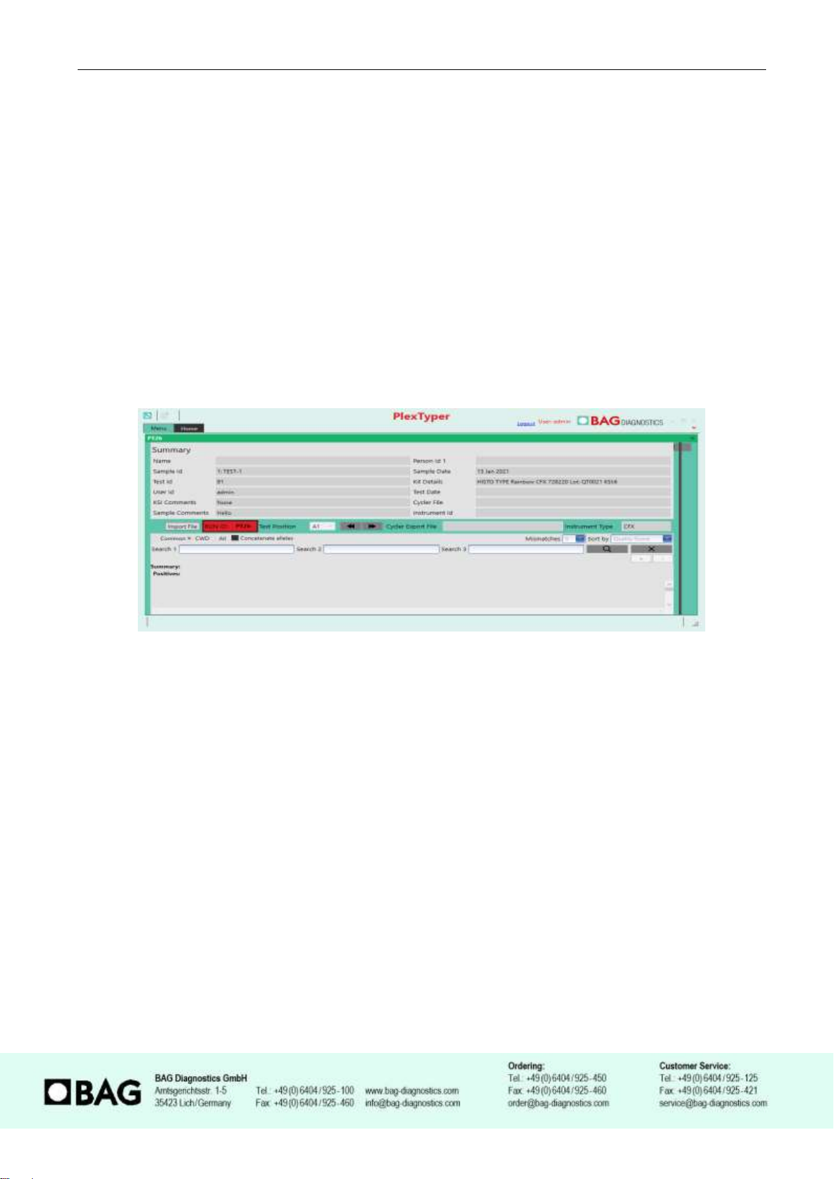
these Cq ratios (CqR) vary from reaction to reaction and hence the PlexTyper®software is required
for the analysis of amplification data.

Instruction for Use HISTO TYPE Rainbow QS6 Version: 02/2021
Page 3 of 16
4. MATERIAL
4.1 Contents of the HISTO TYPE Rainbow QS6 kit
•10x 230 µl Plex Mix, ready to use, contains dNTPs, Taq Polymerase, reaction buffer.
•10x HISTO TYPE Rainbow QS6 plates for HLA-typing. The pre-pipetted and dried reaction
mixtures in each well contain HLA-specific primers and probes as well as HGH-specific
internal amplification control primers and probes (oligomixes).
•10x qPCR Seal
4.2 Additionally required reagents and devices
•Reagents for DNA isolation (validated extraction kits see 6.2)
•Real time-PCR Cycler (validated cycler see 4.3)
•Plate holder QS6 (REF 726321)
•Variable pipettes (0.5 –1000 µl) and pipette tips
•Application spatula for qPCR Seal
•Molecular grade DNAse free water
•Suitable plate centrifuge
•For QuantStudio™ 6 calibration please use BAG RT-PCR Universal Custom Dye Calibration
Kit REF 728260
4.3 Validated real time-PCR Cycler
Real time-PCR Cycler
QuantStudio™ 6 Flex System, Applied Biosystems / Thermo Fisher Scientific (QS6)
The following fluorophores are used in the HISTO TYPE Rainbow QS6 kits.
Fluorophore
Wavelength in nm
TAMRA
Excitation: 557
Emission: 583
FAM
Excitation: 495
Emission: 520
O560 (CAL Fluor®Orange 560)
Excitation: 538
Emission: 559
R610 (CAL Fluor®Red 610)
Excitation: 590
Emission: 610
Q670 (Quasar®670)
Excitation 647
Emission: 670

Instruction for Use HISTO TYPE Rainbow QS6 Version: 02/2021
Page 4 of 16
5. STORAGE AND STABILITY
The kits are shipped on blue ice. All reagents must be stored at ≤-20°C in temperature-controlled
devices. The expiry date is indicated on the label of each reagent. The expiry date indicated on the
outer label refers to the reagent with the shortest stability contained in the kit. The freeze-thaw cycle
testing has shown that up to 12 cycles for the Plex Mix has no detrimental effects on the quality of
the kit.
6. TEST PROCEDURE
6.1 Precautions and special remarks
Molecular genetic techniques are extremely sensitive methods and should only be performed by
qualified personnel with experience in molecular genetic techniques.
Special precautions must be met to avoid contamination and thus false reactions:
◆
Wear suitable gloves (preferably powder-free) during work.
◆
Use tips with filter insert or integrated stamp.
◆
Work in two different areas for pre-amplification (DNA-isolation, preparation of the reactions)
and post-amplification (detection); use two separate rooms if possible.
◆
Use devices and other materials only at the respective workplaces and do not exchange them.
6.2 DNA Isolation
The specimen material for the isolation of the genomic DNA must be sent in appropriate collection
systems. For genomic material from whole blood use only EDTA or citrate anticoagulants. The
presence of heparin may potentially inhibit the PCR reaction (2), therefore such collection systems
are not suitable and must not be used. It is recommended to use IvD certified kits for the DNA
isolation.
Validated DNA Extraction Kits:
•Qiagen QIAamp DNA Blood Kits (columns)
Both, the manual isolation and automated DNA isolation (QIAcube) are validated.
If the standard method established in the laboratory shall be applied for isolation of gDNA without
using one of the specified test kits, it must be validated by the user.
The HISTO TYPE Rainbow QS6 test requires 10 –20 ng DNA per well. The purity indices must be
in the following range:
•OD260 / OD280 = 1,5 and < 2,0
Higher values are an indicator for the presence of RNA, lower values
indicate protein contamination.
•OD260 / OD230 = > 1,8
Lower values indicate contamination with carbohydrates, salts or
organic solvents.

Instruction for Use HISTO TYPE Rainbow QS6 Version: 02/2021
Page 5 of 16
6.3 Entering sample information into PlexTyper®Software
It is mandatory to use PlexTyper®Software to analyse HISTO TYPE Rainbow data. It is advisable
to enter the sample information into PlexTyper®before setting up the PCR amplification to obtain
the unique RUN ID. All import files must be prefixed with a PT number.
When saving a test in PlexTyper®, the software assigns a Run ID to the test which is displayed in
red in the summary window shown below. This is a consecutive number prefixed with PT (PT1, PT2,
PT3….) which is linked to the sample identity and is used as a prefix on the PCR machine export file
to correctly link the PCR export to the sample identity.
This Run ID should be used when setting up the file identity in the PCR machine. If the PCR excel
export file is not pre-labelled with the RUN ID the file can be manually prefixed with the correct PT
Run ID before import into PlexTyper®.
6.4 Amplification
A pre-mix consisting of Plex Mix, water and DNA is made for the amplification that is subsequently
dispensed in the wells 1-95. In well 96 there is the negative control (no template control = NTC)
which should only contain water and Plex Mix. For other DNA concentrations the pre-mix has to be
modified accordingly (see below).
•The reaction volume for each PCR preparation is 10 µl
•For a single well the following reagents must be pipetted into a reaction tube:
2 µl Plex Mix
1 µl DNA specimen
7 µl Molecular grade water
A negative control (NTC) should be performed. Therefore prepare a PCR reaction with molecular
grade water instead of DNA.
2 µl Plex Mix
8 µl Molecular grade water

Instruction for Use HISTO TYPE Rainbow QS6 Version: 02/2021
Page 6 of 16
DNA concentration 10-20 ng/µl
•Add 805 µl molecular grade water to the vial with 230 µl Plex Mix and mix (vortex briefly 1-3
sec).
•After mixing, pipette 10 µl of the mix into the NTC well (well 96; position H12 -see also
Figure 1 and 2).
•Then pipette 115 µl DNA into the vial with the remaining Plex Mix-water-mixture and mix
(vortex briefly 1 –3 sec).
•Distribute 10 µl of the DNA/Plex Mix/water solution into each of the wells 1-95 of the HISTO
TYPE Rainbow QS6 plate (please note figure 1 and 2). The NTC well (well 96; position H12)
must not be filled with the DNA mix as this will cause the NTC to be positive and can
invalidate the test!
DNA with other concentrations
For DNA of different stock concentrations use the dilution table below to adjust the DNA to working
concentration.
•Add 8 µl Molecular grade water and 2 µl Plex Mix to the NTC (well H12). The NTC well (well
96; position H12) must not be filled with the DNA mix!
•Pipette the DNA and the molecular grade water into the remaining 228 µl Plex Mix according
to the following table and mix (vortex briefly 1-3 sec)
Depending on the concentration of DNA, pipette the applicable volumes in the table below to the
228 µl Plex Mix remaining in the vial after setting up the NTC:
Concentration of the DNA [ng/µl]
Molecular grade water [µl]
DNA volume [µl]
2
342
570
5
684
228
50
889
23
80
898
14
100
901
11
150
904
8
200
906
6
250
907
5
300
908
4
500
910
2
•Distribute 10 µl of the DNA-Plex Mix-water solution into each of the wells 1-95 of the HISTO
TYPE Rainbow QS6 plate.

Instruction for Use HISTO TYPE Rainbow QS6 Version: 02/2021
Page 7 of 16
Please note: When pipetting into the PCR wells it is important not to allow the pipette tip to contact
the dried mix (dyed blue) in the bottom of the well. It is advisable to pipette to the side of the well to
allow the 10 µl to mix by gravity with the dried mix (see Figure 1).
Figure 1
Figure 2: HISTO TYPE Rainbow QS6 plate. In wells A1 to G12 are the dried specific mixes (blue coloured). In well H12
there is the dried NTC (also blue).
Seal the PCR plate with the supplied qPCR seal and briefly spin the liquid down. Make sure that the
plate is completely sealed, particularly at the edge of the plate. Make sure that the liquid has
contacted the dried mix and there are no bubbles or air gaps in the reaction wells. If bubbles
appear, gently tap the tubes on the laboratory bench to remove them, or preferably careful spin it
down (10 sec).
Place the sealed PCR plate in the QS6 instrument. Please use the correct plate holder and
orientation as per the notes below.
Note: Please make sure that you place the reaction plate into the cycler in the correct orientation
as shown in Figure 2 above where the NTC well is in the bottom right corner, position H12.
Note: For the HISTO TYPE Rainbow plates a special plate holder for the QuantStudio™ 6 Flex
System is required, which is included in the first starter package (REF 726321). For more
information please contact BAG Diagnostics.

Instruction for Use HISTO TYPE Rainbow QS6 Version: 02/2021
Page 8 of 16
PCR-Program
Please note: HISTO TYPE Rainbow uses custom fluorophores. Prior to use instruments must be
calibrated with Custom Dye Calibration Kit available from BAG Diagnostics, REF 728260.
Following the manufacturer’s user guide for the QuantStudio™ 6 Flex System, set up and save a
PCR protocol with the following parameters:
HISTO TYPE RAINBOW QS6 PCR program set up:
Use the following parameters to set up the PCR program.
Instrument type: QuantStudio™ 6 Flex System
Block type: Fast 96-Well (0.1mL)
Experiment type: Comparative Ct (∆∆Ct)
Reagent type: TaqMan® Reagents
Run properties: Standard
Define Targets:
Please note: Custom dye instrument calibration must be performed prior to initial use of a new
QS6 instrument.
Target Name
Reporter
Quencher
TAMRA
TAMRA
NFQ-MGB
FAM
FAM
NFQ-MGB
Orange560
O560 (Cal Fluor Orange 560)
NFQ-MGB
RED
R610 (Cal Fluor Red610)
NFQ-MGB
Q670
Q670 (Quasar670)
NFQ-MGB
Passive Reference: None
Assign: Assign all targets to each well.
Run Method:
Reaction volume 10 µl
Stage
Cycles
Data Collection
Target
(°C)
Hold
(hh:mm:ss)
Ramp
rate
(°C/s)
Hold Stage
1
Off
96
00:02:00
2.5
PCR Stage
13
Off
98
00:00:05
2.5
68
00:00:25
2.2
PCR Stage
37
Off
On
98
00:00:05
2.5
68
00:00:25
2.2

Instruction for Use HISTO TYPE Rainbow QS6 Version: 02/2021
Page 9 of 16
6.5 Export of results from QuantStudio™ 6 Flex System
After the PCR run is complete there is no requirement for analysis in the Applied Biosystems
software. For analysis in PlexTyper®the QS6 .eds file must be opened and an excel file exported
using either on the machine software or on separate QS6 software.
Export the raw data in excel format with the following steps.
1) Select the .eds file, double click on a .eds file. If the QuantStudio RealTime PCR software is
installed the software will automatically open the .eds file. Navigate to the export button and
select export.
2) In QS6 software (with reference to image below) (A) select the folder location where you
want the export to go, select open folder to save. (B) If required rename the file by prefixing
with the PT appropriate run ID from entering the sample in PlexTyper®prior to running test.
(C) if not selected, select excel format, (D) select export

Instruction for Use HISTO TYPE Rainbow QS6 Version: 02/2021
Page 10 of 16
3) If the excel file has not been set up in the QuantStudio™ 6 Flex System with the PT prefix
number it should now be given the correct prefix obtained when the sample idenity and run
identity was entered into PlexTyper®.
6.6 Evaluation and interpretation of the results
For evaluation and interpretation of the data it is mandatory to use the PlexTyper®software
(available free of charge from BAG Diagnostics) in conjunction with the kit specific data files. The kit
files required for the evaluation are available from the BAG Diagnostics download server
(www.service.bag-diagnostics.com).
Please make a note of the lot number of the kit. The interpretation kit files are product and lot
specific. Use of incorrect kit files could result in incorrect genotyping. For interpretation of the results
the raw data must be transferred from the thermocycler to a computer running the PlexTyper®
software (e.g. with a suitable USB drive).
HISTO TYPE Rainbow QS6 is a 5-channel, 96 well multiplex genotyping test. It is possible to view
data on the amplification machine software, but it is not possible to interpret without use of the
PlexTyper®software. For QuantStudio™ 6 Flex System analysis colour compensation must be
applied.

Instruction for Use HISTO TYPE Rainbow QS6 Version: 02/2021
Page 11 of 16
6.7 Import of the results file into the PlexTyper®software
Please follow the instructions for use for the PlexTyper®software for interpretation of the
data.
Open the PlexTyper®software. From the home screen choose View plates with no associated
results under Plates. A list opens with all tests that are not linked with raw data yet. There is a
global Search field at the top of the table to search the whole table.
Double click on the required test to be interpreted; this opens the results summary window . Select
Import File and select the excel file with the correct PT RUN ID prefix (e.g. PT999.xls) exported
from the QuantStudio™ 6 Flex System.
Initial loading of the kit file data takes approximately 45 seconds, unless it is already open, the
bottom left corner of the screen shows a progress bar. After that the results for user review are
presented.
A negative control (NTC) is used as contamination control. If DNA or contaminating amplicon is
inadvertently added to the NTC reaction a positive signal will occur. If the Cq is less than 36 it will be
detected as possible contamination by the PlexTyper®software and a warning message is
generated. Amplification signals above Cq 36 in the NTC are regarded as PCR artefacts and are
disregarded. If PCR contamination is suspected, it is advisable to follow local decontamination
guidelines and to exchange the reagents.
The raw data collected from the cycler-specific software will be imported into the PlexTyper®
software. Based on the Cq values, RFUs (relative fluorescence units), quality scores and the curve
progression the PlexTyper®software determines the molecular genetic HLA pattern of the
specimens used (see instructions for use for PlexTyper®for details). Positive reactions are
determined based on the Cq ratio between the the Cq of the internal amplfication control (IAC) and
the Cq of the HLA allele specific reaction (exception see below).
Special consideration for analysis of HISTO TYPE Rainbow QS6 results:
In these kits a strong allele specific O560 signal may affect the appearance of the TAMRA IAC
amplification due to emission wavelength interference. This can result in sporadic IAC failure due
either to a false late IAC Cq value, or the reaction failing due to the fluorescence values being
negative in the last cycle. Figures below illustrate the possible appearances of the TAMRA IAC
signals when the O560 signal is strong.
Search

Instruction for Use HISTO TYPE Rainbow QS6 Version: 02/2021
Page 12 of 16
If the IAC amplification is deemed negative, or late due to O560 interference this can lead to no
result being found for the affected HLA gene locus. To automatically correct for this the thresholds
relating to the IAC have been modified for reactions where this phenomenon has been observed
(reactions 7, 20, 43, 52, 63, 84 & 85) so that correct genotypes can be generated automatically. The
software generates a message to inform the user about it.
It may still happen that the problem occurs in other reactions as well or that non-optimal shapes of
the amplification curve for other fluorophores result in a failure to generate an automatic result in
PlexTyper® . For a general procedure to deal with failed automatic generation of results please refer
to the PlexTyper® instructions for use (V4.X-1/2021 chapter 7.4.5 How to investigate ‘No results’ at
an HLA locus).
7. WARNINGS AND DISPOSAL INSTRUCTIONS
HISTO TYPE Rainbow QS6 is designed for in vitro diagnostic use. The kit should only be used
by specially trained, qualified personnel. All work should be performed in accordance with Good
Laboratory Practice.
All materials of biological origin used in the test to obtain DNA (e.g. blood) should be considered as
potentially infectious. Therefore, appropriate safety precautions are recommended when handling
biological materials as required by Good Laboratory Practice standards.
Biological materials must be inactivated before disposal (e.g. by autoclaving). Disposable materials
must be autoclaved or incinerated after use.
Spilled potentially infectious material should be removed immediately with an absorbent paper towel
and the contaminated area disinfected with an appropriate disinfectant or 70% Ethanol. Material
used to remove spills must be inactivated before disposal (e.g. by autoclaving).
Disposal of all specimens, unused reagents and waste should be in accordance with the legislation
of the respective country and the local authorities.

Instruction for Use HISTO TYPE Rainbow QS6 Version: 02/2021
Page 13 of 16
Microbial contamination of reagents while taking aliquots should be avoided. The use of sterile
disposable pipettes and pipette tips is recommended. Do not use reagents looking cloudy or
showing signs of microbial contamination.
A Material Safety Data Sheet (MSDS), respectively a declaration on Material Safety Data Sheets is
available for download at www.bag-diagnostics.com .
8. KIT SPECIFICATIONS
The combination of primers and probes allows the determination of human HLA class I and II alleles
according to lot-specific data (low to medium resolution, detection of all alleles with the exception of
single rare alleles). The accuracy and reproducibility of the reactivity of the test kit is checked for
each lot with control specimens with known HLA-alleles. The kit determines the HLA-Loci A, B, C,
DRB1, DRB3, DRB4, DRB5, DQA1, DQB1, DPA1 and DPB1.
8.1 SPECIFIC PERFORMANCE CHARACTERISTICS
8.1.1 Diagnostic sensitivity and specificity
A total of 116 DNA samples pre-typed for the relevant HLA loci or international reference cell lines
were tested with the HISTO TYPE Rainbow QS6 kit to show the correct reactivity. There was a
100% concordance of results with the pre-typings for all loci.
Locus
Number of sample pre-typed for the locus
concordant
% concordance
A
116
116
100%
B
116
116
100%
C
116
116
100%
DRB1
115
115
100%
DRB3
113
113
100%
DRB4
113
113
100%
DRB5
113
113
100%
DQA1
114
114
100%
DQB1
114
114
100%
DPA1
113
113
100%
DPB1
114
114
100%
8.1.2 Analytical sensitivity and measuring range
Validation tests have shown that variation of the amount of DNA from 8 ng to 30 ng per reaction has
no significant effect on the specific detection of HLA alleles.

Instruction for Use HISTO TYPE Rainbow QS6 Version: 02/2021
Page 14 of 16
8.1.3 Analytical specificity / cross reactive substances
Eight substances that might interfere with the assay were tested and the following concentrations
were shown to have no detrimetal effect on the results:
Substance
Maximal non-inhibitory concentration
Protein (BSA)
0.2 mg/ml
TE (Tris/EDTA, pH 8.0)
7 mM Tris, 0.7 mM EDTA
NaCl
20 mM
Ethanol
1%
Haemoglobin
0.01 mg/ml
Sodium Citrate
7 mM
DNA extraction buffer 1 (Qiagen QIAamp DNA Blood Kits)
1%
DNA extraction buffer 2 (Qiagen QIAamp DNA Blood Kits)
2%
9. LIMITS OF THE METHOD
During DNA isolation, special attention must be paid to the fact that the RT-PCR method reacts very
sensitively to cross-contaminations. Special care should be taken to avoid contamination of kit
reagents and other laboratory materials with amplicons or DNA.
The performance of a negative control without DNA in well H12 is strongly recommended. No
fluorescence signal below 36 Cq should be detected in the NTC (H12) with molecular grade water.
In the case of signal development in the negative control, the PCR laboratory workplace may have
to be decontaminated from DNA and the reagents exchanged if necessary.
All devices (e.g. pipettes, real-time cyclers) must be calibrated according to the manufacturer’s
specifications.
10. INTERNAL QUALITY CONTROL
Internal quality controls for new lots can be performed with a combination of DNA specimens with
known HLA type. An internal control to verify successful amplification is included in the dried
oligomixes.
Performance of negative controls (well H12) to detect possible contaminations is recommended. For
this purpose, prepare a test without DNA (NTC), see section 6.4 Amplification.

Instruction for Use HISTO TYPE Rainbow QS6 Version: 02/2021
Page 15 of 16
11. TROUBLESHOOTING
For interpretation trouble shooting please see the Instructions for Use for PlexTyper®Software.
Symptom
Possible Reason
Potential Solution
TAMRA signals
weak or absent
across whole
plate
Presence of an inhibitor in the DNA.
Try a different extraction or sample.
Insufficient DNA in the reaction.
Repeat test with correct amount of DNA.
Wrong amplification parameters.
Check PCR program.
Contaminated or degraded DNA.
Check concentration / quality of DNA.
Check DNA on a gel.
Repeat DNA isolation.
Degraded Fluorescent probes or
primers.
Avoid exposure to light and frequent thawing and
freezing.
Observe storage conditions.
Poor or no
TAMRA signal
in individual
wells
Bubbles in the reaction / residual
liquid at the inner wall of the tube.
Careful pipetting.
Spin down PCR plate.
User error.
Ensure all wells receive the required volume of
reagents.
Evaporation of the reagents due to
incorrect closing of the PCR tubes.
Make sure that the PCR tubes are closed properly.
Caution with adhesive foils in the edge area.
Strong O560 signal supressed
TAMRA signal resulting in potential
false negative allele specific results.
Use data review in PlexTyper®to correct results.
Signal in the
Negative
Control
Contamination with DNA or amplicon
in the negative control.
Repeat the test.
Decontaminate the workplace.
Amplification due to PCR artefact.
Review after import into PlexTyper®, if the signal may
be below the thresholds or the data may in fact be OK
(please note the PlexTyper®Instructions for Use).
12. TRADE NAMES USED
QuantStudio™ 6 Flex System is a tradename of Applied Biosystem (Thermo Fisher Scientific)
TaqMan®is a tradename of Roche Molecular Systems Inc.
® Cal Fluor & Quasar Dyes are the registered trademark of LGC Biosearch Technologies

Instruction for Use HISTO TYPE Rainbow QS6 Version: 02/2021
Page 16 of 16
13. EXPLANATION OF THE SYMBOLS USED ON THE LABELS
Sufficient for n tests
Storage temperature / lower limit of temperature
Consult to instruction for use
Manufacturer
Use by
CONT
Content, contains
eIFU
Electronic instructions for use
HLA TYPING
Intended use: HLA typing
IVD
For in vitro diagnostic use
LOT
Batch code
PCRFOIL
PCR foil
PCRPLATE
PCR plates
PLEX MIX
Plex Mix: Mastermix, contains dNTPs, Taq polymerase, reaction buffer
REACTIONMIX
Reaction mixes
REF
Catalogue number
RTU
Ready to use
14. LITERATURE
1. Mack, S.J. et al., 2013. Tissue Antigens 81, 194–203
2. Beutler, E. et al., 1990. BioTechniques 9:166
15. REVIEW HISTORY
Version: 01/2021 / Issue: 2021-04: Initial version
Version: 02/2021 / Issue: 2021-06: Page 3: Adjustment of the naming of the fluorophores TAMRA
and FAM.
Page 8: Under "Define targets" "Target Name" the indications
changed to TAMRA, FAM, Orange560 and RED.
Pages 11-12: Text added to explain ‘Special consideration for
analysis of HISTO TYPE Rainbow QS6 results’.
Page 15: Trouble shooting table modified to advise on
additional possibility of IAC amplification detection failure.
For further information please refer to our website www.bag-diagnostics.com or contact us directly
Table of contents