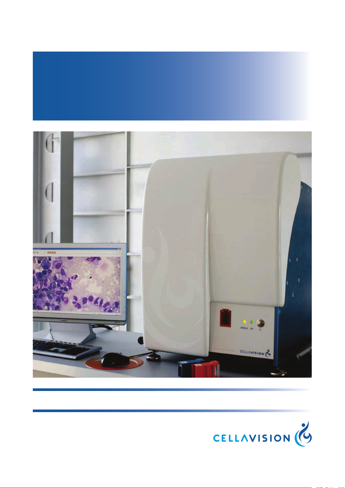
CellaVision DM1200 User manual

CellaVision®DM1200
USER’S MANUAL 6.0


CellaVision®DM1200
Preface
CellaVision®is a trademark of CellaVision AB.
All other trademarks used in this document are property of their respective owners.
No part of this document or the products it describes may be reproduced or transmitted by any
means or in any form without prior consent in writing from CellaVision AB.
U.S. patents no. 6268611, 6341180, 7034883, 7327901, 7450762, 8914255
Swedish patents no. 0101319, 9902863, 1350568-0
German patents no. 60045076, 602004008471, 602007037624, 602008017260,
602009011617, 602010005069, 60231032, 60231032
French patents no. 1210634, 1646964, 2204686, 2310892
U.K. patents no. 1210634, 1646964, 1986046, 2204686, 2310892
Japanese patents no. 5198476, 5215474, 5461630, 5539489
Chinese patents no. 200980153358.5, 201210433235.0
Other patents pending.
Caution!
US federal law restricts this device to sale by or on the order of a physician (or
properly licensed medical practitioner).
Note:
The Advanced RBC application is not available on all markets.
Note:
The CellaVision®Image Capture System is not available on all markets.
Article No.: PM-10829-01
Revision: 2015-07-10
Copyright © 2015 CellaVision AB. All rights reserved.
Headquarters
CellaVision AB
Ideon Science Park
SE-223 70 Lund
Sweden
Phone +46 (0) 46 286 44 00
Fax +46 (0) 46 286 44 70
User’s Manual PM-10829-01 2015-07-10 i

CellaVision®DM1200
1 INTRODUCTION .................................................................................................................. 1
1.1 About this User's Manual.......................................................................................... 1
1.2 Warnings and precautions ....................................................................................... 1
1.2.1 Safety alerts in this document ...................................................................... 1
1.2.2 Symbols on the system ................................................................................. 2
1.2.3 General safety information............................................................................ 3
1.3 Intended use of CellaVision®DM1200 ................................................................... 4
1.3.1 Peripheral Blood Application ........................................................................ 4
1.3.2 Body Fluid Application................................................................................... 4
1.4 General description of CellaVision®DM1200........................................................ 4
1.4.1 Peripheral Blood Application ........................................................................ 5
1.4.2 Advanced RBC Application .......................................................................... 6
1.4.3 Body Fluid Application................................................................................... 7
1.5 Components and mechanical operation ................................................................ 8
1.6 References to scientific literature.......................................................................... 10
1.7 References to standards ........................................................................................ 10
2 OPERATING PROCEDURES .......................................................................................... 11
2.1 Starting the system.................................................................................................. 11
2.2 System control view ................................................................................................ 11
2.3 Magazines and magazine ID ................................................................................. 12
2.4 Slides and order ID (slide ID)................................................................................. 12
2.5 Loading slides into a magazine ............................................................................. 13
2.6 Processing slides..................................................................................................... 13
2.6.1 Adding magazine ......................................................................................... 14
2.6.2 Starting the slide processing ...................................................................... 15
2.6.3 Magazine status ........................................................................................... 16
2.6.4 Slide status.................................................................................................... 16
2.7 Ejecting magazine ................................................................................................... 17
2.8 Shutting down the system ...................................................................................... 17
3 QUALITY CONTROL......................................................................................................... 18
3.1 Cell location test for peripheral blood ................................................................... 18
3.1.1 Explanation of markings.............................................................................. 21
3.1.2 Manual correction......................................................................................... 23
3.2 Cell location test for body fluids............................................................................. 23
3.2.1 Explanation of markings (body fluid) ......................................................... 25
3.3 Self tests ................................................................................................................... 26
4 VERIFYING PROCESSED SLIDES................................................................................ 27
4.1 Peripheral blood....................................................................................................... 27
4.1.1 White blood cell classification..................................................................... 27
4.1.2 Red blood cell characterization.................................................................. 35
ii PM-10829-01 2015-07-10 User’s Manual

CellaVision®DM1200
4.1.3 Red blood cell characterization using the Advanced RBC
Application............................................................................................... 39
4.1.4 Estimating platelets...................................................................................... 45
4.1.5 Comments..................................................................................................... 49
4.1.6 Pathology review.......................................................................................... 50
4.1.7 Order data ..................................................................................................... 52
4.1.8 Signing a slide .............................................................................................. 52
4.2 Body fluids ................................................................................................................ 54
4.2.1 Body fluids differential ................................................................................. 54
4.2.2 Body fluids overview image ........................................................................ 56
4.2.3 Comments..................................................................................................... 59
4.2.4 Pathology review.......................................................................................... 59
4.2.5 Order data ..................................................................................................... 59
4.2.6 Signing a slide .............................................................................................. 59
5 REPORTING RESULTS.................................................................................................... 60
5.1 Merging slides.......................................................................................................... 60
5.2 Report preview......................................................................................................... 62
5.3 Signing an order (result) ......................................................................................... 63
5.4 Sending an order to the LIS ................................................................................... 63
5.5 Canceling an order .................................................................................................. 63
6 DATABASE ......................................................................................................................... 64
6.1 Processed orders .................................................................................................... 64
6.1.1 Order list ........................................................................................................ 65
6.1.2 Slide list ......................................................................................................... 68
6.1.3 Opening an order or a slide ........................................................................ 69
6.1.4 Protecting an order ...................................................................................... 69
6.1.5 Searching for an order or a slide................................................................ 70
6.1.6 Deleting an order or a slide......................................................................... 70
6.1.7 Exporting orders ........................................................................................... 70
6.1.8 Save cell images to disk.............................................................................. 71
6.1.9 Printing orders .............................................................................................. 72
6.1.10 Worklist........................................................................................................ 72
6.2 Pending orders......................................................................................................... 73
6.3 Usage log.................................................................................................................. 74
6.3.1 Statistics ........................................................................................................ 75
6.3.2 Specification.................................................................................................. 75
6.3.3 Audit trail........................................................................................................ 75
6.4 Export log files.......................................................................................................... 76
6.5 Backup and recovery of the database.................................................................. 76
7 DIGITAL SLIDES................................................................................................................ 77
7.1 Scanning a slide ...................................................................................................... 77
User’s Manual PM-10829-01 2015-07-10 iii

CellaVision®DM1200
7.1.1 Slide requirements ....................................................................................... 77
7.2 Scan overview image.............................................................................................. 77
7.2.1 Overview........................................................................................................ 78
7.2.2 Navigating ..................................................................................................... 79
7.2.3 Tagging regions of interest.......................................................................... 80
7.2.4 Copying regions of interest to disk............................................................. 80
7.2.5 Adding comments ........................................................................................ 80
7.2.6 Order data ..................................................................................................... 81
7.3 Database view ......................................................................................................... 81
7.3.1 Processed orders ......................................................................................... 81
7.3.2 Pending orders ............................................................................................. 81
8 SYSTEM INFORMATION.................................................................................................. 83
9 CUSTOMIZING THE SYSTEM......................................................................................... 84
9.1 Database settings.................................................................................................... 84
9.1.1 Manage databases ...................................................................................... 85
9.1.2 Manage database size ................................................................................ 87
9.1.3 Database compression ............................................................................... 90
9.2 User account settings ............................................................................................. 91
9.2.1 Options for restricted users......................................................................... 93
9.3 Analysis settings...................................................................................................... 93
9.3.1 Default processing settings ........................................................................ 94
9.3.2 RBC precharacterization settings .............................................................. 94
9.3.3 RBC analysis area settings for the Advanced RBC Application............ 98
9.3.4 Reclassification settings.............................................................................. 98
9.3.5 PLT settings................................................................................................... 99
9.3.6 BF analysis settings...................................................................................101
9.3.7 Worklist setting ...........................................................................................103
9.4 Report settings.......................................................................................................103
9.5 Order signing settings...........................................................................................105
9.6 Standard comment settings .................................................................................105
9.7 Reference cells settings .......................................................................................106
9.8 E-mail settings .......................................................................................................107
9.9 Digital slides settings ............................................................................................108
9.10 Language settings...............................................................................................109
10 MAINTENANCE ............................................................................................................. 110
10.1 Weekly maintenance .......................................................................................... 110
10.1.1 Clean objectives and LED table............................................................. 110
10.1.2 Clean hood................................................................................................ 112
10.1.3 Clean bottom tray..................................................................................... 112
10.1.4 Delete unsigned orders........................................................................... 113
10.1.5 Restart system computer........................................................................ 113
10.2 Preventive maintenance..................................................................................... 113
iv PM-10829-01 2015-07-10 User’s Manual

CellaVision®DM1200
10.3 Remedial maintenance....................................................................................... 113
10.3.1 Change immersion oil pack .................................................................... 113
10.3.2 Remove stuck slide from the gripper..................................................... 116
10.4 Database performance....................................................................................... 116
10.4.1 Check database size ............................................................................... 117
11 TROUBLESHOOTING .................................................................................................. 118
11.1 General processing problems............................................................................ 118
11.2 LIS errors ..............................................................................................................120
11.3 Cell location problems.........................................................................................121
11.4 Barcode problems ...............................................................................................123
11.5 Staining problems................................................................................................124
11.6 General startup problems...................................................................................125
11.7 Error message list................................................................................................126
APPENDIX A SPECIFICATIONS......................................................................................133
A.1 System specification.............................................................................................133
A.1.1 Climate specification .................................................................................133
A.1.2 Physical specification SSU.......................................................................133
A.1.3 Electric specification (monitor not included) ..........................................134
A.1.4 Noise specification.....................................................................................135
A.2 Performance specification ...................................................................................136
A.3 Materials specification..........................................................................................140
A.3.1 Immersion oil ..............................................................................................140
A.3.2 Stain.............................................................................................................140
A.3.3 Slides...........................................................................................................141
A.3.4 Magazines ..................................................................................................141
A.3.5 Barcodes.....................................................................................................142
APPENDIX B STORAGE AND HANDLING ...................................................................145
B.1 Storage ...................................................................................................................145
B.2 Transporting the system.......................................................................................145
B.3 Disposal information .............................................................................................145
APPENDIX C BUTTONS AND INDICATORS ................................................................146
APPENDIX D RECOMMENDED WORKFLOW .............................................................149
D.1 Recommended settings .......................................................................................149
D.2 Workflow for single slides and confirm cell counter results ............................150
D.3 Workflow for duplicate slides...............................................................................151
APPENDIX E USER ACCESS LEVELS..........................................................................152
APPENDIX F SLIDE PREPARATION GUIDELINES ....................................................156
F.1 Slide preparation for peripheral blood ................................................................156
F.1.1 Sample.........................................................................................................156
F.1.2 Preparing blood films .................................................................................157
F.2 Example smears ....................................................................................................158
User’s Manual PM-10829-01 2015-07-10 v

CellaVision®DM1200
F.3 Recommended staining recipes ..........................................................................159
F.3.1 MGG stain ...................................................................................................160
F.3.2 Wright stain .................................................................................................161
F.3.3 Wright-Giemsa stain ..................................................................................161
F.4 Slide preparation for body fluids ..........................................................................162
F.4.1 Sample.........................................................................................................162
F.4.2 Preparing the sample.................................................................................162
F.4.3 Staining ........................................................................................................162
APPENDIX G GLOSSARY ................................................................................................163
INDEX....................................................................................................................................165
vi PM-10829-01 2015-07-10 User’s Manual

CellaVision®DM1200 Introduction
1 Introduction
1.1 About this User's Manual
This User's manual will guide you step-by-step through the activity sequence of
normal use of CellaVision®DM1200 (also referred to as the system), aiming to
give you a good understanding of the system and its features. References are
made to appendices providing additional information.
Note:
The applicability of some sections in this document depends on the applications
installed on the system. Contact your local vendor for more information.
1.2 Warnings and precautions
Study the meaning of symbols and safety alerts carefully and always use the
system in the safest possible manner. Read all instructions carefully before
starting to use the system. Using it without being suitably qualified, or in a
manner not specified in this User's manual, may damage or deteriorate the
system, cause misleading results or even lead to injury.
1.2.1 Safety alerts in this document
These safety alerts are used throughout this document. Make sure that you
always read, understand, and obey all safety alerts.
Alert Explanation
Warning! May cause injury.
Caution! May cause damage to the system.
Important! May cause misleading results.
User’s Manual PM-10829-01 2015-07-10 1

Introduction CellaVision®DM1200
1.2.2 Symbols on the system
These symbols can be found on the system. Make sure that you understand the
meaning of each symbol.
Symbol Explanation
Caution.
Indicates the need for the user to consult the instructions
for use for important cautionary information such as
warnings and precautions that cannot, for a variety of
reasons, be presented on the medical device itself.
In vitro diagnostic medical device.
Indicates a medical device that is intended to be used as
an in vitro diagnostic medical device.
Temperature limitation.
Indicates the temperature limits to which the medical
device can be safely exposed.
Batch code
Indicates the manufacturer's batch code so that the batch
or lot can be identified.
Catalogue number.
Indicates the manufacturer's catalogue number so that
the medical device can be identified.
Serial number
Indicates the manufacturer's serial number so that a
specific medical device can be identified.
Manufacturer
Indicates the medical device manufacturer, as defined in
EU Directives 90/385/EEC, 93/42/EEC and 98/79/EC.
This symbol is only valid in the European Community
and indicates separate disposal of waste of electrical and
electronic equipment
Standby button
2PM-10829-01 2015-07-10 User’s Manual

CellaVision®DM1200 Introduction
Symbol Explanation
Consult user’s manual.
Indicates the need for the user to consult the user’s
manual.
The EAC mark shows that the products conforms to all
technical regulations of the Eurasian Customs Union
assessment procedures.
1.2.3 General safety information
Warning!
The system should be serviced by authorized personnel only.
Warning!
If the equipment is used in a manner not specified by the manufacturer,
the protection provided by the equipment may be impaired.
• Make sure that the system is placed on a steady table and not exposed to
bumps or vibrations, excessive temperature variations, or direct sunlight.
• Make sure that there is at least 20 cm clearance around the system for
ventilation purposes.
• If the system is moved, authorized personnel must perform the installation
of the system in the new location according to the Installation Instructions.
• Don’t install or run any software not supplied with the system. It is allowed
to install an anti virus program, but it is not recommended to scan the
database files.
• To maintain electromagnetic compatibility, always use original CellaVision®
listed spare parts and their specified components.
• Spillage of fluid on the surfaces of the system may cause malfunctions or
deterioration. Wipe off spilled fluids immediately with a soft tissue.
User’s Manual PM-10829-01 2015-07-10 3

Introduction CellaVision®DM1200
1.3 Intended use of CellaVision®DM1200
DM1200 is an automated cell-locating device.
DM1200 automatically locates and presents images of blood cells on peripheral
blood smears. The operator identifies and verifies the suggested classification
of each cell according to type.
DM1200 is intended to be used by skilled operators, trained in the use of the
device and in recognition of blood cells.
1.3.1 Peripheral Blood Application
The peripheral blood application (PB) is intended for differential count of white
blood cells, characterization of red blood cell morphology and platelet
estimation. The system automatically locates and presents images of blood cells
on peripheral blood smears. The operator identifies and verifies the suggested
classification of each cell according to type.
1.3.2 Body Fluid Application
DM1200 is an automated system intended for in-vitro diagnostic use.
The body fluid application is intended for differential count of white blood
cells. The system automatically locates and presents images of cells on
cytocentrifuged body fluid preparations. The operator identifies and verifies the
suggested classification of each cell according to type.
DM1200 is intended to be used by skilled operators, trained in the use of the
device and in recognition of blood cells.
1.4 General description of CellaVision®DM1200
CellaVision®DM1200 consists of an optic unit consisting of a microscope and
camera (referred to as a slide scanning unit) and a computer system containing
the acquisition and classification software CellaVision DM Software. It is
important that slide preparation is performed according to standardized
methods (see Appendix F Slide preparation guidelines).
General functionality of the system:
• Receives order information from and sends results to the LIS.
• Scans a user-defined area on a slide.
• Locates and presents images of every located cell or object found on the
smear.
• Stores images and results in a database.
• Presents an overview image of a user-defined area on a slide.
4PM-10829-01 2015-07-10 User’s Manual

CellaVision®DM1200 Introduction
1.4.1 Peripheral Blood Application
The Peripheral Blood application is included in the CellaVision DM Software.
General functionality
• Presents an image on a screen of every located cell or object;
• Organizes and suggests cell classification (preclassification) for white blood
cells;
• Makes it possible to identify, confirm or modify (reclassification) the
suggested classification of white blood cells;
• Presents and suggests morphological characteristics (precharacterization) in
an overview image of red blood cells;
• Makes it possible to confirm or modify the precharacterization of red blood
cell morphology;
• Presents an overview image and facilitates platelet estimation.
WBC preclassification
The system preclassifies the following WBC classes: Band neutrophils,
Segmented neutrophils, Eosinophils, Basophils, Lymphocytes, Monocytes,
Promyelocytes, Myelocytes, Metamyelocytes, Blast cells, Lymphocytes variant
forms and Plasma cells.
The system preclassifies the following non-WBCs: Erythroblasts (NRBC),
Giant thrombocytes, Thrombocyte aggregations, Smudge cells and Artefacts.
Non-WBCs are reported as number of cells or objects /100 WBCs.
Unidentified is a class for cells and objects which the system has preclassified
with a low confidence level.
WBC reclassification
Besides the cell classes mentioned above, the operator can reclassify cells into
the following classes: Immature eosinophils, Immature basophils,
Promonocytes, Prolymphocytes, Large granular lymphocytes, Hairy cells,
Sézary cells, Other, Megakaryocytes, Not classed and 15 user defined cell
classes.
Other should be used for cells which the operator identifies as a WBC, but of a
type other than those listed. WBCs put here will be included in the differential
count.
Not classed should be used for cells and objects which the operator cannot
identify and wants to exclude from the differential count.
RBC precharacterization
The system precharacterizes the following RBC morphology characteristics in
an overview image: Polychromatic cells, Hypochromatic cells, Anisocytosis,
Microcytes, Macrocytes, and Poikilocytosis.
User’s Manual PM-10829-01 2015-07-10 5

Introduction CellaVision®DM1200
RBC characterization
Besides the RBC morphology characteristics mentioned above, the operator can
characterize to Target cells, Schistocytes, Helmet cells, Sickle cells,
Spherocytes, Elliptocytes, Ovalocytes, Tear drop cells, Stomatocytes,
Acanthocytes, Echinocytes, Howell-Jolly bodies, Pappenheimer bodies,
Basophilic stippling, Parasites, and 10 user defined characteristics.
Platelet estimation
The operator counts or estimates platelets in an overview image.
Sample preparation
To perform a peripheral blood differential count a thin blood film is wedged on
a glass slide (a blood smear) from a peripheral blood sample and stained with
Romanowsky stain (see Appendix F Slide preparation guidelines for
recommended staining recipes).
1.4.2 Advanced RBC Application
The Advanced RBC Application is an optional application which replaces the
standard RBC functionality.
General functionality
• Presents and suggests morphological characteristics (precharacterization) in
an overview image of red blood cells on a screen;
• Makes it possible to confirm or modify the precharacterization of the red
blood cell morphology;
• Organizes, presents and suggests a morphology for every individual red
blood cell image on a screen;
• Makes it possible to confirm or modify the suggested classification of the
red blood cells.
Precharacterization
The system precharacterizes the following RBC morphology characteristics in
an overview and individual cells image: Polychromatic cells, Hypochromatic
cells, Anisocytosis, Microcytes, Macrocytes, Poikilocytosis, Target cells,
Schistocytes, Helmet cells, Sickle cells, Spherocytes, Elliptocytes, Ovalocytes,
Tear drop cells, Stomatocytes, Acanthocytes, Echinocytes, Howell-Jolly bodies,
Pappenheimer bodies, Basophilic stippling and Parasites.
Characterization
Besides the RBC morphology characteristics mentioned above, the operator can
characterize to 10 user defined characteristics.
6PM-10829-01 2015-07-10 User’s Manual

CellaVision®DM1200 Introduction
1.4.3 Body Fluid Application
The Body Fluid application is an optional application.
General functionality
• Presents an image on a screen of every located cell or object;
• Organizes and suggests cell classification (preclassification) for the located
nucleated cells;
• Makes it possible to identify, confirm or modify (reclassification) the
suggested classification of the located cells;
• Presents an overview image.
Preclassification
The system preclassifies the following WBC classes: Neutrophils, Eosinophils,
Lymphocytes, Macrophages (including Monocytes) and Other. Cells
preclassified as Basophils, Lymphoma cells, Atypical lymphocytes, Blasts and
Tumor cells are automatically forwarded to the cell class Other.
Unidentified is a class for cells and objects which the system has preclassified
with a low confidence level.
The system preclassifies the following non-WBCs: Smudge cells and Artefacts.
Non-WBCs are reported as number of cells or objects /100 WBCs.
WBC reclassification
Besides the cell classes mentioned above, the operator can reclassify cells into
the following cell classes: Not classed and 15 user defined cell classes.
Not classed should be used for cells and objects which the operator cannot
identify and wants to exclude from the differential count.
Sample preparation
Body fluid samples are prepared by using a cytocentrifuge and a staining unit.
The sample is centrifuged onto a glass slide and stained with Romanovsky stain
(see Appendix F Slide preparation guidelines).
User’s Manual PM-10829-01 2015-07-10 7

Introduction CellaVision®DM1200
1.5 Components and mechanical operation
Major parts of the system
The system comprises the following major units:
• System computer
• Slide scanning unit (SSU)
– Motorized microscope
– Digital color camera
– Immersion oil unit
– Robot gripper unit
– Barcode reader
– Control unit
– Casing
System computer
The system computer is a PC running Microsoft Windows and CellaVision DM
Software.
Slide scanning unit
1. Motorized microscope
2. Digital color camera
(inside)
3. Immersion oil unit
4. Robot gripper unit
5. Barcode reader
6. Magazine holder
7. Control unit
8. Casing
9. Power switch
10.Status LED
11.Power-on LED
12.Hood
8PM-10829-01 2015-07-10 User’s Manual

CellaVision®DM1200 Introduction
Warning!
Never tamper with sensors or other safety devices. These make sure that
the system can operate without any risk of personal injury.
Motorized microscope
The motorized microscope is an upright light microscope with a LED
illumination system. It has one 10x objective and one 100x objective and
intermediate optics switching between 1.0x and 0.5x magnification which
combined yields images with 5x, 10x, 50x, or 100x magnification.
Digital color camera
The camera is a high-quality progressive-scan CCD color camera, for
maximum image quality and high speed image acquisition.
Immersion oil unit
The unit automatically applies drops of immersion oil to a slide. An optical
drop counter controls the procedure.
Robot gripper unit
The robot gripper unit enables fully automated focusing and positioning of the
slide during slide processing. It also transports the slide from the magazine and
back again after the slide has been processed.
Barcode reader
The barcode reader scans the barcode of both the slide and the magazine. For
more information on barcodes, see A.3.5 Barcodes.
Control unit
The control unit controls motors, sensors, oil applying and illumination. It
functions as a subordinate computer to the system computer via an unshared
Ethernet connection.
Casing
The casing comprises a metal cabinet and the main hood.
User’s Manual PM-10829-01 2015-07-10 9

Introduction CellaVision®DM1200
1.6 References to scientific literature
Bain, B. J., Bates, I., Laffan, M. A., & Lewis, S. M. (2012). Dacie and Lewis
Practical Haematology (11th ed.). London: Churchill Livingstone.
Galagan, K. A., Blomberg, D., Cornbleet, P. J., & Glassy, E. F., (Eds.). (2006).
Color Atlas of Body Fluids, An Illustrated Field Guide Based on Proficiency
Testing. Northfield, Illinois: College of American Pathologists.
Glassy, E. F. (Ed.). (1998) Color Atlas of Hematology, An Illustrated Field
Guide Based on Proficiency Testing, College of American Pathologists
Hematology and Clinical Microscopy Resource Committee. Northfield, Illinois:
College of American Pathologists.
Gulati, G. (2009). Blood Cell Morphology, Grading Guide. Chicago, Illinois:
American Society for Clinical Pathology Press.
Gulati, G., & Caro, J. (2014). Blood Cells, Morphology & Clinical Relevance
(2nd ed.). Chicago, Illinois: American Society for Clinical Pathology Press.
O'Connor, B. H. (1984). A Color Atlas and Instruction Manual of Peripheral
Blood Cell Morphology. Baltimore, Maryland: Williams & Wilkins.
1.7 References to standards
NCCLS. Evaluation of Precision Performance of Quantitative Measurement
Methods; Approved Guidelines—Second Edition. NCCLS document EP5-A2
[ISBN 1-56238-542-9]. NCCLS, 940 West Valley Road, Suite 1400, Wayne,
Pennsylvania 19087-1898 USA, 2004.
NCCLS. Method Comparison and Bias Estimation Using Patient Samples;
Approved Guideline—Second edition. NCCLS document EP9-A2 [ISBN
1-56238-472-4]. NCCLS, 940 West Valley Road, Suite 1400, Wayne,
Pennsylvania 19087-1898 USA, 2002.
NCCLS. Reference Leukocyte Differential Count (Proportional) and
Evaluation of Instrument Method. NCCLS document H20-A [ISBN
1-56238-131-8]. NCCLS, 940 West Valley Road, Suite 1400, Wayne,
Pennsylvania 19087-1898 USA, 1992
Clinical and Laboratory Standards Institute. Reference Leukocyte (WBC)
Differential Count (Proportional) and Evaluation of Instrumental Methods;
Approved Standard—Second Edition. CLSI document H20-A2 [ISBN
1-56238-628-X]. Clinical and Laboratory Standards Institute, 940 West Valley
Road, Suite 1400, Wayne, Pennsylvania 19087-1898 USA, 2007.
Clinical and Laboratory Standards Institute. Body Fluid Analysis for cellular
Composition; Approved Guideline. CLSI document H56-A [ISBN 1-56238-
614-X]. Clinical and Laboratory Standards Institute, 940 West Valley Road,
Suite 1400, Wayne, Pennsylvania 19087-1898 USA, 2006.
10 PM-10829-01 2015-07-10 User’s Manual

CellaVision®DM1200 Operating procedures
2 Operating procedures
2.1 Starting the system
The system computer is configured with a Windows policy restricting access to
the operating system for the normal user. When starting the system computer
the user will automatically be logged on to Windows and then the software
logon window will be displayed.
Start the system as follows:
1. Switch on the slide scanning unit.
2. Switch on the system computer.
3. Wait until the status lamp on the slide scanning unit is flashing or
continuously lit (see picture in 1.5 Components and mechanical operation).
4. In the Log On dialog, type username, password and select the desired
database. The following icons indicate available database types:
Processing database
Export database
Scan database
5. Click OK.
For more information on database types and how to create and manage
databases, see 9.1 Database settings.
2.2 System control view
The System Control view shows the ongoing slide processing, gives an
overview of the preclassification and presents a log of processed slide holders.
The layout of the System Control view depends on the type of application.
Click System Control view in the toolbar.
User’s Manual PM-10829-01 2015-07-10 11

Operating procedures CellaVision®DM1200
Information in toolbar
System indicators show oil level, hood position etc. For a description of these
indicators, see Appendix C Buttons and indicators.
System status is shown as a text: Idle,Analyzing,Stopped,Paused or Error.
1. System status
2. System indicators
2.3 Magazines and magazine ID
The barcode on the magazine is the magazine ID. When the system has no LIS
connection, the magazine ID determines the analysis type.
• Orange magazines with barcodes consisting of prefix “PB” followed by at
least three digits - a peripheral blood analysis will be performed.
• Blue magazines with barcodes consisting of the prefix “BFS” followed by at
least three digits - a body fluid analysis will be performed.
Note:
A LIS order will override the magazine barcode in determining analysis type.
Note:
The magazines are supplied with barcode labels. If you replace the barcode
label, the new barcode must not contain the '/' character.
2.4 Slides and order ID (slide ID)
Important!
Only slides labeled with barcodes can be processed. Use only barcode
formats appropriate for the system (see A.3.5 Barcodes).
The order ID (slide ID) is the barcode printed on the slide's label.
12 PM-10829-01 2015-07-10 User’s Manual
Other manuals for DM1200
1
Table of contents
Other CellaVision Laboratory Equipment manuals
Popular Laboratory Equipment manuals by other brands

Insphero
Insphero Incubox product manual

Monmouth Scientific
Monmouth Scientific Cleanroom Kit 4M installation guide

Keysight
Keysight 81950A Getting started guide

Showa Denko
Showa Denko Shodex IC NI-424 Operation manual

Drummond
Drummond Pipet-Aid XL 4-000-105 Operation manual

DrBrowns
DrBrowns AC178-INTL Instructions for use

Thermo Scientific
Thermo Scientific EVOS AMEP4947 user manual

Wrap Tite
Wrap Tite NCTIPFHF01 user manual

Precision
Precision 51221076 Installation & service manual

Qiagen
Qiagen PyroMark Q24 quick start guide

Bibby Sterilin
Bibby Sterilin Electrothermal ATS10232E Instruction book

Diesse
Diesse cube 30 touch Service manual