Siemens Healthcare MULTIX Impact C User manual


Table of contents
Other Siemens Healthcare Medical Equipment manuals
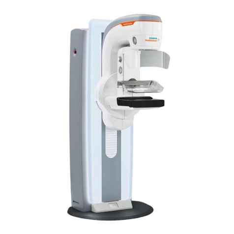
Siemens Healthcare
Siemens Healthcare MAMMOMAT Revelation User manual

Siemens Healthcare
Siemens Healthcare epoc Firmware update
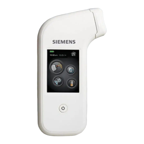
Siemens Healthcare
Siemens Healthcare XPRECIA STRIDE User manual
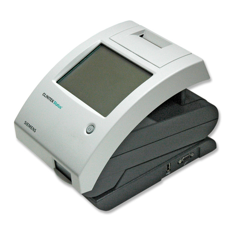
Siemens Healthcare
Siemens Healthcare Clinitek Status Connect System Manual
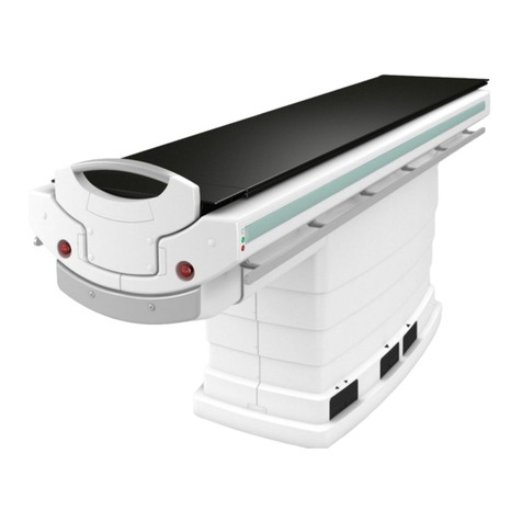
Siemens Healthcare
Siemens Healthcare SOMATOM User manual
Siemens Healthcare
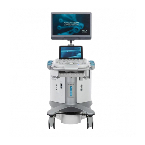
Siemens Healthcare ACUSON S Series User manual
Popular Medical Equipment manuals by other brands

C-Aire
C-Aire HELiOS Series user manual
Bionet
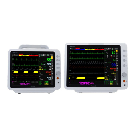
Bionet BM Elite Series Instructions for use
B. Braun
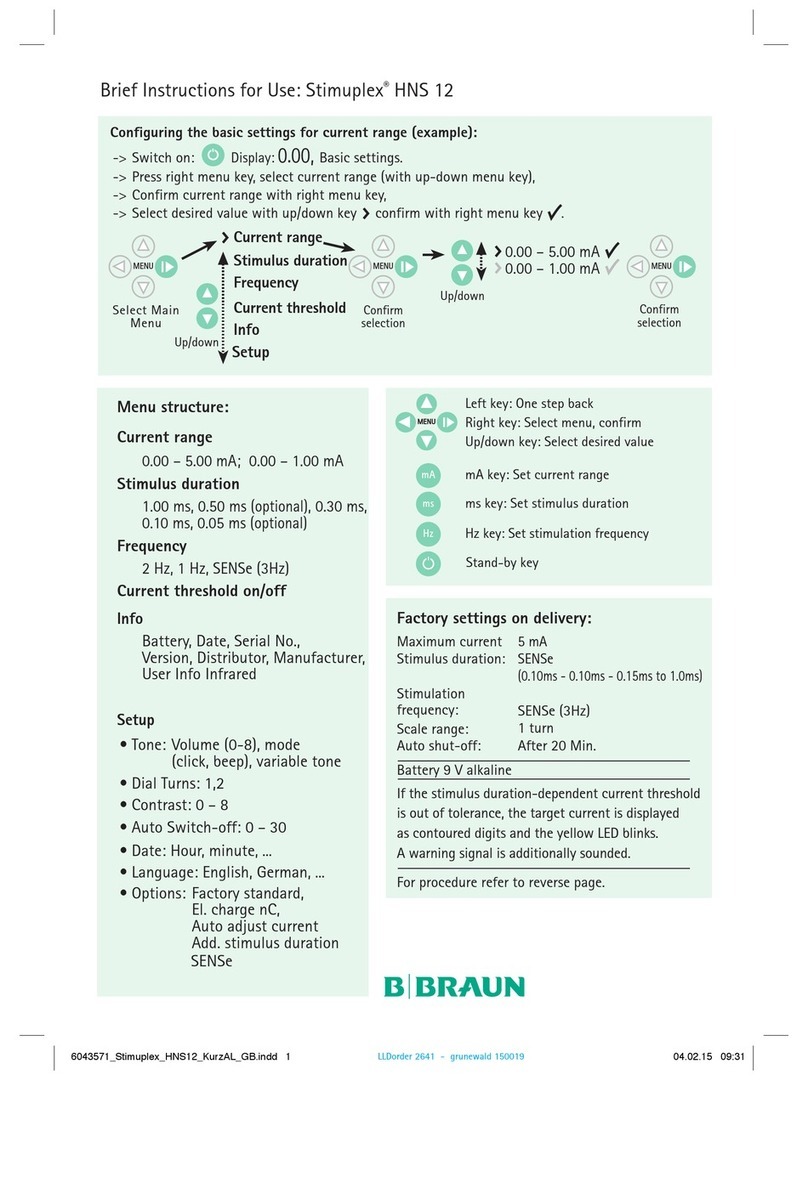
B. Braun Stimuplex HNS 12 Quick instructions
Hillrom
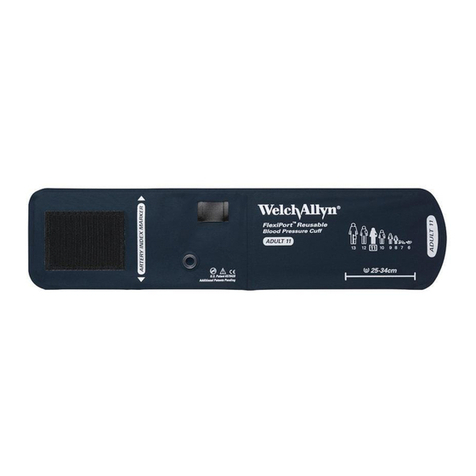
Hillrom Welch Allyn FlexiPort Blood Pressure Cuffs Instructions for use

Etac
Etac Immedia In2Sheet Instructions for use
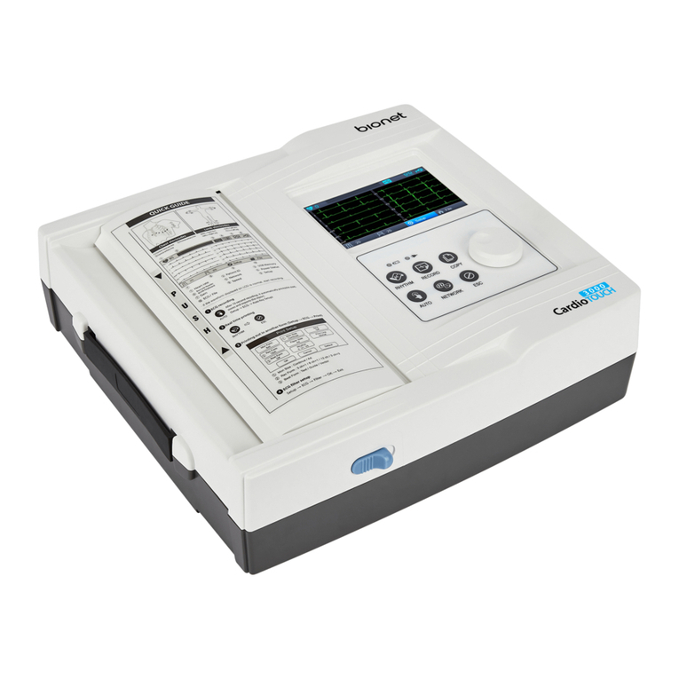
Bionet
Bionet CardioTouch3000 Operation manual
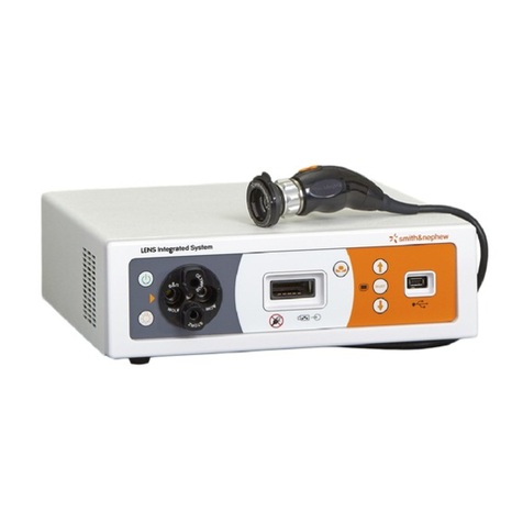
Smith & Nephew
Smith & Nephew LENS Integrated System Operation & service manual
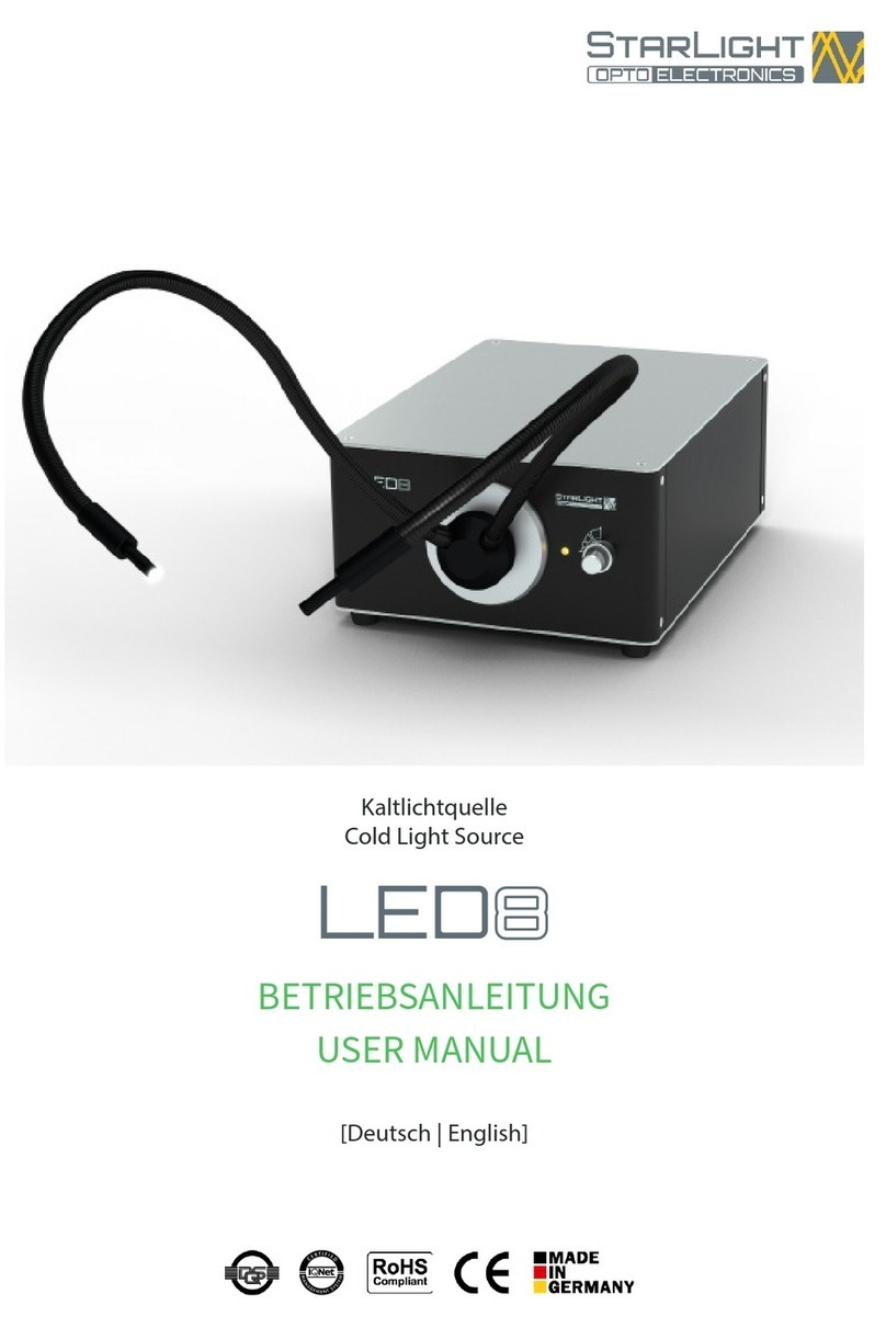
starlight
starlight LED8 user manual
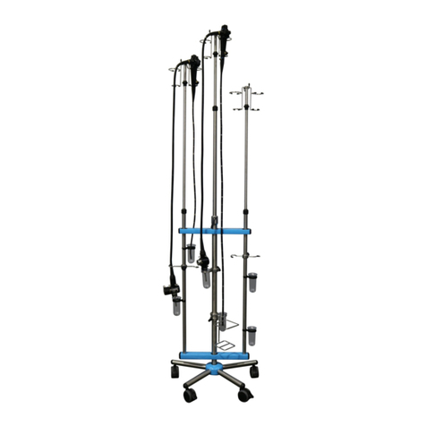
Healthmark
Healthmark EndoDolly Instructions for use
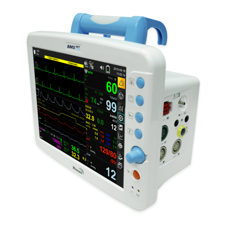
Bionet
Bionet BM5 user manual
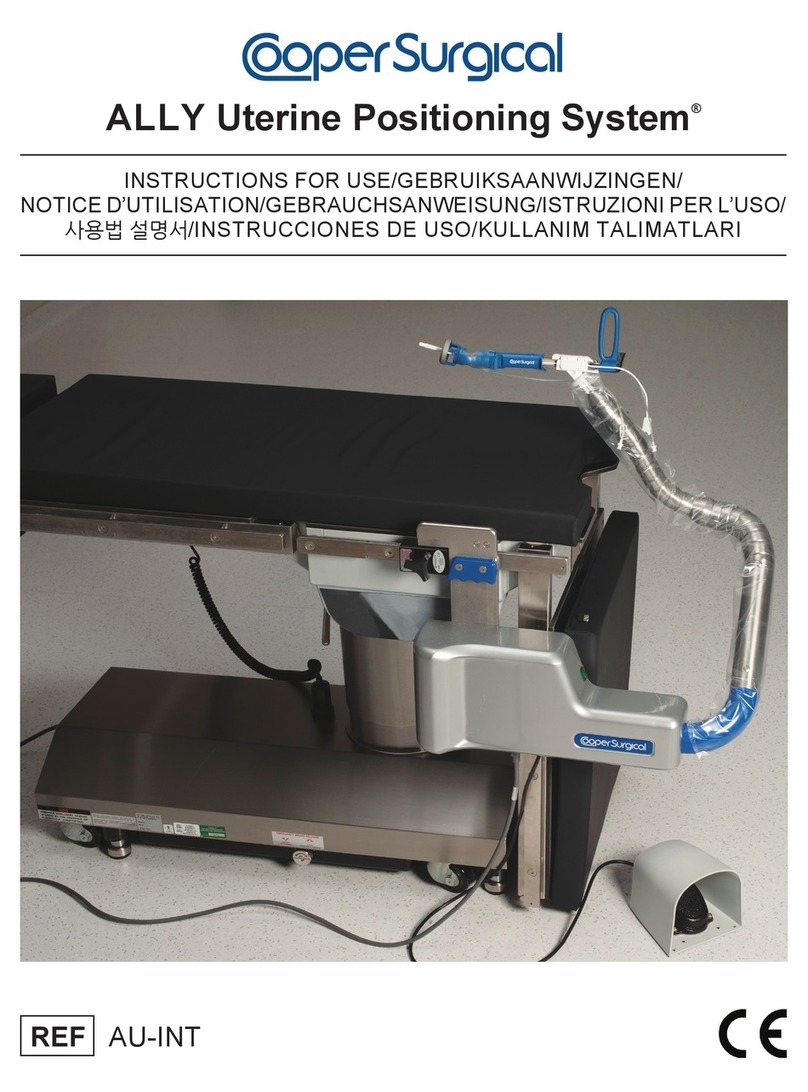
Cooper Surgical
Cooper Surgical ALLY Uterine Positioning System Instructions for use
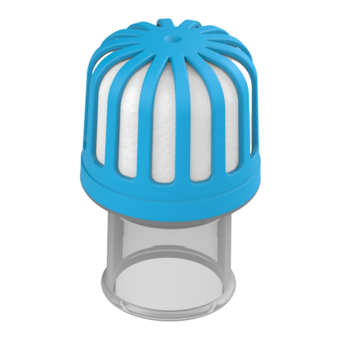
Atos
Atos Freevent XtraCare Mini How to use
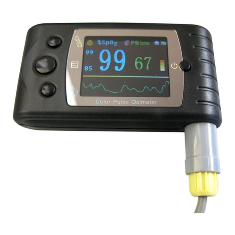
Contec
Contec CMS60C user manual
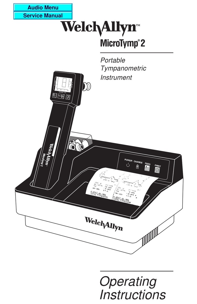
Welch Allyn
Welch Allyn MicroTymp 2 operating instructions

Envision
Envision Scrip Talk Station user guide

Etac
Etac Immedia SatinSheet 4Direction Instructions for use
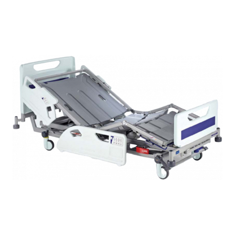
ARJO HUNTLEIGH
ARJO HUNTLEIGH Enterprise 8000 series Instructions for use

Care Fusion
Care Fusion Snowden-Pencer Indications for Use