6
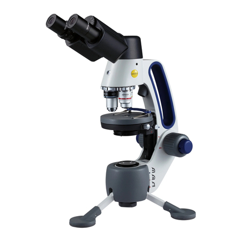
Step 1: Loosen the stage ring thumbscrew on the right side of the stage ring. Insert the stage plate into the stage ring and
secure it in place by tightening the thumbscrew.
Step 2: Loosen the stage position thumbscrew on the right side of the stage to move the stage assembly to its uppermost
position. The red dot underneath the stage position thumbscrew should be lined up with the blue dot on the right side of
the microscope arm near the coarse focus knob.
Step 3: Select the bottom (transmitted) illuminator by pressing the light source selector switch on the back of the
microscope’s arm to the bottom position.
Step 4: Turn on the illumination by rotating the light on/off & intensity control dial towards the bottom illuminator. (Note:
Please notice that that the dial will "click" when turning on the light. When turning the unit off, please ensure that the dial
is rotated all the way back until it "clicks" off to save power and prolong LED lifespan.)
Step 5: Place the slide on the stage, securing it with the stage clips. Center the specimen in the optical path.
Step 6: After securing and moving the slide into position, rotate the nosepiece to place the lowest power 4XD objective
into position over the specimen. Be sure the objective “clicks” into position. The iris diaphragm should be adjusted at this
time to about a ¼ inch (5 mm) open.
Step 7: (M3-B & M3-F only) Adjust the Siedentopf binocular head (by moving the eyepiece tubes up and down in an arc-like
motion, similar to adjusting binoculars) until one perfect circle is seen in the field of view
Step 8: While viewing through the eyepiece(s), rotate the coarse focus knob slowly and carefully to bring the specimen into
focus. The specimen may require some centering in the field of view at this time. By using the fine focusing knob, slowly
and carefully refine the focus to clearly observe the fine details of the specimen. Now you can turn the nosepiece to the
higher magnification micro objectives. The objectives are parfocalled so that once the 4x objective is focused; only a slight
turn of the fine focus is required to refine the focus when changing to higher power objectives.
Step 9: (M3-B & M3-F only) Set the diopter adjustment which is designed to help compensate for the difference between
the user’s eyes. To adjust, first bring the specimen into perfect focus by using the coaxial focusing knobs while looking
through the eyepiece with the right eye only (close your left eye). Now, using your left eye only (close the right eye) turn
the left eye diopter only (don’t touch the focus controls) to obtain a crisply focused image. The diopter adjustment is now
set and no further adjustment will be needed until a new operator uses the scope.
Please note: A smaller diaphragm aperture (opening) increases the contrast in the image while a larger aperture decreases
the contrast. (The diaphragm is not intended for controlling the brightness of the illumination). A good procedure to follow
in selecting the proper opening is to start with a large aperture and reducing it until the fine detail of the specimen is in
exact focus. Using an inappropriate aperture results in a “washing out” of the image. Care must be exercised not to reduce
the aperture too much to gain high contrast, as then the fine structure in the image of the specimen will be destroyed.
Reducing the aperture does increase contrast and depth of focus, but it also reduces resolution and causes diffraction. The
aperture for the 10X objective will not be the same as for the 40XRD objective, since the angle of the required light is
determined by the numerical aperture (N.A.) of the objective. The proper aperture of the diaphragm can be easily achieved
after minimal experience with the microscope.
