4
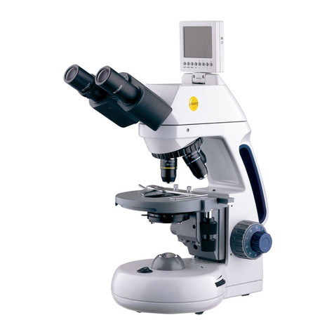
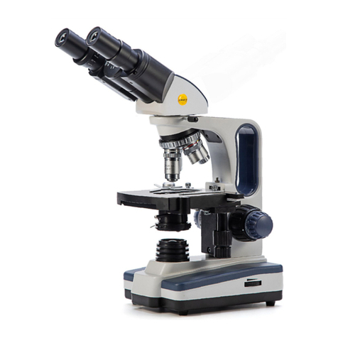
IRIS DIAPHRAGM – a multi-leaf round shaped device which is controlled
by a lever. It is similar to a camera shutter, and is installed under the
condenser. By moving the lever back and forth, the iris diaphragm opens
and closes, increasing and decreasing the contrast of the specimen. If the
image is “washed out” the iris diaphragm is opened too wide. If the image
is too dark the iris is not open wide enough.
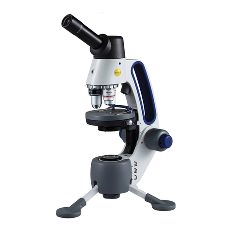
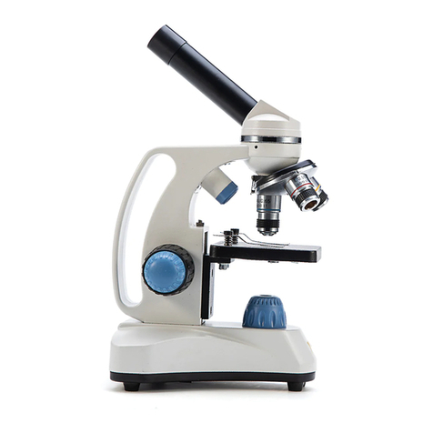
NOSEPIECE – the revolving turret that holds the objective lenses,
permitting changes in magnification by rotating different powered
objective lenses into the optical path. The nosepiece must “click” into
place for the objectives to be in proper alignment.
OBJECTIVES – the infinity plan optical system which magnifies the
primary image of the instrument. Magnifications are typically 4X, 10X,
40X and 100X.
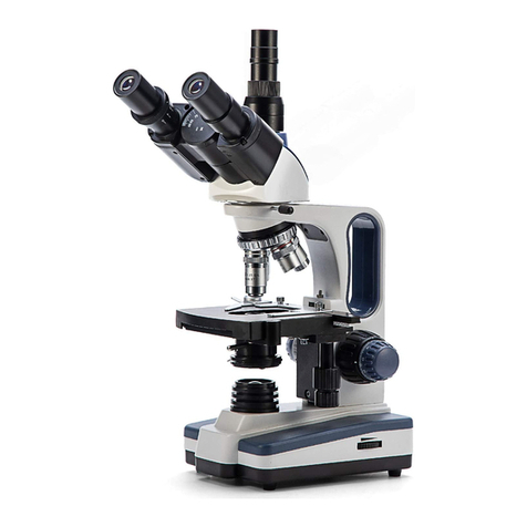
SIEDENTOPF – a binocular head design where the interpupillary
adjustment (increasing or decreasing the distance between the
eyepieces) is achieved by twisting the eyepiece tubes in an up and down
arc motion similar to binoculars.
STAGE – the table of the microscope where the slide is placed for
viewing. This component moves upward and downward when the
focusing knobs are turned. The stage of the M10 has a built-in mechanical
stage with a below-stage ergonomic “X” and “Y” axis controls. A finger
clip holds the slide securely and is designed to be a slow return holder to
provide protection to the specimen.
IMPORTANT TERMINOLOGY
“COATED” LENS – in attempting to transmit light through glass, much of
the light is lost through reflection. Coating a lens increases the light
transmission by reducing or eliminating reflection, thus allowing more light
to pass through.
COMPOUND MICROSCOPE – a microscope having a primary magnifier
(the objective) and a second (the eyepiece) to both conduct light, amplify
magnification and convert the image into a field of view easily seen by the
human eye.
COVER GLASS – thin glass cut in circles, rectangles, or squares, for
covering the specimen (usually a thickness of 0.15 to 0.17mm). The
majority of specimens should be protected by a cover glass, and must be
covered when using 40XRD or 100XRD objectives.
