LED FLUORESCENCE ILLUMINATOR
ACCU-SCOPE 73 Mall Drive, Commack, NY 11725 • 631-864-1000 • www.accu-scope.com 10
TROUBLESHOOTING
Image does not fill the field of view.
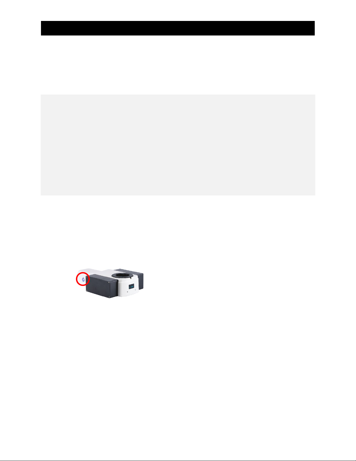
1. Ensure that the fluorescence channel selector is fully
seated in a position. Push in or pull out to confirm.
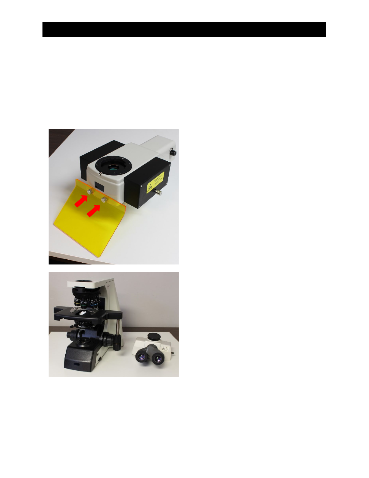
2. Ensure the illuminator is fully aligned with the
microscope frame dovetail receiver and the viewing
head is fully aligned with the dovetail receiver on the
top of the illuminator.
3. Check that the eyepieces are fully inserted and the
diopters are set appropriately.
Field of view is too bright and
overwhelms the sample.
1. Perform fluorescence observation under dark
surroundings. Avoid stray light from entering the light
path (e.g., eyepiece, camera adapter and other
adapters which do not have light barrier) and from
lighting the sample.
2. If the microscope has a trinocular head and no camera
is attached, cover the trinocular tube with non-
reflective black paper.
3. Check that the microscope transmitted light for
brightfield has been turned off.
4. Lower the condenser position and close the condenser
aperture diaphragm. This will reduce or eliminate
autofluorescence from the transmitted light
lamphouse.
If possible, use a piece of non-reflective black paper to
cover the condenser aperture diaphragm.
1. Change microscope observation to brightfield to
confirm the specimen image is clear and sharp.
Cannot focus on the sample.
1. Change microscope observation to bright field to
check if it is work well, focusing system has been
adjusted to correct position.
No fluorescence excitation light coming
from the illuminator (shining onto the
sample).
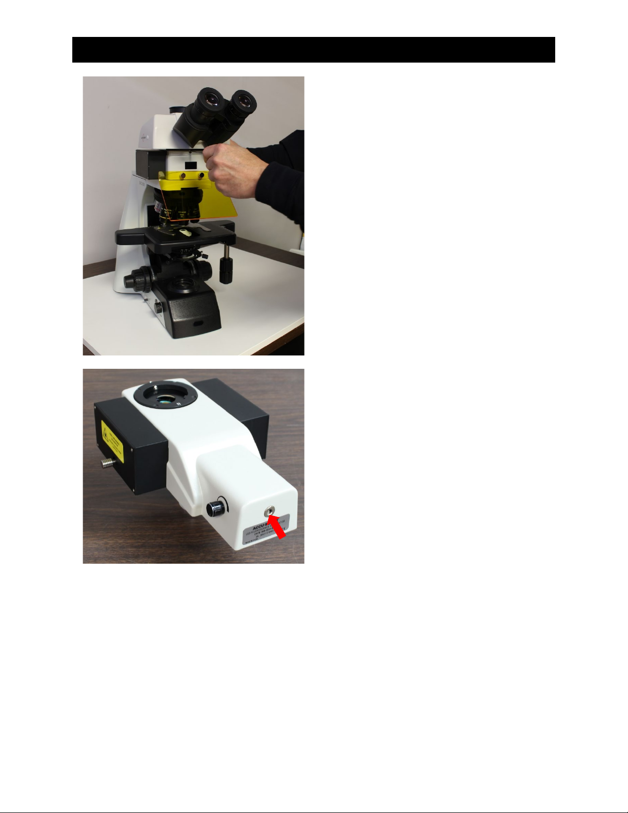
1. Rotate LED illumination control knob.
2. Check the front LED display is illuminated and does not
say “Off”. If it says “Off”, depress the LED intensity
control knob, confirm that the LED display no longer
says “Off”, then turn the LED illumination control knob
to adjust intensity.
3. Confirm the fluorescence channel selector is in
position (it should “click” into each channel position).
4. Confirm the illuminator is secure to the microscope
frame, and the viewing head is secure to the
illuminator body.
5. The objective is in position.