Caption AI Quick Reference Guide
PRESCRIPTIVE GUIDANCE AND PROBE MOVEMENT
MOVEMENT: ROCKING
Rocking is angling along the probe’s indicator axis.
Rock
towards
indicator -
along the
indicator
axis
Rock
away from
indicator -
along the
indicator
axis
ANGLING MOVEMENTS
Probe
tail
Non-indicator
axis of probe
Indicator axis
of probe
Probe
indicator
Blue line depicts view’s
indicator direction
Version: 1.1
●The yellow text and icons indicate the movements you need to make to
reach a diagnostic image. Slowly follow guidance until it disappears.
●Prescriptive Guidance (PG) is more likely to appear when probe is steady
with structures visible.
●Note: All guidance is in relation to the patient. E.g., “Slide Lateral” = slide
towards patient’s left, within the intercostal space.
●If you slide over a rib, you may lose the image. If this happens, simply re-
establish the image in that intercostal space.
●For rotation and angling movements (rocking & tailing), make slow, fine
movements with the probe planted on the patient’s body.
MOVEMENT: TAILING
Tailing is angling along the non-indicator axis of the probe.
Tail up
(towards head)
Tail down
(towards feet)
Tail medial
(towards midline)
Tail lateral (away
from midline)
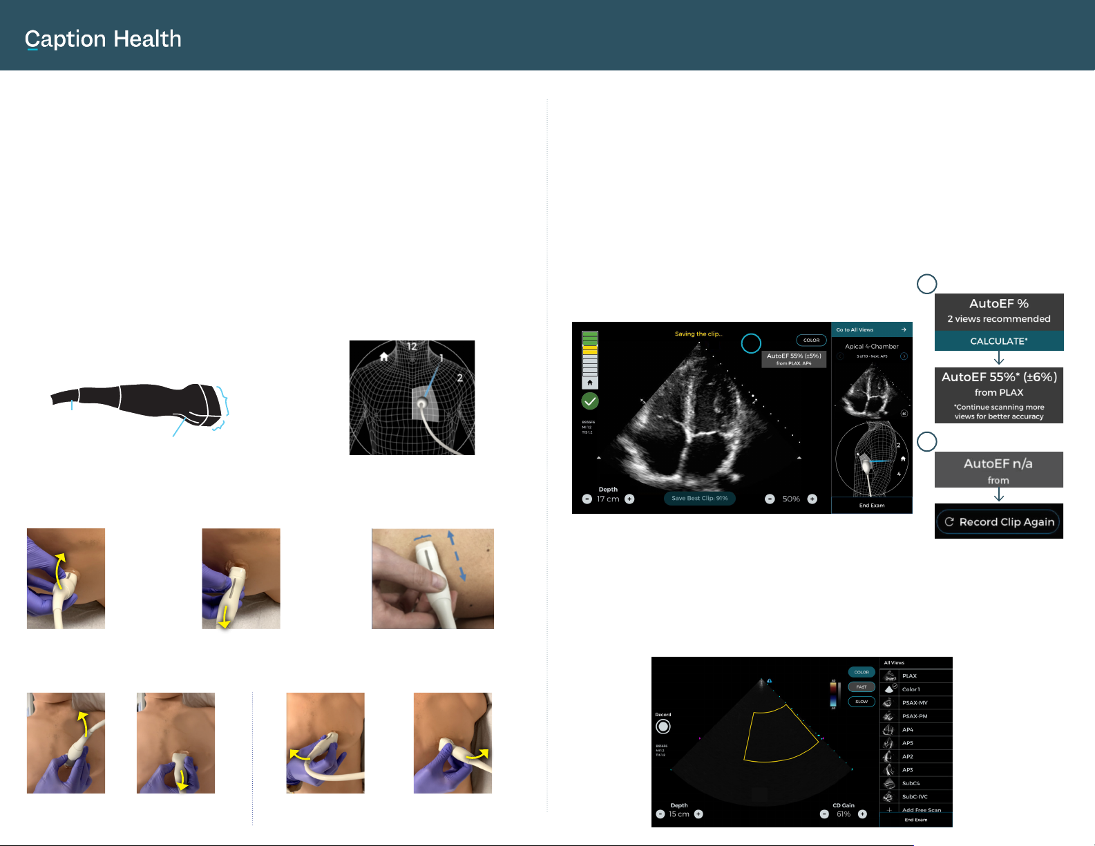
AUTOMATED EJECTION FRACTION CALCULATION
Caption Interpretation automatically calculates ejection fraction (EF) from
PLAX, AP4, and AP2 views of sufficient quality.
1.
EF can be calculated from a single view or multiple views. 2 or more views
are recommended for better accuracy. If a user wants to calculate EF from a
single view, they will be prompted to confirm before they are shown the EF.
2.
EF will not be produced in cases where the software does not deem
captured view(s) to be of sufficient quality. You will have the option to
acquire a new image (“Record Clip Again”).
3.
1
USING CAPTION AI FOR ADVANCED APPLICATIONS
More experienced users can choose to activate Color Doppler and/or Free Scan
modes. AI guidance is disabled when scanning in these modes.
2
3
Acquire
manually
using the
“Record”
button.
From “All
Views”, users
can choose to
enter Free Scan
mode to acquire
additional
cardiac or non-
cardiac views.
Select “Color” to enable Color Doppler. Flow can be optimized for Fast or Slow.


