EYE TECH CARE EyeOP1 User manual

User Manual MUT_RD_009H_GB Page 1 of 43
EyeOP1 & associated EYEOP-PACK
USER MANUAL
Version H of 2020/11/17
English
Date of CE-marking affixing: 05/2011
Printed by: Manufacturer:
EYE TECH CARE
2871, Avenue de l’Europe
69140 RILLIEUX-LA-PAPE, FRANCE
Tel. +33 (0)4.78.88.09.00
Fax. +33 (0)4.78.97.45.11
support@eyetechcare.com

User Manual MUT_RD_009H_GB Page 2 of 43
TABLE OF CONTENTS
SECTION 1: INTRODUCTION ___________________________________________________ 3
1.1 Use of the manual_________________________________________________________ 3
1.2 Icons and symbols_________________________________________________________ 4
1.3 Abbreviations ____________________________________________________________ 4
2. SECTION 2: GENERAL INFORMATION ________________________________________ 4
2.1 Intended use and indications________________________________________________ 4
2.2 Contra-indications ________________________________________________________ 5
2.3 Poor indications __________________________________________________________ 5
2.4 Adverse effects ___________________________________________________________ 6
2.5 User profile ______________________________________________________________ 6
2.6 Treatment session ________________________________________________________ 7
2.7 Anesthesia_______________________________________________________________ 7
2.8 Pre- and post-treatment____________________________________________________ 7
2.8.1 Recommendations concerning hypotensive and anti-inflammatory medications______________ 7
2.8.2 Recommendations concerning post-treatment follow-up ________________________________ 7
3. SECTION 3: SAFETY AND PRECAUTIONS FOR USE ______________________________ 8
3.1 General safety____________________________________________________________ 8
3.2 General warnings _________________________________________________________ 9
3.2.1 Warnings on the medical device_____________________________________________________ 9
3.2.2 Main warnings on treatment ______________________________________________________ 10
3.3 Protection against electric shocks ___________________________________________ 11
3.4 Labelling _______________________________________________________________ 11
3.4.1 Signage plate of the EyeOP1 Control Unit ____________________________________________ 12
3.4.2 EYEOP-PACK labels ______________________________________________________________ 12
3.4.3 Electromagnetic compatibility tables (EMC) __________________________________________ 14
3.5 Safety system and precautions _____________________________________________ 18
3.5.1 Characteristics and roles of the general system safeties_________________________________ 18
3.5.2 Dysfunction ____________________________________________________________________ 19
3.5.3 Emergency stop device ___________________________________________________________ 19
4. SECTION 4: DESCRIPTION OF THE EyeOP1 DEVICE AND EYEOP-PACK CONSUMABLE _ 19
4.1 General presentation and focused ultrasound principle _________________________ 19
4.2 Description of the device elements __________________________________________ 20
4.2.1 The EyeOP1 Control Unit: _________________________________________________________ 20
4.2.2 The single-use EYEOP-PACK consumable _____________________________________________ 22
4.3 Description of the system installation and treatment configuration________________ 24
5SECTION 5: SWITCHING ON THE EyeOP1 CONTROL UNIT _______________________ 25
6SECTION 6: DESCRIPTION OF THE TREATMENT PROCEDURE_____________________ 25

User Manual MUT_RD_009H_GB Page 3 of 43
6.1 General operation________________________________________________________ 25
6.2 General rules and conventions______________________________________________ 26
6.2.1 Screens and navigation bar________________________________________________________ 26
6.2.2 Data entry _____________________________________________________________________ 27
6.3 Detailed operation _______________________________________________________ 27
6.3.1 Step 1. Information for the physician-ophthalmologist-user _____________________________ 28
6.3.2 Step 2. Patient information________________________________________________________ 28
6.3.3 Step 3. Selection of the eye and of the number of sectors to treat ________________________ 29
6.3.4 Step 4. Equipment preparation ____________________________________________________ 30
6.3.5 Step 5. Treatment _______________________________________________________________ 35
6.3.6 Step 6. Treatment completion _____________________________________________________ 37
6.4 Other settings ___________________________________________________________ 38
6.4.1 Adjusting the screen brightness and sound volume _____________________________________ 38
6.4.2 Adjusting automatic treatment report printing________________________________________ 39
6.4.3 System information______________________________________________________________ 39
6.4.4 Setting the date and time _________________________________________________________ 39
6.4.5 Language selection ______________________________________________________________ 40
6.4.6 Checking, replacing and repositioning the printer paper ________________________________ 40
7. SECTION 7: ERRORS AND WARRANTY ________________________________________ 41
7.1 Warranty_______________________________________________________________ 41
7.2 Management of interruptions in the treatment sequence________________________ 41
7.3 Breakdowns and errors ___________________________________________________ 42
8. SECTION 8: MAINTENANCE AND CLEANING__________________________________ 43
9. SECTION 9: TRANSPORTING, MOVING AND STORING THE MEDICAL DEVICE________ 43
9.1 Transportation __________________________________________________________ 44
9.2 Storage and use conditions of the Control Unit and EYEOP-PACK __________________ 44
9.3 Lifetime ________________________________________________________________ 44
10. INFORMATION ON USER MANUAL UPDATES_______________________________ 44
The original version of this manual was written in French.
This device is CE-marked, in compliance with the directives 93/42/CEE and 2007/47/CE on medical devices.
1. SECTION 1: INTRODUCTION
1.1 Use of the manual
The present manual contains the instructions for use of the EyeOP1 electro-medical device (Control Unit) and
the associated EYEOP-PACK consumable (sterile single-use device) composed of a therapy probe and a
coupling cone equipped with a liquid trap. The system is indicated for clinical ophthalmologic use enabling
cyclocoagulation of the ciliary body with focused ultrasound for the non-invasive treatment of glaucoma.

User Manual MUT_RD_009H_GB Page 4 of 43
The manual provides the duly trained users with complete and detailed instructions for use. Use of the
medical device described in this manual must always be prescribed by a qualified physician.
The manual is divided into several parts covering all aspects of the use of the medical device and also safety
aspects of focused ultrasound. Read the manual thoroughly before using the device and keep it for further
use.
The term ‘Medical device’ used in this manual refers to the combination of the EyeOP1 control unit and
associated EYEOP-PACK consumables.
1.2 Icons and symbols
In order to make reading and understanding of the manual easier, the following symbols will be used:
= Information
This symbol is used when specific or additional information is added by
the writer.
= Caution
This symbol is used to indicate an important recommendation regarding
the use of the product.
= Prohibited
This symbol informs the user of a ban or potential hazard for the patient
health or of a problem liable to occur with the product.
1.3 Abbreviations
Abbreviations
Complete name
Abbreviations
Complete name
IOP
Intraocular pressure
MHz
Megahertz
M
Meter
GHz
Gigahertz
mm
Millimeter
W
Watt
mm Hg
Millimeter of mercury
V
Volt
°C
Degree Celsius
VA
Voltampere
°F
Degree Fahrenheit
kg
Kilogram
Hz
Hertz
g/L
Gram per liter
hPa
HectoPascal
atm
Atmosphere
2. SECTION 2: GENERAL INFORMATION
2.1 Intended use and indications

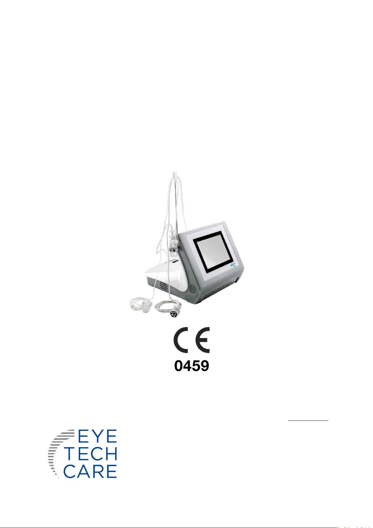
User Manual MUT_RD_009H_GB Page 5 of 43
This medical device (EyeOP1 control unit and associated EYEOP-PACK consumables) is intended to treat
glaucoma. This device allows for non-invasive treatment of glaucoma via coagulation of part of the ciliary
body by using focused ultrasound in order to reduce the production of aqueous humor and thereby to
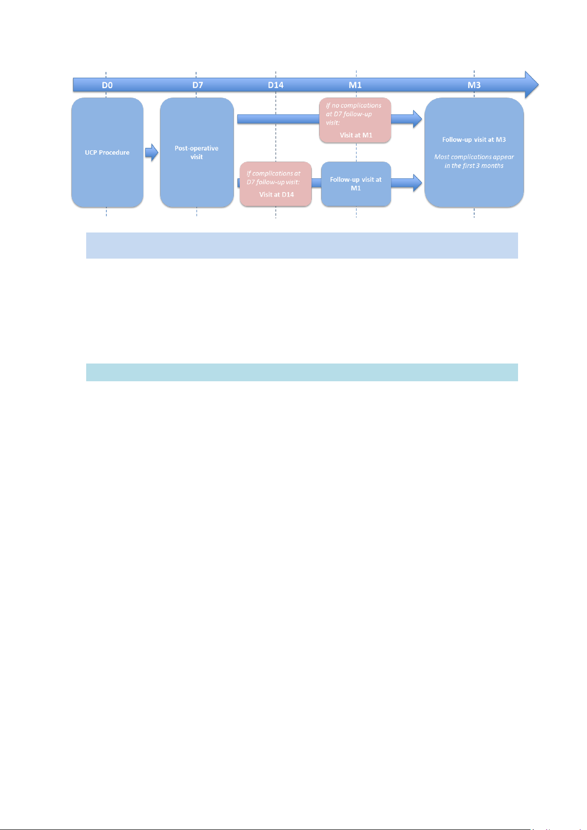
decrease the intraocular pressure (IOP). This treatment is called a UCP Procedure (“Ultrasound Cyclo Plasty”).
This type of treatment is indicated for adult patients (over the age of 18) who have glaucoma characterized
by ocular hypertension greater than or equal to 21 mmHg (for 6-sectors protocol) and greater than or equal
to 30 mmHg (for 8-sectors protocol).
Expected clinical benefits:
-Reduced production of aqueous humor leading to decreased intraocular pressure.
-Non-invasive procedure enabling to perform the treatment, in one step, without opening the eyeball,
thereby reducing the risk of infection or bleeding complications.
2.2 Contra-indications
Contra-indications are as follows:
-Glaucoma with normal pressure,
-Scleral thinning or ectasia,
-Eye tumour,
-Eye infection,
-Anatomy of the eye non-compatible with
the positioning of the device on the
patient’s eye (too narrow palpebral eyelid,
buphthalmic eye, etc),
-Thyroid orbitopathy,
-Choroidal hematoma,
-Aphakic patient,
-Presence of a valve, tube, or any other
element on the eye surface preventing
proper positioning on the patient’s eye (ex:
chemosis caused by the anesthesia, scaring
filtration bleb after filtering surgery, etc).
-Patient’s history of retinal detachment,
macular edema, choroidal hematoma,
and/or uveitis.
Practitioner is asked to not treat under these conditions.
Precautions:
➢A second treatment may be necessary to reach the targeted IOP below 21 mmHg.
➢There are no contraindications to conducting a second ultrasound treatment procedure. No increased
occurrence of side effects related to retreatment has been reported. The interval between two
treatments is left to the judgment of the qualified practitioner.
2.3 Poor indications
Pathologies or patient’s history for which the treatment with the medical device may have a limited efficacy
and/or may induce higher risk of complications are as follow:
-Diabetic patient with or without diabetic -
retinopathy,
-Retinal vein occlusion,
-Vitrectomy,
-Multiple intra vitreous injections,
-Age-related Macular Degeneration (AMD),
especially the exudative form,
-High myopia.
Note:
Knowing the results of the clinical studies on secondary glaucoma (neovascular, pigmentary, pseudo-
exfoliative, etc), the patient’s response rate is lower than for primary open angle and angle closure glaucoma.
The responsibility to treat patients with these poor indications with the medical device is left to the judgement
of the qualified practitioner.

User Manual MUT_RD_009H_GB Page 6 of 43
2.4 Adverse effects
Possible adverse effects or complications during treatment and/or clinical follow-up (expected adverse
effects) are:
Anterior chamber:
-Mild / moderate intraocular inflammation of the anterior chamber of the treated eye (tyndall effect …)
-Severe intraocular inflammation (uveitis, etc.).
-Hyphema.
Intraocular pressure:
-Transient hypertonia (> 10 mmHg compared to the initial IOP, at less than 3 months).
-Chronic hypertonia (> 10 mmHg compared to the initial IOP, at more than 3 months).
-Transient major hypotonia defined as intraocular pressure strictly less than 6 mmHg.
-Persistent major hypotonia defined as intraocular pressure strictly less than 6 mmHg.
-Phthisis of the eyeball
Eyelid:
-Blepharitis
-Bleeding of the internal lining.
Cornea:
-Superficial corneal complications (superficial
punctate keratitis, ulcers).
-Deep corneal complications (edema, opacity,
decompensation, corneal dystrophy).
Crystalline lens:
-Induced cataract (clouding of the lens)
-Progression of existing cataract.
-Phacodonesis (tremulousness or vibration of
the lens with eye movement, often due to lens
subluxation).
-Subluxation of the lens
Visual acuity –Refraction:
-Change in refraction (induced astigmatism, etc.)
-Transient (< 3 months) or permanent (> 3
months) decrease in visual acuity
-Transient monocular diplopia
Retina –Choroid –Vitreous humor:
-Retinal complications (retinal tear, retinal
detachment)
-Choroidal detachment
-Macular edema
-Bleeding of choroid, vitreous humor
Sclera and conjunctiva:
-Scleral thinning and/or spots on sclera.
-Conjunctival ulceration/erosion.
-Scleral ulceration.
-Limbal-scleral perforation.
-Conjunctival hyperemia, chemosis, conjunctival
bleeding.
-Subconjunctival haemorrhage, conjunctivitis.
Iris:
-Burning of the iris.
-Lens and corneal synechiae.
-Iris and corneal synechiae.
-Goniosynechiae.
-Paresis.
-Mydriasis.
-Corectopia.
Pain:
-Transient acute ocular pain.
-Chronic ocular pain.
-Headaches.
Infection of the eyeball:
-Infection of the eyeball.
2.5 User profile
This medical device (EyeOP1 Control Unit and associated EYEOP-PACK consumables) is not intended to be
used by a layperson to perform a treatment.

User Manual MUT_RD_009H_GB Page 7 of 43
It is intended for use by public or private sector ophthalmologists who specialize in the treatment of glaucoma
or cataract (for combined cataract/glaucoma treatment). The ophthalmologist is trained for the use of the
appropriate protocols.
In addition, the present User Manual supplied the EyeOP1 Control Unit is available to the user, detailing the
treatment procedure.
The trained ophthalmologist may be assisted by a nurse or intern in performing the treatment preparation
and monitoring during the treatment procedure.
2.6 Treatment session
A treatment session corresponds to the treatment of one eye, the therapy probe being set up with
appropriate parameter configuration. The treatment must be prescribed by a qualified physician.
2.7 Anesthesia
Anesthesia (general, local or topical) must be performed on the patient before the treatment session.
The patient must lie on his back (supine position) and remain still throughout the treatment so as not to shift
the coupling cone and therapy probe out of position.
The physician or operator using the medical device must be present throughout the treatment and carefully
supervise the patient.
2.8 Pre- and post-treatment
2.8.1 Recommendations concerning hypotensive and anti-inflammatory medications
2.8.2 Recommendations concerning post-treatment follow-up

User Manual MUT_RD_009H_GB Page 8 of 43
3. SECTION 3: SAFETY AND PRECAUTIONS FOR USE
This section concerns the safety and precautions for use of the medical device (EyeOP1 control module, its
accessories and associated EYEOP-PACK consumables). These precautions must be taken in order to ensure
optimal product performance, treatment efficacy, as well as patient and operator safety.
3.1 General safety
The EyeOP1 control module and associated EYEOP-PACK consumables form a medical device that generates
focused ultrasound via the therapy probe. Consequently, several precautions and general safety measures must
be taken:
-It is essential to thoroughly read and understand the contents of the present manual before using the
EyeOP1 control unit, its associated accessories and EYEOP-PACK consumables.
-The medical device is not intended for use other than that described in the present manual and for
which it was designed.
-The EyeOP1 control unit and EYEOP-PACK consumables presented herein have been developed and
tested for a specific use within the frame of glaucoma treatment by coagulation of the patient’s ciliary
body, upon prescription of a qualified physician, and upon selection of parameters specific to each
therapy probe. No other use shall be made of this device.
-Installation of the medical device must be carried out by a physician approved by EYE TECH CARE.
-Only accessories and options authorized by EYE TECH CARE shall be used with the medical device
described in the present manual. The mains supply cable must not be replaced by the user. Use of any
other equipment shall be subject to authorization upon request to EYE TECH CARE.
-The various elements of the EYEOP-PACK consumable are intended for single-use. Consequently, they
must not be reused. They must be discarded in appropriate containers after use (contaminated waste).
-The medical device shall never be left unattended when in operation. The operator shall be present
throughout the treatment.
-All technical software supplied with the medical device are exclusively intended for usage and
indications initially defined by the manufacturer. Any other use must be excluded.

User Manual MUT_RD_009H_GB Page 9 of 43
-No other software shall be installed on the device without written authorization from EYE TECH CARE.
Doing so could induce a risk of failure of the equipment, or even a risk for the patient safety or
environment.
-Do not expose the medical device to a material that could affect its operation, such as magnetic fields,
external electrical influences, electrostatic effects, etc.
-Do not use the medical device in the presence of flammable or explosive gases.
-The users shall comply with the maintenance guidelines provided by EYE TECH CARE in order to preserve
the safety and performance of the medical device. That is why a periodic maintenance (annual) must be
done by EYE TECH CARE or person qualified by EYE TECH CARE in order to do the necessary maintenance
and calibration operations.
3.2 General warnings
3.2.1 Warnings on the medical device
It is essential to read and understand these warnings before going further into the manual:
Only a properly trained and qualified users shall use the EyeOP1 control unit, its associated
accessories, EYEOP-PACK consumables and mains supply cables described in the present
manual.
The EyeOP1 control unit is only compatible with the associated EYEOP-PACK consumables.
Consequently, only EYEOP-PACK sterile consumables can be used with the EyeOP1 control
unit. Likewise, EYEOP-PACK sterile consumables should not be used with a device other than
the EyeOP1 control unit.
Use of the medical device, or any part thereof, other than as described in this manual will,
under no circumstances, be construed as the responsibility or liability of EYE TECH CARE.
Use of the medical device described herein must always be prescribed by a qualified physician
and shall consequently never occur under other conditions.
EYE TECH CARE shall not be held responsible if the user does not comply with all procedures
or recommendations described in the present manual and/or if he uses the device to other
aims than the claimed intended use.
The room where the device is installed shall meet the requirements on electric equipment.
EYE TECH CARE cannot be held responsible if these requirements are not met.
Any technical interventions on the medical device other than those authorized by EYE TECH
CARE can induce partial or total suspension of the warranty.

User Manual MUT_RD_009H_GB Page 10 of 43
The maintenance operations shall be done by technicians trained and qualified by EYE TECH
CARE. EYE TECH CARE shall not be held responsible if the user refuses maintenance operations
by a trained and qualified technician, or if he does it by himself.
Switch off the electrical mains and disconnect the system from the mains supply before
cleaning or inspection.
CE 0459 identification is placed on the medical device to indicate that the medical device was
produced in compliance with the current European Directives on medical devices
The EyeOP1 medical device is not intended for domestic use. If it is not installed in compliance
with the instructions for use, it can cause interference that could damage other more sensitive
equipment or be disrupted by their possible excessive radio-electric emissions. It may be
necessary to take mitigation measures, such as reorientation or installation of the EyeOP1
control unit in another room, or shielding of the location.
Moreover, cellular phones and other mobile communication equipment must not be used
close to the medical device, since they could affect it. Any other device used close to the
EyeOP1 device must comply with the IEC 60601-1-2 standard.
When an error message appears, it is recommended to read and understand the message, to
write it down in order to make the technical diagnostic easier, to discontinue treatment if the
procedure is not described in the present manual, and to rapidly inform EYE TECH CARE (or
the local distributor).
If unusual events persist or if unidentified problems occur, do not try to dismantle the medical
device. Take note of the problem and contact the technical department of EYE TECH CARE at
support@eyetechcare.com, or the local representative
The procedure to be followed for collecting, discarding, destroying or recycling the
equipment, its sub-assemblies, associated accessories and single-use devices, shall comply
with the regulation in force in the country and/or hospital where it is being used (if necessary,
contact the local agent or distributor).
The EYEOP-PACK single-use consumable must be discarded immediately after use in the
appropriate container (contaminated waste).
The end-of-life EyeOP1 Control Unit being considered as an electric/electronic
waste (WEEE), EYE TECH CARE will recover and dispose of it according to the
WEEE waste collection and recycling procedure. To this end, contact the local
representative. By doing so, the user acts positively for the environment,
preservation of natural resources and health protection.
3.2.2 Main warnings on treatment
Only a properly trained and qualified users shall use the EyeOP1 control unit, its associated
accessories and EYEOP-PACK consumables described in the present manual.

User Manual MUT_RD_009H_GB Page 11 of 43
The EYEOP-PACK consumable, containing the therapy probe and coupling cone, is intended
for single-use. Consequently, it must not, under any circumstance, be reused.
The EYEOP-PACK elements are ethylene-oxide sterilized. Before use, check the sterility of the
product by making sure the packaging is undamaged.
Do not resterilize the EYEOP-PACK elements, before or after use. In case of sterility problems
due to damaged packaging, do not use the product.
Check for leakage of the coupling cone tubing, this could prevent sufficient suction.
An inappropriate re-sterilization by the user, not authorized by EYE TECH CARE, could damage
the elements of the EYEOP-PACK consumable, such as the probe’s transducers, and could lead
to under-treatment and/or undesired treatment on surrounding tissues.
The parameters of the therapy probe are specific to each probe and determine the ultrasound
energy delivered to the patient. They are automatically detected by the Control Unit once the
therapy probe is connected to it.
Proper positioning of the coupling cone and therapy probe is essential, so the patient must
remain still in order not to displace them. If these recommendations are not complied with,
this may cause improper treatment to be delivered, or even patient injury.
The operator must be present at all times during the treatment and carefully supervise the
medical device and the patient.
3.3 Protection against electric shocks
In order to protect the persons against electric shocks, the following recommendations must be
followed:
-To avoid any risk of electric shock, this device must be connected to an earthed mains network.
-Always comply with the rules and specifications on electrical installations.
-Never open the EyeOP1 Control Unit. Only qualified personnel are authorized to undertake such an
operation.
-Keep the Control Unit away from liquids and never spray water or other liquids on it.
-In case of breakdown or prolonged disuse, it is recommended to disconnect the device.
-If the EyeOP1 control unit is moved, check that the electric plug is suitable for the equipment.
-In case of fire and risk for a device, disconnect the EyeOP1 control unit from the mains network and
contact the relevant staff.
3.4 Labelling
Before going further into this manual, read and understand the information appearing on the EyeOP1
Control Unit and EYEOP-PACK labels.

User Manual MUT_RD_009H_GB Page 12 of 43
3.4.1 Signage plate of the EyeOP1 Control Unit
The signage plate of the EyeOP1 device is located on the rear side. It bears the following information:
1. Address and telephone number of
manufacturer (EYE TECH CARE).
2. Device model and item code (ETC_XXXX_YY).
3. Date of manufacture (year and month) and
serial number of the unit.
4. Type and characteristics of changeable fuses.
5. Supply voltage (V), frequency (Hz), and
maximum absorbed power (VA).
6. Nominal power (acoustic W) and nominal
output frequency (MHz) of the therapy
probe.
7. Weight of the Control Unit.
a. "Manufacturer" symbol accompanied by
manufacturer's address on label (1.)
b. Warning: Essential to refer to instructions for
use.
c. Applied part of the type BF medical device
d. Temperature of storage
e. CE marking
f. Storage in a dry place
g. The control unit to be discarded is WEEE
(waste electrical and electronic equipment).
Return it to an appropriate collection point
for processing, recovery, and recycling of this
type of waste or to a distributor.
3.4.2 EYEOP-PACK labels
The EYEOP-PACK contains several labels affixed in various locations on the primary and secondary
packaging.
3.4.2.1 Tyvek covering and label of the PET primary packaging
The various elements of the EYEOP-PACK are packaged in a PETg (polyethylene
terephthalate glycol) blister, a recyclable plastic material closed with a Tyvek covering,
the content of which has been ethylene-oxide sterilized. This primary package is
contained in an individual carton box. The printed tyvek covering contains important
information. Moreover, an adhesive label is affixed to it.

User Manual MUT_RD_009H_GB Page 13 of 43
The information shown on these labels is described and explained in the table below and must be fully
understood by the user:
Adhesive label affixed to the EYEOP-PACK lid:
1. Item code of consumable
2. Lot number
3. Date of manufacture
4. Size of the therapy probe and coupling
cone
5. CE marking
6. Expiration date
EYEOP-PACK printed lid:
a. Upper and lower temperature limits for
use and storage
b. Do not re-sterilize.
c. Do not reuse/single use.
d. ‘Manufacturer’ symbol accompanied by
manufacturer’s address on the label.
e. Sterilization by ethylene-oxide.
f. Warning: Imperatively refer to
instructions for use.
g. Do not use if packaging is damaged.
h. Storage conditions: store in a dry place,
away from light.
i. Symbol which identifies the sterile
barrier system (covered blister).
3.4.2.2 Adhesive label of the individual carton box
A label is affixed on the carton box containing the PETG blister. Part of this label is folded over the lid
of the box thereby enabling to check that the equipment has not been opened. It contains detailed
information about the contents of the EYEOP-PACK (see § 4.2.1).

User Manual MUT_RD_009H_GB Page 14 of 43
3.4.2.3 Sheet of detachable labels
The individual box contains a sheet of 4 identical labels to be used by the operator/user in order to
ensure traceability of the product according to the procedures in force in the establishment. The
information contained is similar to that present on the adhesive label affixed on the box lid.
3.4.3 Electromagnetic compatibility tables (EMC)
The medical device (EyeOP1 control unit and associated EYEOP-PACK consumables) requires specific
precautions to be taken with respect to the EMC and must be installed and operated according to the EMC
information provided in the tables below.
Cellular phones and other mobile communication devices must not be used close to the medical device,
since they could affect its operation. Any other device used close to the medical device must comply with
the IEC 60601-1-2 standard. Any incident related with electromagnetic compatibility must be reported to
the manufacturer.
Exposure of patients equipped with active implantable medical devices (pacemaker…) to the EyeOP1
control unit and associated EYEOP-PACK consumables must be supervised by a cardiologist.

User Manual MUT_RD_009H_GB Page 15 of 43
This symbol means that any equipment emitting radio waves must be OFF when close to the device.
This symbol means that the equipment features a RF transmitter.
Directives and manufacturer’s claim – electromagnetic emissions
The medical device is intended for use in the electromagnetic environment described below. The
customer or medical device user must ensure that it is used in such an environment.
Emission trials
Compliance
Electromagnetic environment - directives
RF emissions
CISPR 11
Group 1
The medical device uses RF energy only for internal functions.
Consequently, its RF emissions are very low and not liable to
provoke interferences in any neighbouring electronic devices.
RF emissions
CISPR 11
Class A
The medical device is suitable for use in all premises other than
domestic and those directly linked to the public low voltage
supply system feeding domestic use premises.
Harmonic emissions
IEC 61000-3-2
Class A
Voltage fluctuations/
Flicker
IEC 61000-3-3
Complies
Directives and manufacturer’s claim – electromagnetic immunity
The medical device is intended for use in the electromagnetic environment described below. The customer
or medical device user must ensure that it is used in such an environment.
Immunity testing
Test level
IEC 60601
Compliance
level
Electromagnetic environment -
directives
Electrostatic discharge
(ESD)
IEC 61000-4-2
± 6 kV in contact
± 8 kV in the air
Complies
Complies
The floor must be made of wood,
concrete or thin set tile. If the ground is
covered with synthetic material, the
relative humidity should be at least 30%.
Electrical fast transient
burst
IEC 61000-4-4
± 2 kV for electrical
feed lines
± 1 kV for
input/output lines
Complies
Complies
The quality of the mains supply system
must be that of a typical commercial or
hospital environment.
Transient voltage
surges
IEC 61000-4-5
± 1 kV between
phases
± 2 kV between
phase and ground
Complies
Complies
The quality of the mains supply system
must be that of a typical commercial or
hospital environment.
Voltage dips, short
interruptions and
voltage variations on
<5 % UT
(>95 % dips of UT)
during 0.5 cycle
Complies
The quality of the mains supply system
must be that of a typical commercial or
hospital environment. If the medical
device user demands continuous

User Manual MUT_RD_009H_GB Page 16 of 43
input electrical feed
lines
IEC 61000-4-11
40 % UT
(60 % dips of UT)
during 5 cycles
70 % UT
(30 % dips of UT)
during 25 cycles
<5 % UT
(>95 % dips of UT)
during 5 s
Complies
Complies
Complies
operation during the mains supply
failure, it is recommended to feed the
EyeOP1 control unit from a failure-free
energy supply or from a battery.
Magnetic field at the
mains supply
frequency (50/60 Hz)
IEC 61000-4-8
3 A/m
Complies
The magnetic fields at the mains supply
frequency must feature the specific
levels of a representative location in a
typical commercial or hospital
environment.
Note: UTis the voltage of the AC power supply before application of the test level.
Directives and manufacturer’s claim –electromagnetic immunity
The medical device is intended for use in the electromagnetic environment described below. The
customer or medical device user must ensure that it is used in such an environment.
Immunity
testing
Test level
as per IEC
60601
Compliance
level
Electromagnetic environment - directives
Conducted
RF emissions
IEC 61000-4-
6
Radiated RF
emissions
IEC 61000-4-
3
3 Veff
from 150
kHz to 80
MHz
3 V/m
from 80
MHz to
2.5 GHz
3 V
3 V/m
The RF communication mobile devices should not be
used closer to any part of the medical device (including
cables) than the recommended separation distance,
calculated from the equation applicable to the
transmitter frequency.
Recommended separation distance:
d = 1.17 √𝑃
d =1.17 √𝑃 80 MHz to 800 MHz
d = 2.34√𝑃 from 800 MHz to 2.5 GHz
where P is the transmitter maximal power output in
watts (W), according to the transmitter manufacturer
and d is the recommended separation distance in meters
(m).

User Manual MUT_RD_009H_GB Page 17 of 43
It is important that fixed RF transmitter field intensity,
determined by an electromagnetic investigation on sitea,
be less than the compliance levels, for each frequency
rangeb
Interferences may occur close to the device marked with
the following symbol:
Note 1: at 80 MHz and at 800 MHz, the highest frequency range applies.
Note 2: These directives may not apply in all cases. Electromagnetic propagation is affected by
absorption and reflection of structures, objects and persons.
a The field intensities of fixed transmitters, such as base stations for radiotelephones (cellular) and
terrestrial mobile radios, amateur radio, MA and MF radio, and TV diffusion, cannot be theoretically
predicted with accuracy. To assess the electromagnetic environment produced by the fixed RF
transmitters, an electromagnetic investigation must be conducted on site. If the field intensity,
measured at the location where the medical device is being used, exceeds the above applicable RF
compliance level, the medical device must be checked for normal operation. If abnormal performances
are observed, it may be necessary to take additional measures, such as reorienting or repositioning the
medical device.
bAs pertains to the frequency range from 150 kHz to 80 MHz, the field intensities must be less than 3
V/m.
Recommended separation distances between RF communication
mobile devices and medical device
The medical device is intended for use in an electromagnetic environment in which RF radiated
emissions are controlled. The customer or medical device user can contribute to preventing
electromagnetic interferences by maintaining a minimal distance between the mobile device/RF mobile
communication device and medical device, as recommended below, in function of the maximal
emission power of the mobile communication device.
Maximum rated
output power of
the transmitter
(in W)
Separation distance depending on the transmitter frequency (in m)
from 150 kHz to 80 MHz
d = [3.5/3]√𝑃
from 80 MHz to 800
MHz
d = [3.5/3]√𝑃
from 800 MHz to 2.5
GHz
d = [3.5/3]√𝑃
0.01
0.12
0 .12
0.23
0.1
0.37
0.37
0.74
1
1.17
1.17
2.34
10
3.7
3.7
7.4
100
11.7
11.7
23.4
For transmitters whose maximum rated output power is not indicated in the above table, the
recommended separation distance din meters (m) can be assessed by using the equation applicable to
the transmitter frequency, where Pis the transmitter maximal output power in watts (W), according to
the transmitter’s manufacturer.
Note 1: at 80 MHz and at 800 MHz, the separation distance for the highest frequency range applies.

User Manual MUT_RD_009H_GB Page 18 of 43
Note 2: These directives may not apply in all cases. Electromagnetic propagation is affected by
absorption and reflection of structures, objects and persons.
3.5 Safety system and precautions
3.5.1 Characteristics and roles of the general system safeties
Safety checks are constantly being performed by the medical device software throughout the various
phases of its use. The following table lists the checks and describe their role and consequences in case of
occurrence of the problem they are meant to solve:
Definition
Description
Activated by
Action on
When
ON/OFF button
Starts/Stops the
device
User
(equipment)
Controls general power
supply and
consequently all sub-
assemblies
Continuously
Fuses
Switches off the
mains supply
Electrical
overcurrent
(equipment)
Turns off the device and
protects the sub-
assemblies
Continuously
Firing pedal
Starts the firing, and
immediately stops it
in case of pedal
release
User
(equipment)
Turns off power supply
to the RF electronic
board of the generator
Continuously
Watchdog
communication
Displays a message
when communication
between MMI and RF
generator is lost
Device
(equipment +
software)
Displays a user message
Start screen
Suction release
Displays a message
when suction is lost
(vacuum decay)
Device
(equipment +
software)
Displays a user message
During
treatment
Adaptation failure
Detects an adaptation
failure on the line of
transducers
Device
(equipment +
software)
Turns off power supply
to the RF electronic
board (RF generator)
During
treatment
Power failure
Stops firing
Device
(equipment +
software)
Turns off power supply
to the RF electronic
board (RF generator)
During
treatment
Control of power
output
Control of emitted RF
power by Wattmeter
Device
(equipment +
software)
Turns off power supply
to the RF electronic
board (RF generator).
During
treatment
RF shooting
indication/
Ultrasound
Visualization of the
ultrasound/RF
shooting power (user
information)
Device
(equipment +
software)
Activates the
ultrasound beam on the
sector being shot
During
treatment

User Manual MUT_RD_009H_GB Page 19 of 43
Definition
Description
Activated by
Action on
When
Off-time
Imposes a time
interval between two
shots
Device
(software)
Sets a time interval
between two shots and
displays remaining time
before next shot. Sound
signal during waiting
period.
During
treatment
Probe detection
Checks integrity of the
probe data and
compatibility of the
probe model.
Device
(software)
Displays an error
message asking the user
to change probe
At probe
connection &
before start of
treatment phase
3.5.2 Dysfunction
When the software detects an error or abnormality, the user is informed by an error message displayed on
the Control Unit screen.
When an error message appears, it is highly recommended that the user:
-Read and understand the message,
-Write down the message in order to make technical diagnostic easier, if necessary,
-Discontinue treatment if, necessary,
-Rapidly inform the EYE TECH CARE technical department at suppor[email protected]
or the local representative.
3.5.3 Emergency stop device
Should the treatment need to be discontinued in an emergency, the operator must release pressure from
the firing pedal (otherwise depressed during treatment), which will automatically stop the treatment
procedure. If needed, stop aspiration by pressing on the suction button (on top of the firing pedal). Remove
the therapy probe and the coupling cone from the patient’s eye.
To start treatment again, if needed and appropriate, follow the procedure described in part 2. Management
of interruptions in the treatment sequence of Section 7.
4. SECTION 4: DESCRIPTION OF THE EyeOP1 DEVICE AND EYEOP-
PACK CONSUMABLE
4.1 General presentation and focused ultrasound principle
The EyeOP1 control unit, combined with the sterile single-use EYEOP-PACK consumable, is intended for
clinical use in ophthalmology, enabling to treat glaucoma by cyclo-coagulation of the ciliary body with
focused ultrasound delivered by the therapy probe, thereby allowing for a reduction of the intraocular
pressure (IOP).
Ultrasound beams, generated by the concave piezoelectric transducers present in the therapy probe,
propagate easily through the non-transparent biological tissues without damaging them and can reach the
focal zone, i.e. the targeted ciliary body. The radiation penetrates the eye with an effective intensity (IEeff)
of 60 ± 11 W/cm2. At the focal zone, the delivered energy allows for quasi-instantaneous thermal build-up
which will destroy the targeted cells through coagulation necrosis.

User Manual MUT_RD_009H_GB Page 20 of 43
Pressure at the focal zone generated by the focused ultrasound shot by a transducer of the therapy probe
The medical device is composed of several functional elements:
-The EyeOP1 Control Unit which enables to set up the parameters and control the procedure
throughout the treatment.
-The therapy probe, the active element which is connected to the EyeOP1 Control Unit and delivers
the focused ultrasound via the six transducers.
-The coupling cone, a non-active part in contact with the eye, which ensures proper positioning of
the therapy probe on the eye.
4.2 Description of the device elements
The system is composed of the following elements:
5. The EyeOP1 Control Unit
6. The sterile single-use EYEOP-PACK consumable, containing a therapy probe and a coupling cone
4.2.1 The EyeOP1 Control Unit:
It comprises the following elements:
✓Touch screen,
✓ON/OFF button,
✓Mains supply feeding cable,
✓Connections located at the rear (connections for the therapy probe cable and for the suction tubing
of the coupling cone, etc.),
✓Cable and liquid trap holder,
✓Printer,
✓Dual function foot pedal (IPX8 classification) with a 3-m cable: large square pedal = firing pedal
(treatment) and small round pedal = suction pedal.
Table of contents