STEINDORFF®
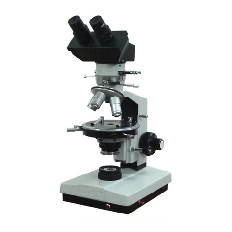
The Observation of Orthoscope
Usually to observe a tissue section by Microscope, the condenser should be lower down and the Bertrand lens is taken away from
the light path. Doing like thisway, the optical nature of minerals beabletobeobserved bymeansof getting in visual field of transmitting
parallel light.
How to Use
Polarizer and Analyzer is to be set on the orthogonal crossed position.
Slide out the analyzer from the light path.
Bertrand lens is also to slide out from the light path.
Without any slide, adjust the Sub Stage Condenser as to become the samebrightnessineachobjectiveswhileobserving
throughthe microscope.
To set with 10X eyepiece and low power Objective.
Insert the Analyzer into the light path, then the field becomes dark.
To rotate the rotating stage having on a slide with specimen, to be observed through the Microscope.
Insert the Gypsum-Plate in order to change a wave length of specimen in 1/4 degree, if required.
Subject to these steps, we may be able to know a figure and color or refractive index of minerals by orthoscope.
Adjustment of Centripetal
The center of the object & rotating stage have to accord with the crossed point of specimen in visual field always. If it is not
correct, it should be adjusted by the centering screws, while rotating the stage.Focus thespecimen sharply,find a marked feature in
field view and make it situated at the center of the eyepiece cross line.
The Observation of Conoscope
Set the eyepiece & object, insert an analyzer and polarizer in the light path and adjust the condenser to observe the Conoscope or
interference figure by transmitting light with various magnifications.
How to use
Adjust the Condenser to Upper position.
Insert the Bertrand lens in the light path.
To set with 5X eyepiece & 40X Objective.
To put a specimen on the stage and adjust the focus.
Adjust the intensity of the light/through diaphragm of the condenser.
