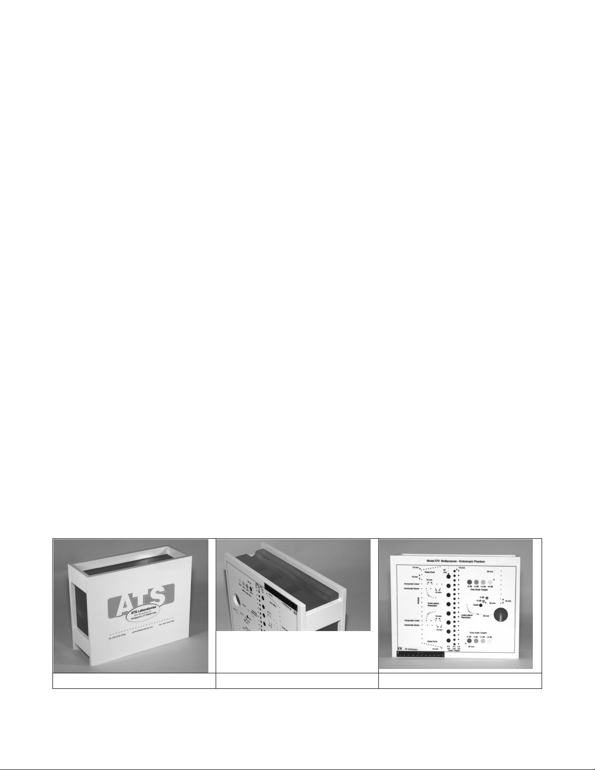
ATS 570 User manual

2
TABLE OF CONTENTS
Section
Page
Introduction
2
Sound Velocity of Rubber Based TM Material
2
Effect of Temperature
2
Tissue Mimicking Materials 3
Baseline Values
3
General Guidelines for Setting up the Phantom for Scanning
4
Phantom Re-Certification
4
Product Description
4
Tests Performed
5
Model 570 Target Diagram
5
Model 570 Specifications
6
Dead Zone
6
Vertical Measurement Calibration
7
Horizontal Measurement Calibration
7
Axial – Lateral Resolution
8
Focal Zone
8
Sensitivity (Maximum Depth of Penetration)
9
Functional Resolution and Image Uniformity
9
Gray Scale & Displayed Dynamic Range
10
References
10
Care of Rubber-Based Phantoms
11
Warranty
11
Introduction
Tissue-mimicking phantoms are used to evaluate the performance of diagnostic ultrasound imaging systems. The
phantoms mimic the acoustic properties of human tissue and provide target structures within the simulated
environment. They are essential to detect the performance changes that occur through normal aging and
deterioration of system components. Routine equipment performance monitoring can reduce the number of
repeat examinations, the duration of examinations, and maintenance time. Phantoms are employed in the areas
of; Clinical Quality Assurance, Preventative Maintenance Programs, Field Service Testing, Research and
Development, Manufacturing, Teaching and Sales & Marketing.
Sound Velocity of Rubber based TM Material
The sound velocity of most diagnostic imaging systems is calibrated to 1,540 meters per second (mps), the
assumed average velocity of sound through human soft tissue. The rubber-based tissue-mimicking material used
in the Model 570 has a sound velocity of 1450 m/s when measured at room temperature (22-24°C). The line
targets and anechoic target structures have been physically positioned to compensate for the differences in the
speed of sound, assuring accuracy of measurements.
Effect of Temperature
The acoustic properties of all biologic and non-biologic materials are affected by temperature variations. Most
diagnostic imaging systems and tissue-mimicking phantoms are calibrated at room temperature, commonly
referred to as 23°C. ATS has affixed a thermometer strip to the outside surface of the phantom housing, room
temperature is reflected on the strip, not the interior of the phantom. Therefore, if a rubber-based phantom is
exposed to extreme temperatures for several hours, it will take the equivalent amount of time to reach room

3
temperature once again. Leaving the phantom in your car overnight during a typical New England winter, while
the phantom will not be damaged, you will need to wait approximately 24 hours for it to reach room temperature.
Tissue Mimicking Materials
ATS offers a choice between our standard hydrogel-based (water) or rubber-based tissue-mimicking material. As
with most things in life, the choice between rubber-based or Hydrogel TM material is a trade off of benefits. We
have provided a listing of the features and benefits of both material.
Features
Hydrogel
TM Material
Rubber-Base
TM Material
Speed of Sound
1540 mps
Yes
No
Comment:
Line targets & target structures have been
physically moved to compensate for the difference
in the assumed average speed of sound of soft
tissue of 1540 mps and the speed of sound of the
phantom. As the attenuation increases so does
the speed of sound in the rubber. Attenuation of
0.5 dB/cm/MHz = 1450 mps (measured at 3.5
MHz and 23°C
Rate of Desiccation
.00005 gm/day/sq. cm
Not affected
Warranty
One-year
Lifetime (estimated to be 10 years)
Estimated Usable life
2-3 years
Greater than 10 years
Consistency of the
measurements with time
Changes begin to occur as
desiccation progresses, this
depends on the climate,
storage conditions and the
care taken of the phantom.
No change throughout the usable life have been
noted to date.
Exposure to temperatures
above 49°C or below 0°C
Phantom will be will severely
damaged or destroyed
Allow the phantom time to reaches room
temperature before using
Accidental Dropping
causing cracks in the
housing
Damage is usually beyond
repair, requiring replacement
Repairable
Baseline Values
The baseline represents the instrument’s peak performance. Ideally, the baseline values are established
immediately following the installation and acceptance of a new imaging system. If this is not possible, immediately
after preventive maintenance and servicing by a qualified service engineer.
Scan the Model 570 Multipurpose Endoscopic phantom, while adjusting the controls to produce the best possible
image. If the bottom of the phantom is visualized, adjust the depth of penetration until only the lower targets are
visualized without artifacts produced from the work surface. One should take care to avoid over emphasizing a
particular area of the image. Make the display monitor’s brightness and contrast settings and the room lighting
conditions reflect a clinical environment.
When an acceptable image has been obtained with a particular scanner-transducer pair, the system settings must
be accurately documented on the quality assurance record. The settings that should be included; dynamic range,
gray scale level, power level, gain level, and time gain compensation (TGC).
Remember, the accuracy or the baseline values obtained and recorded are extremely important. These values will
become the basis for all future performance testing. Some systems have the ability to save the baseline values.

4
When recalled the system is automatically setup to reflect the programmed baseline values, thereby reducing the
potential for errors.
General Guidelines for Setting Up the Phantom for Scanning
1. Place the phantom on a clean, flat surface with scan surface positioned for use.
2. Apply an adequate amount of low viscosity gel or water to the scan surface. If water is used, fill the
scanning well slowly to avoid introduction of air bubbles.
3. Set-up the imaging system-transducer pair in accordance with the established baseline values. Record
these settings on the quality assurance record.
Phantom Re-certification
Re-certification of an ATS rubber-base phantom can be performed at any time in accordance with an individual
institution’s quality assurance procedure. ATS produces a test block of TM material for every rubber-based
phantom sold. The test block is used to measure the acoustic properties of the phantom as part of the products
final quality assurance procedure. The test block is retained at the company for future reference and testing
during the re-certification process.
Procedure for phantom re-certification is simple. The phantom is returned to our facility with all shipping charges
paid by the customer. We perform the following steps:
1. Upon arrival, the phantom is inspected for physical damage.
2. The test block is retrieved from the storage library and the acoustic properties are measured.
3. The phantom is than scanned repeating all of the original tests.
4. Results are than compared with the original results on file.
5. A new Quality Assurance certification document similar to the one originally received when the phantom was
purchased will be issued. The phantom is then returned to our customer.
Product Description
The Model 570 Multipurpose & Endoscopic phantom is an easy, comprehensive means of evaluating imaging
systems with an operating frequency range of 2.25 to 7.5 MHz. The phantom is designed with a combination of
monofilament line targets for distance measurements and tissue mimicking target structures of varying sizes and
contrasts. Due to the acoustic similarity of the background material and the target structures, artifacts caused by
distortion, shadowing and enhancement have been eliminated. Four gray scale targets ranging in contrast from
+6 to -3 dB are provided to evaluate the system's displayed dynamic range and gray scale processing
performance.
The Model 570 offers a new and improved scan surface design to easily accommodate linear, sector, endoscopic
probes and mechanical sector probes such as used for rectal scanning.
Scan Surfaces
Standard Scan Surface
Endoscopic Scan Surface
Scanning Well

5
Tests Performed
•Dead Zone or Ring-Down (A1 & A2)
•Functional Resolution (F1, F2)
•Horizontal Measurement Calibration Linear
(C1&C2)
•Focal Zone (B1, B2 or F)
•Horizontal Measurement Calibration Sector (C1 &
C2)
•Axial & Lateral Resolution (E1, E2, E3)
•Sensitivity/Penetration (F)
•Contrast, Gray Scale (G1, G2, G3)
•Vertical Measurement Calibration (B1 & B2)
•Image Uniformity
Standard Scan
Endoscopic Scan Surface

6
Specifications
General
General
Tissue Mimicking Material
Overall Dimensions
270 x 215 x 96 cm
Type
Urethane rubber
Housing Material
PVC
Freezing Point
< -40°C
Scan Surfaces
2
Melting Point
> 100°C
Scan Wells
1
Attenuation Coefficient
0.5 dB/cm/MHz ± 5.0%
Weight
4.55 Kg (10 lb)
Speed of Sound
1450 m/s ±1.0% at 23°
Line Targets
Line Targets
Line Targets
Line Targets
Line Targets
Line Targets
Material Diameter
t Nylon
Monofilame
Monofilamennt Nylon
.12 mm
.12 mm
±0.1 mm
Dead Zone Groups
Lateral Displacement
Interval Spacing
Scan Surface Depth
Standard
Endoscopic
A1
5 mm
1 mm
2-10 mm
A2
5 mm
1 mm
2-10 mm
Vertical Groups
Number of Targets
Interval Spacing
Depth
B1
17
10 mm
10-160 mm
B2
8
10 mm
10-80 mm
Horizontal Linear
Groups
Number of Targets
Interval Spacing
Scan Surface Depth
Standard
Endoscopic
C1
5
10 mm
40 mm
C2
5
10 mm
40 mm
Axial-Lateral
Resolution Groups
Number of Targets
Interval Spacing
Scan Surface Depth
E1
Standard
E2
Endo-
scopic
E3
Scan
Well
Horizontal Sector
Groups
Number of Targets
Interval Spacing
Scan Surface Depth
Standard
Endoscopic
D1
5
10 mm
50 mm
D2
5
10 mm
50 mm
Axial-Lateral
Resolution Groups
Number of Targets
Interval Spacing
Scan Surface Depth
6
6
6
Horizontal Sector
Groups
Number of Targets
Interval Spacing
Scan Surface Depth
Standard
Endoscopic
D1
5
10 mm
50 mm
D2
5
10 mm
50 mm
Axial-Lateral
Resolution Groups
Number of Targets
Interval Spacing
Scan Surface Depth
5, 4, 3, 2, 1 mm
5, 4, 3, 2, 1 mm
5, 4, 3, 2, 1 mm
Horizontal Sector
Groups
Number of Targets
Interval Spacing
Scan Surface Depth
Standard
Endoscopic
D1
5
10 mm
50 mm
D2
5
10 mm
50 mm
Axial-Lateral
Resolution Groups
Number of Targets
Interval Spacing
Scan Surface Depth
40 mm
60 mm
40 mm
Horizontal Sector
Groups
Number of Targets
Interval Spacing
Scan Surface Depth
Standard
Endoscopic
D1
5
10 mm
50 mm
D2
5
10 mm
50 mm
Anechoic Target
Structures
F1
F2
Gray Scale Target
Structures
G1
Standard
G2
Endo-
scopic
G3
Scan
Well
Type
Non-echogenic, cylindrical
Type
Echogenic, Cylindrical
Size
8, 4, 2 mm
6 mm
Diameters
10 mm
10 mm
6 mm
Number of Targets
27
1
Scan Surface Depth
30 mm
20 mm
30 mm
Depth
1 0 – 170 mm
30 mm
Number of Targets
4
4
2
Interval Spacing
10 & 20 mm
NA
Contrast relative to
background material
(dB)
+6, +3, -3, -6
+6, +3, -3, -6
+3, -3
*Nominal dimensions
DEAD ZONE (A1 & A2)
Description and Reason For Testing
The dead zone is the distance from the front face of the transducer to the first identifiable echo at the phantom/
patient interface. The dead zone occurs because an imaging system cannot send and receive data at the same
time. Therefore, no clinical data can be collected in this region. However if artifacts are noted within the dead
zone, they may indicate fluctuations in the input power to the system. The depth of the dead zone depends upon
the frequency and performance of the transducer and the pulsing/receiving section of the system.
Testing Procedure

7
1. Scan the phantom until the dead zone target group is clearly displayed. Freeze this image.
2. This group is composed of 9 line targets. The first target is positioned 2 mm below the scan surface.
Subsequent targets are spaced 1 mm apart, to a depth of 10 mm.
3. Using the electronic calipers, measure the distance between the first target imaged and the echo produced by
the scan surface. The resulting value will be the depth of the dead zone.
4. Document the depth measurement on the quality assurance record.
Results
The system's dead zone should remain consistent from week to week when using the same instrument settings
and Model 570 phantom. Compare the test results obtained from the baseline records. If the current image
demonstrates changes in the system's ability to resolve these targets, corrective action should be considered.
VERTICAL MEASUREMENT CALIBRATION (B1 & B2)
Description and Reason For Testing
Vertical distance measurements are obtained along the axis of the sound beam. Accurate representation of the
size, depth and volume of a structure is a critical factor in a proper diagnosis. Most imaging systems use depth
markers and/or electronic calipers to obtain these measurements. The phantom is scanned and a distance
measurement obtained using the timing markers and/or electronic calipers. The resulting measurement is then
compared to the known distance between the line targets in the phantom. The accuracy of vertical distance
measurements depends on the integrity of the timing circuitry of the imaging system.
Testing Procedure
1. Position the transducer over the vertical group of line targets until a clear image is obtained. Freeze the
display.
2. Using the electronic calipers or the timing markers measure the greatest distance that can be clearly imaged
between line targets.
3. Document the measurement obtained on the quality assurance record.
Results
The system's vertical distances measurements should remain consistent from week to week when using the same
instrument settings and Model 570 phantom. Compare the test results obtained from the baseline records. If the
current image demonstrates changes in the system's ability to resolve these targets, corrective action should be
considered.
HORIZONTAL MEASUREMENT CALIBRATION
Linear Horizontal Group (C1 & C2)
Sector Horizontal Group (D1 & D2)
Description and Reason For Testing
Horizontal distance measurements are obtained perpendicular to the axis of the sound beam. Proper diagnosis
depends on the accurate representation of the size and volume of a structure being examined. Most imaging
systems use distance markers and/or electronic calipers to obtain these measurements. The phantom is scanned
and a distance measurement obtained. The resulting measurement is then compared to the known distance in the
phantom. The accuracy of the horizontal distance measurements depends on the integrity of the transducer
scanning assembly, the output intensity and the resolution of the imaging system.
Testing Procedure
Note: The Model 570 General & Small Parts phantom provides two scanning surfaces used to evaluate horizontal
measurement calibration. Due to the geometry and variety of sector scan transducers a separate set of horizontal
line targets are provided to evaluate lateral resolution. Please refer to the specification page for the location of
these groups.

8
1. Position the transducer over the horizontal group of line targets until a clear image is obtained. Freeze the
image.
2. Using the electronic calipers or the timing markers measure the greatest distance that can be clearly imaged
between line targets displayed.
3. Note: Some sector scanners have distance markers on the outside edges of the sector image with no other
indicators available. Hand-held calipers must be used for distance measurements within the image on the
monitor.
4. Document all of the measurements on the quality assurance record.
Results
The system's horizontal distance measurements should remain consistent from week to week when using the
same instrument settings and Model 570 phantom. Compare the test results obtained from the baseline records.
If the current image demonstrates changes in the system's ability to resolve these targets, corrective action
should be considered.
AXIAL – LATERAL RESOLUTION ARRAYS (E1, E2 & E3)
Description and Reason For Testing
Resolution is the minimum reflector separation between two closely spaced objects which can be imaged
separately along the axis of the beam, whereas lateral resolution defines the system’s ability to image objects
separately that lie perpendicular to the axis of the sound beam. If a system has poor resolution capabilities, small
structures lying close to each other will appear as one image, causing improper interpretation of the ultrasound
findings. Axial Resolution depends on the transducer’s center frequency, damping characteristics and pulse
length. Generally, the higher the frequency the better the system’s axial resolution. Lateral Resolution depends
on the beam width, focusing characteristics of the transducer, number of displayed scan lines and the system’s
sensitivity and gain settings.
Testing Procedure
The locations in the phantom are referenced from the first axial target.
The line targets are spaced at 5.0, 4.0, 3.0, 2.0, 1.0 mm intervals both axially and laterally. The last point of the
axial array target group is also the first target point in the lateral array group.
1. Position the transducer over the axial-lateral resolution group of line targets on the phantom until a clear
image is obtained. Freeze this image.
2. Examine the image to determine if all of the line targets within the group are clearly displayed as separate
target points. Record the closest spaced target points which can be imaged (refer to specification drawing).
Obtain a hard copy of the display.
3. Document all observations made on the quality assurance record.
Results
The system's ability to resolve the array targets at given depths should remain consistent from week to week
when using the same instrument settings and Model 570 phantom. Compare the test results obtained from the
baseline records. If the current image demonstrates changes in the system's ability to resolve these targets,
corrective action should be considered.
FOCAL ZONE (B1 & B2)
Description and Reason For Testing
The focal zone is the region surrounding the focal point in which the intensity and the lateral resolution is the
greatest. Clinically, structures examined within the focal zone will provide the best diagnostic information
obtainable. The focal zone can be affected by changes in the pulsing/receiving section of the imaging system or
damage to the transducer.
Testing Procedure
1. Position the transducer over the vertical group of line targets on the phantom, until a clear image is obtained.
A line rather than a dot is produced on the display. The length of the line is indicative of the width of the beam.
Therefore, targets inside the focal zone form a shorter line than those outside of the focal zone. Adjustments

9
in the gain settings will change the length of the line targets displayed. Freeze the display and obtain a hard
copy.
2. For a variable focused transducer, scans with several different focal zone settings should be performed.
Dynamically focused transducers may not display changes in the width of the line targets. However a change
in the intensity can be observed upon adjustment of the transmitting focus of the transducer.
3. Using the hard copy, draw a line connecting the ends of the echoes received from the line targets (both
sides), the line should form a smooth curve. This will illustrate the shape of the sound beam. Now locate the
narrowest portion, this is the focal zone. Measure the width of the beam and the depth at this point.
4. Document the depth of the focal zone and the measurement of the focal width on the quality assurance
record.
Results
The system's focal zone should remain consistent from week to week when using the same instrument settings
and Model 570 phantom. Compare the test results obtained from the baseline records. If the current image
demonstrates changes in the system's ability to resolve these targets, corrective action should be considered.
SENSITIVITY (MAXIMUM DEPTH OF PENETRATION) (F1)
Description and Reason For Testing
The ability of an imaging system to detect and display weak echoes from small objects located at specified depths
(penetration) is referred to as sensitivity. Clinically, weak reflecting echoes are commonly produced from internal
structures of organs. Definition of these structures can be extremely important in the interpretation of the
ultrasound findings. Sensitivity can be affected by the pulser/receiver section of the system, the degree of
focusing of the transducer, attenuation of the medium, depth and shape (geometry) of the reflecting object, and
electromagnetic interference from the local surroundings. A system’s maximum depth is limited by output power,
TGC, gain, transducer frequency, focal depth, number of scan lines and electrical noise.
Testing Procedure
1. Position the transducer over the 8 mm group of anechoic targets.
2. Freeze image and obtain a hard copy.
3. Examine the image to determine the last or deepest target structure displayed. Using the electronic calipers
or the timing markers measure the depth of this target.
4. This test should also be performed with output levels set at the highest and lowest settings. This enables any
changes in output to be more easily detected.
5. Document the depth measurement on the quality assurance record.
Results
The system's depth of penetration should remain consistent from week to week when using the same instrument
settings and Model 570 phantom. Compare the test results obtained from the baseline records. If the current
image demonstrates changes in the system's ability to resolve these targets, corrective action should be
considered.
Functional Resolution and Image Uniformity (F1)
Description and Reason For Testing
Functional resolution is an imaging system's ability to detect and display the size, shape, and depth of the non-
echogenic target structures within the TM matrix of the test phantom. The targets should appear circular with
sharp clearly defined edges, indicating an abrupt transition from the echogenic to the non-echogenic region. The
targets are anechoic and should be free of any internal echoes or fill-in.
Bright artifacts may be observed at the top and bottom of the targets, these are normal specular reflections and
do not present a problem. However, observable shade of gray within the anechoic target, usually is indicative of
internal system noise and/or the presence of side lobes. Should the targets appear flattened, a geometric
distortion problem should be considered. In practice, the data obtained will give a direct indication of the smallest
diameter target the system is capable of resolving at a given depth. The functional resolution capabilities of a
system can be affected by side lobes in the transducer beam, electrical noise, and problems in the imaging
processing hardware.

10
These artifacts can be the result of transducer malfunction, poor electrical contacts, failure in the image
processing and/or system’s software, and poor acoustic coupling between the transducer/patient interface
causing the introduction of reverberations artifacts. Generally, horizontal bands are often caused by circuitry and
focusing problems while vertical bands indicate a damaged transducer element.
Testing Procedure
1. Position the transducer over the anechoic target structures until a clear image is obtained.
2. Freeze image and obtain a hard copy.
3. Examine the image to determine the first and last target in each size group displayed. Record the
range of depths visualized for each group. Due to the configuration of the sound beam small targets
in the near field may not be imaged.
4. Scan this region to determine if there are any areas of non-uniformity. If the initial image
demonstrates non-uniformity or artifacts of this type, repeat the scan at a different location using the
same phantom to rule out a defect in a particular region of the phantom. If the artifacts are still
present, note the gain settings, gray scale level and focal setting and document with a photograph.
Repeat the scan using a different gain and focal setting.
5. Document all findings on the quality assurance record.
Results
The system's functional resolution and image uniformity should remain consistent from week to week when using
the same instrument settings and Model 570 phantom. Compare the test results obtained from the baseline
records. If the current image demonstrates changes in the system's ability to resolve these targets, or major
areas of image non-uniformity are observed, corrective action should be considered.
GRAY SCALE & DISPLAYED DYNAMIC RANGE
Description and Reason For Testing
Gray scale or gray scale processing uses the amplitude of the echoes received to vary the degree of brightness
of the displayed image. The adjustment of the echo signal required to go from a just noticeable (lowest gray
scale level) echo to the maximum echo brightness is referred to as the displayed dynamic range. Clinically, gray
scale processing and displayed dynamic range allow echoes of varying degrees of amplitude to be displayed in
the same image.
Test Procedure
1. Position the transducer over the gray scale target group until a clear image is obtained.
2. Freeze image and obtain a hard copy.
3. Examine the image. The targets should appear circular in shape, with clear sharp edges and vary in
the degree of brightness ranging from low to high levels of contrast. The presence or absence of any
shadowing behind the structures should be noted.
4. All findings should be documented on the quality assurance record.
Results
This target group varies in echogenicity and provides a good indication of the performance of the gray scale
processing and displayed dynamic range. The system's gray scale processing should remain consistent from
week to week when using the same instrument settings and Model 570 phantom. Compare the test results
obtained from the baseline records. If the current image demonstrates changes in the system's ability to resolve
these targets, corrective action should be considered.
General References:
Michell M. Goodsitt, Paul Carson; “Real-Time B-mode Ultrasound Quality Control Test Procedures, Report of
AAPM Ultrasound Task Group No. 1,” Medical Physics, 25 (8) August 1998
W. N. McDicken, PhD, “Diagnostic Ultrasonics, Principles and Use of Instruments,” John Wiley & Sons, 1976.
Sandra L. Hagen-Ansert; “Textbook of Diagnostic Ultrasonography,” Mosby, 1989.

11
CARE OF RUBBER-BASED PHANTOMS
For best results the phantom should be kept clean at all times. In particular a build-up of dried coupling
gel on the scan surface should be avoided. The phantom may be cleaned with mild hand soap and warm
water. Particularly stubborn stains and dirt may be removed with a mild household cleaner. The use of
petroleum solvents should be avoided since they may adversely react with the rubber-based material.
WARRANTY
Statement of Warranty
ATS Laboratories, Incorporated warrants this rubber-based phantom for it’s lifetime from the date of
delivery to the purchaser, that the Phantom is free from functional defects in materials and workmanship.
The lifetime of this phantom is estimated to be between 7 to 10 years from the date of manufacturing. If
ATS Laboratories, Incorporated, deems the phantom to be defective, at its sole option, the Phantom will
be repaired or replaced free of charge, in a reasonable amount of time.
ATS shall not be otherwise liable for any damages, including but not limited to incidental damages,
consequential damages, or special damages.
There are no express or implied warranties which extend beyond the warranties as stated below.
Conditions of Warranty
1. The defect must be reported and the Phantom returned within the warranty period.
2. The Phantom must be packaged properly to avoid damage during shipping.
3. All transportation charges will be paid by the purchaser.
Invalidation of Warranty
1. If the phantom has been altered or repaired other than by ATS Laboratories, Incorporated.
2. If the phantom has been subject to abuse, misuse, negligence or accident.
3. If the purchaser has exposed the Phantom to petroleum solvents.

ATS Laboratories Incorporated Tel: (800) 617-1177 Webpage: cirsinc.com Email: admin@cirsinc.com
Ultrasound Performance Testing Record Date ________________
__ Routine Testing
__ Baseline
__ Initial Setup
__ Software Upgrade
__ New Phantom
__ New Transducer
Facility: _______________________________ Department: ____________________
Technician / Sonographer: ________________________________
System Identification
System Manufacturer: _______________ Model: ______________ S/N: _____________
Transducer Type: __________________ Model #: _____________ S/N: _____________
ATS Phantom Model: ________________ S/N: ________________
General Inspection
Pass
Fail
Pass
Fail
Power Cord (cracks, plugs, discoloration)
Transducer (Cable, housing, plug, transducer face)
Dust Filters (clean & dust free)
Scanner console (free of damage)
Controls (Clean, broken knobs & switches)
Wheels (rotate freely, locks hold properly)
Display (clean, free scratches, Brightness/
contrast controls)
Comments
Comments
Comments
System Settings
System Settings
System Settings
System Settings
System Settings
System Settings
Power
dB
Gain
dB
Dynamic Range
dB
Pre-Processing
Post-Processing
Programed Presets
Transmit Focus
cm
Image Magnification
Room Temperature:
Room Temperature:
Geometric Accuracy Testing
Phantom
Distance
Baseline
Measured
Distance
Measured
Error/Change
Vertical Distance Measurements
Electronic Calipers
mm
mm
mm
mm
Display Devices used for interpretation
mm
mm
mm
mm
Horizontal Distance Measurements
Electronic Calipers
mm
mm
mm
mm
Display Devices used for interpretation
mm
mm
mm
mm
Dead Zone (Ring-down ramp)
Electronic Calipers
mm
mm
mm
mm
Display Devices used for interpretation
mm
mm
mm
mm
Spatial Resolution
Phantom
Distance
Baseline
Measured
Distance
Measured
Error/Change
Axial Resolution
Electronic Calipers
mm
mm
mm
mm
Display Devices used for interpretation
mm
mm
mm
mm
Lateral Resolution
Electronic Calipers
mm
mm
mm
mm
Display Devices used for interpretation
mm
mm
mm
mm

ATS Laboratories Incorporated Tel: (800) 617-1177 Webpage: cirsinc.com Email: admin@cirsinc.com
Focal Zone & Sensitivity
System’s Mfg.
Specification
Baseline
Depth
Measured
Depth
Error/Change
Focal Zone
Electronic Calipers
mm
mm
mm
mm
Display Devices used for interpretation
mm
mm
mm
mm
Sensitivity
Electronic Calipers
mm
mm
mm
mm
Display Devices used for interpretation
mm
mm
mm
mm
Functional
Resolution
Range of Depths
Displayed
Range of Depths
Displayed
Target Shape
Target Shape
Target Edges
Target Edges
Change
Yes/No
Target Sizes
(mm)
Baseline
Display**
Baseline
Display**
Baseline
Display**
1.0
mm
mm
2.0
mm
mm
30
mm
mm
4.0
mm
mm
6.0
mm
mm
8.0
mm
mm
10.0
mm
mm
**Display Devices used for interpretation
Image Uniformity - Display Devices used for interpretation
__ No Artifact Detected __ Artifact Detected
Image Uniformity Artifact Detected
Gain Settings
Gray Scale
Level
Focal Setting
Change
Yes/No
First Scan System Settings
Repeat Scan at a different region in the phantom and at different
gain and focal distance settings.
If artifact persists, further investigation and/or corrective action is recommended.
Gray Scale - Displayed Dynamic Range **Display Devices used for interpretation
Range of Contrast
Range of Contrast
Target Shape - Circular
Target Edges - Clear/Sharp
Change
Yes/No
Targets
(dB)
Baseline
Yes/No
Display**
Yes/No
Baseline
Yes/No
Display**
Yes/No
Baseline
Yes/No
Display**
Yes/No
+15
mm
mm
+6
mm
mm
+3
mm
mm
-3
mm
mm
-6
mm
mm
-15
mm
mm
Summary of Results:
Signature:
Table of contents
Other ATS Laboratory Equipment manuals
Popular Laboratory Equipment manuals by other brands

Thermo Scientific
Thermo Scientific VARIOMAG MINI 07 operating manual

Agilent Technologies
Agilent Technologies Cary 8454 installation guide

Agilent Technologies
Agilent Technologies IDP3GBKIT Quick reference guide

Advanced Instruments
Advanced Instruments 4250 user guide

LLG
LLG LLG-uniINCU 20 Cool Operation manual

Raue
Raue Steri 3000 user manual