ATS 532A User manual

Tel: 1-800- 617-1177 Fax: 1-757- 857-0523 Email: [email protected]
Ultrasound Imaging Phantoms
www.atslaboratories-phantoms.com
Model 532A & B
Contrast Resolution Phantom Instructions
Manual
Revised January 2015
Made in USA

INTRODUCTION
Quality assurance tissue-mimicking phantoms are used to evaluate the accuracy and
performance of ultrasound imaging systems. The phantoms mimic the acoustic properties of
human tissue and provide test structures within the simulated environment. They are essential
to detect the performance changes that occur through normal aging and deterioration of system
components. Routine equipment performance monitoring can reduce the number of repeat
examinations, the duration of examinations and maintenance time.
This phantom is constructed of a rubber-based tissue-mimicking material. This material extends
the useful life of the phantom by avoiding problems due to melting, freezing, dehydration and
breakage from dropping which are common with hydrogel (water-based) phantoms. By
eliminating these problems, the durability, quality and reliability of this product is guaranteed for
three years.
The acoustic properties of all biologic and non-biologic materials are affected by temperature
variations. Most diagnostic imaging systems and tissue-mimicking phantoms are calibrated at
room temperature, commonly referred to as 23°C. To ensure measurement accuracy ATS
incorporates a thermometer strip affixed to the outside surface of the phantom.
The sound velocity of most diagnostic imaging systems is calibrated to 1,540 meters per second
(mps), the assumed average velocity of sound through human soft tissue. The rubber-based
tissue-mimicking material has a sound velocity of 1450 and 1473 mps at 0.5dB/cm/Mhz and
0.7dB/cm/Mhz respectively at room temperature (23°C).
PRODUCT DESCRIPTION
The Models 532A and 532B Contrast Resolution phantoms are composed of a rubber-based
tissue-mimicking material. The Contrast Resolution phantoms are designed evaluated an
imaging system's gray scale processing and displayed dynamic range. Both models provide
three target sizes at eight levels of contrast, ranging from -12 to +12 dB/cm/MHz relative to the
background material.
The difference between the models is the target size. The Model 532A contains 2.0, 4.0 and 8.0
mm diameter cylindrical targets. Whereas, the Model 532B contains cylindrical targets of 5.0,
10.0, 20.0 mm.
TESTS PERFORMED
·Functional Resolution
·Definition and fill-in
·Gray Scale
·Displayed Dynamic Range

SPECIFICATIONS
General
Overall dimensions: 28.5 x 14 x 14.5 cm*
Weight: 4.4 Kg*
Scanning Surfaces: 2
Dimensions: 27 x 11 cm*
Tissue Mimicking Material
Type: Urethane rubber
Freezing Point: < - 40°C
Melting Point: > 100°C
Background Attenuation:** 0.5 dB/cm/Mhz measured at 3.5 Mhz
Speed of Sound: 1450 mps @ 23°C
Targets
Type: Echogenic, cylindrical
Number of targets: 24
Target groups: 3
Size (mm):
Diameter Length
Model #532A: 2.0 47
4.0 50
8.0 43
Model #532B: 5.0 50
10.0 50
20.0 40
Horizontal spacing: 3.0 cm
Depth: 7.5 cm
3.5 cm
Contrast relative to the
background material
+12 dB, + 9 dB, +6dB, +3dB
-12 dB, - 9 dB, -6dB, -3 dB
*Nominal dimensions
**Other attenuations available upon request.!

"
3.0
cm
4.7
cm
5.0
cm
2.8
cm
12.5
cm
28.5 cm
2 mm 4 mm 8 mm
MODEL 532A
Contrast Resolution Phantom
-12-9-6-3+3+6+9+12

"
Model 532B
Contrast Resolution Phantom
12.5
cm
5.1
cm
4.9
cm
2.0
cm
12.5
cm
7.5
cm
+12 +9 +6 +3 -3 -6 -9 -12
3.0
cm
28.5 cm
5 mm 10 mm 20 mm

CONTRAST RESOLUTION
Reason for Testing
Contrast resolution is defined as the system's ability to distinguish between regions of
differing levels of brightness. An imaging system's degree of contrast resolution refers to
it's ability to distinctly display two nearly equal degrees of brightness in the same view.
The gray scale processing section of the imaging system controls the level of contrast by
the adjusting the amplitude of the echoes received to vary the degree of brightness of
the displayed image. The adjustment of the echo signal required to go from a just
noticeable (lowest contrast) displayed echo to the maximum echo brightness is referred
to as the displayed dynamic range. Clinically, the ability of a system to distinguish and
display separately structures of similar levels of contrast is extremely important. Some
cystic or solid masses, located within an organ, having very similar levels of brightness,
potentially could go undetected.
Test Procedure
1. Place the phantom on a clean, flat surface with the appropriate scanning surface
positioned for use.
2. Fill the scanning well with water. Attempts should be made to avoid introducing air
bubbles while filling.
3. Adjust the instrument settings (TGC, output, etc.) to establish baseline values for
"normal" liver scanning. If the penetration is such that the bottom of the phantom is seen,
the gain settings are reduced such that the image fades and goes entirely black. These
setting should be noted on the quality assurance record and used for subsequent
testing.
4. Position the transducer over the cylindrical target group, until a clear image is
displayed. Freeze image and obtain a hard copy.
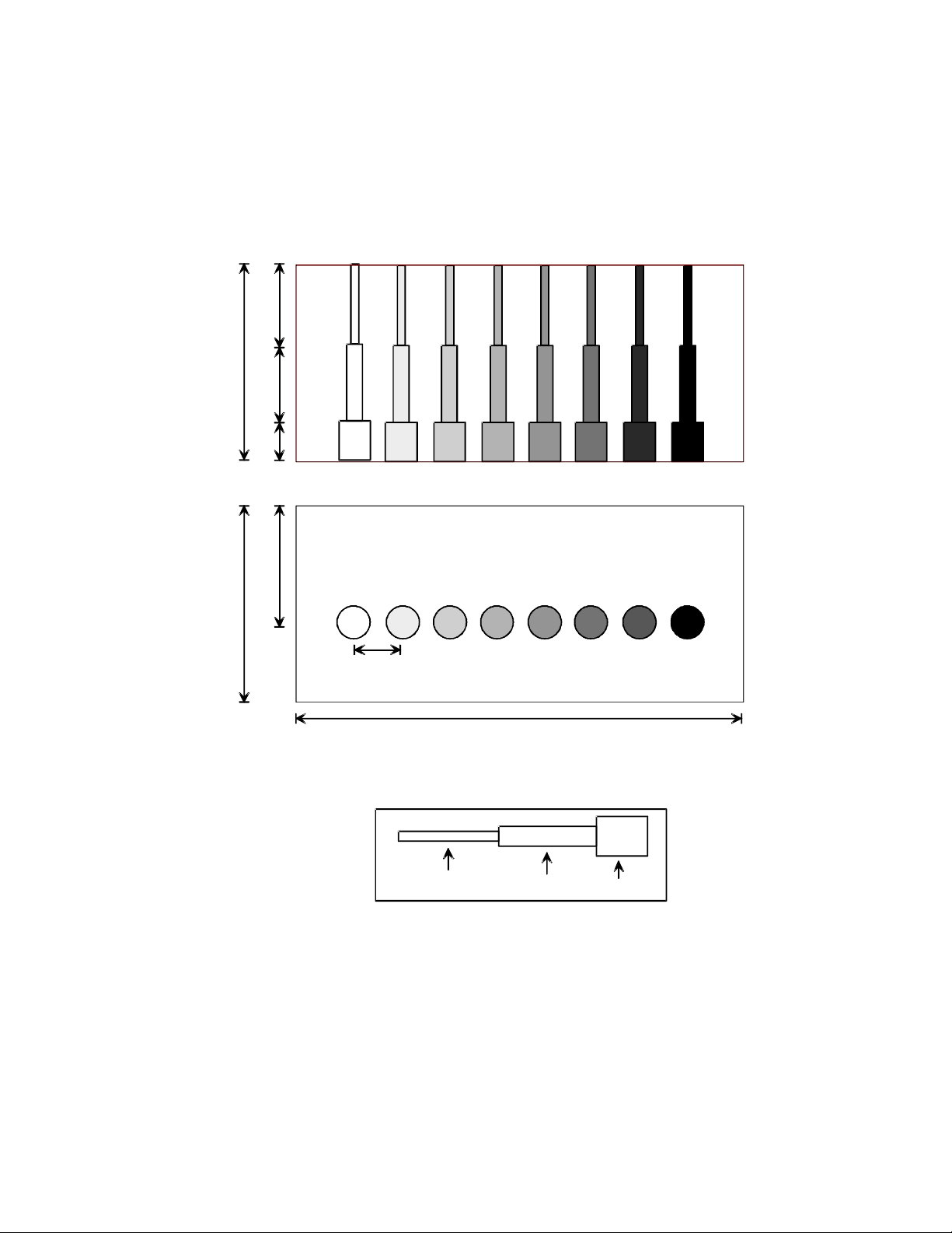
5. The targets should appear circular in shape, with clear sharp edges and vary in the
degree of brightness ranging from low to high levels of contrast. Examine the image to
determine the smallest diameter target which has been displayed. Using this target
group, determine if all eight levels of contrast, are distinctly displayed as varying levels of
brightness. The presence or absence of any shadowing behind the structures should be
noted.
6. Document all observation on the quality assurance record.
Results
The Model 532A or 532B provides a good indication of the performance of the gray scale
processing and displayed dynamic range. The test should be compared with a baseline
test using the same instrument settings, to determine if any change in the shape of the
targets has occurred or the systems ability to distinguish between varying degrees of
contrast.

CARE RUBBER-BASED PHANTOMS
For best results the phantom should be kept clean at all times and stored at room
temperature. In particular a build-up of dried coupling gel on the scan surface should be
avoided. The phantom may be cleaned with warm water using a lint free cloth.
Particularly stubborn stains and dirt may be removed with a mild household cleaner. The
use of petroleum solvents should be avoided since they may adversely react with the
rubber-based material.
WARRANTY
Statement of Warranty:
ATS Laboratories, Incorporated warrants this rubber-based phantom for it’s lifetime from
the date of delivery to the purchaser, that the Phantom is free from functional defects in
materials and workmanship. The lifetime of this phantom is estimated to be 10 years
from the date of manufacturing. If ATS Laboratories, Incorporated, deems the phantom
to be defective, at its sole option, the Phantom will be repaired or replaced free of
charge, in a reasonable amount of time.
ATS shall not be otherwise liable for any damages, including but not limited to incidental
damages, consequential damages, or special damages.
There are no express or implied warranties which extend beyond the warranties as
stated below.
Conditions of Warranty:
1. The defect must be reported and the Phantom returned within the warranty period.
2. The Phantom must be packaged properly to avoid damage during shipping.
3. All transportation charges will be paid by the purchaser.
Invalidation of Warranty:
1. If the phantom has been altered or repaired other than by ATS Laboratories,
Incorporated.
2. If the phantom has been subject to abuse, misuse, negligence or accident.
3. If the purchaser has exposed the Phantom to petroleum solvents.
This manual suits for next models
1
Table of contents
Other ATS Laboratory Equipment manuals