HARVARD CMA 7 User manual
TECHNICAL INFORMATION
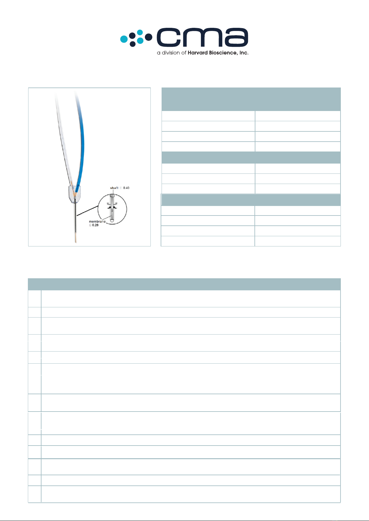
Membrane
Material Polyethersulfone (PES)
Molecular Cut-O 500kDa, 2MDa
Outer Diameter 0.28 mm
Length 1 and 2 mm
Probe Shaft
Material Stainless-steel
Diameter 0.40 mm
Length 14 mm
CMA 755 kDa & 2MDa Microdialysis Probe User’s Manual
Instructions for CMA 7 500 kDa & 2MDa Microdialysis Probes
1.
2.
3.
4.
5.
6.
7.
8.
9.
10.
Note: The 500 kDa and 2 MDa Cut Off membrane has to be use in a Push-Pull system to avoid loss of fluid out to the
surrounding tissue (ultra-filtration). Here the use of a CMA syringe pump and REGLO ICC Pump is recommended.
Load the peristaltic pump with the "FEP Tubing Connector Peristaltic Kit". Adjust the lengths of the FEP tubing if needed for a
lower dead volume; recalculate the outlet volume if needed (1.2 µl/100 mm length)
Run the pump to make sure that liquid leaves the tip of the syringe cannula.
Fill a microsyringe with perfusion fluid and mount it in the CMA Syringe Pump. The Perfusion Fluid must be clean, at
room temperature and preferably degassed.
Place the inlet end of the “FEP Tubing Connector Peristaltic Kit” in a beaker with Perfusion Fluid and flush the pump to fill all
tubing with perfusion fluid. Make sure there is no air bubbles in the complete tubing set.
Set the pump to the required perfusion flow, usually 1 – 5 µL/min.
Prepare a desired length of FEP tubing with a Tubing Adapter on both ends and connect the tubing to the inlet of the
probe. Remove the protection tube from the probe carefully. Blue = Inlet, Transparent = Outlet. Tubing Adapters and FEP
tubing should be used for all connections. To facilitate the handling of Tubing Adapters, they should be soaked in Ethanol
70% for minimum 10 minutes.
Mount the probe in a Probe/Guide Clip on the CMA 130 In Vitro Stand. Put the probe membrane into a vial filled with
perfusion fluid. Connect the inlet tubing of the probe to the syringe cannula by sliding the Tubing Adapter over the cannula.
Flush the probe with perfusion fluid at 8-10µL/min for 4-5 min to wash out air. Knock on the shaft of the clip to help the air to
flush away. At this process the Ultra High Cut Off membrane will look as leaking but this is due to ultra-filtration of fluid
through the membrane.
Set the pump to the required perfusion flow, usually 1 – 5 µL/min. Make sure both pumps have the same flow rate.
Connect the inlet “FEP Tubing Connector Peristaltic Kit” at the peristaltic pump to the outlet of the probe
11.
12.
13.
Lift up the probe from the vial and check that the membrane doesn’t ultrafiltrate or dry out; it should look filled out but
not sweating. Lower the probe into the vial and control the outlet volume for the complete system.
The system with the probe is now ready for use.
When changing sample vials, remember to consider the internal volume in the system (see TECHNICAL INFORMATION).
This causes a delay that must be calculated when using low perfusion rates and short sampling times
Internal Volume
Inlet Volume 0.06 µL
Outlet Volume 0.3 µL
200 mm Inlet Tubing (blue) 3.5 µL
200 mm Outlet Tubing (transparent) 3.5 µL

WARRANTY
The probes manufactured by CMA Microdialysis are warranted to be free from defects in material and workmanship for
a period of two years from the manufacturing date if stored in the original package.
Claims should be forwarded without delay to CMA Microdialysis or to your local distributor.
ORDER INFORMATION
CMA 7 500 kDa Microdialysis Probe, 1 mm, 3/pkg*
CMA 7 500 kDa Microdialysis Probe, 2 mm, 3/pkg*
CMA 7 Guide Cannula, 3/pkg
CMA 7 Guide Cannula, 30/pkg
The CMA 7 Microdialysis Probe is not intended for use in humans. It is only suitable for laboratory research in animals.
CMA Microdialysis only guarantees single usage of CMA 7 Microdialysis Probes
CMA Microdialysis AB
Head Office, Sweden
Torshamnsgatan 30A, SE-164 40 Kista, Sweden
Tel: +46 8 470 10 00
E-mail: [email protected]
Harvard Apparatus
US Office
84 October Hill Road
Holliston, MA 01746 USA
Phone Orders: 800-232-2380 • Fax: 508-429-5732
E-mail: [email protected]
www.microdialysis.com 8011680A
*β –Irradiated Probes are available as Custom Probes
Tubing Adapter, 10/pkg
FEP Tubing, 1 m, 1/pkg
FEP Tubing, 1 m, 10/pkg
FEP Tubing Connector Peristaltic Kit, 3/pkg
CMA 7 & 8 Probe Clip
CMA 7 2MDa Microdialysis Probe, 1 mm, 3/pkg*
CMA 7 2MDa Microdialysis Probe, 2 mm, 3/pkg*
Microsyringe 1 mL
Microsyringe 2.5 mL
Ref No.
CMA 8012421
CMA 8012422
CMA P000137
CMA P000138
CMA 3409500
CMA 3409501
CMA 8409501
CMA 8012518
CMA P000136
CMA 8012423
CMA 8012424
CMA 8309020
CMA 8309021
OPTIONAL ACCESSORIES Ref No.
CMA 4004 Syringe Pump
CMA 402 Microdialysis Pump with Accessory Kit
CMA 400400
CMA 8003100
CMA 402 Microdialysis Pump
CMA 110 Liquid Switch
CMA 8003110
CMA 8308200
CMA 130 In-Vitro Stand with CMA 7 & 8 Clips
CMA 142 Microfraction Collector 230V
CMA 8309104
CMA 8381142
CMA 142 Microfraction Collector 115V
CMA 470 Refrigerated Fraction Collector
CMA 8381143
CMA 8002770
For other probes and microdialysis accessories please call
your local CMA Microdialysis dealer.
Other HARVARD Medical Equipment manuals


Popular Medical Equipment manuals by other brands


Getinge
Getinge Arjohuntleigh Nimbus 3 Professional Instructions for use

Mettler Electronics
Mettler Electronics Sonicator 730 Maintenance manual

Pressalit Care
Pressalit Care R1100 Mounting instruction

Denas MS
Denas MS DENAS-T operating manual

bort medical
bort medical ActiveColor quick guide

AccuVein
AccuVein AV400 user manual


