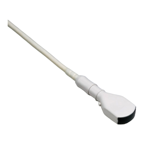
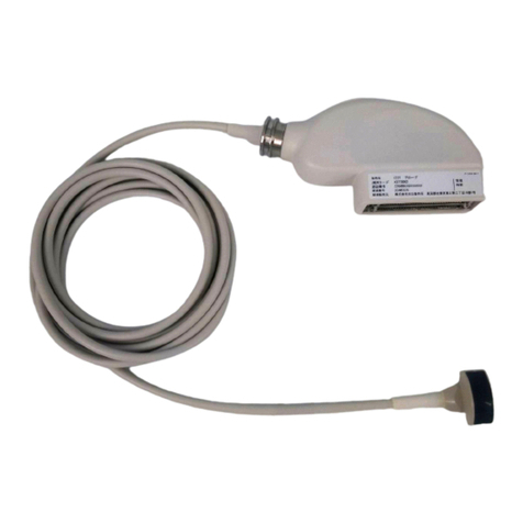
Hitachi C41V1 User manual

C41V1 Probe
INSTRUCTION MANUAL
Tokyo , Japan
Q1E-EP1432-7
© Hitachi, Ltd. 2013,2017. All rights reserved.
Notes for operators and responsible maintenance personnel
★ Please read through this Instruction Manual carefully prior to use.
★ Keep this Instruction Manual together with the system with care to
make it available anytime.
0123

( 1 ) Q1E-EP1432
Manufacturer: Hitachi, Ltd.
2-16-1, Higashi-Ueno, Taito-ku, Tokyo,110-0015,
Japan
+81-3-6284-3668
http://www.hitachi.com/businesses/healthcare/
index.html
European
Representative: Hitachi Medical Systems GmbH
Otto-von-Guericke-Ring 3 D-65205 Wiesbaden,
Germany
EU Importer: Hitachi Medical Systems Europe Holding AG
Address: Sumpfstrasse 13 CH-6300 Zug, Switzerland
Local Distributor:

( 2 ) Q1E-EP1432
About this manual
This instruction manual contains safety precautions, the inspection,
the operation procedure and the reprocessing procedure of C41V1 Probe.
Please read this manual thoroughly to ensure the safety operation.
If you have any questions concerning the operation of the probe, please
contact a service support.
The following conventions are used throughout the manual to denote
information of special emphasis.
WARNING: “Warning” indicates the presence of a hazard which may
result in severe personal injury, substantial property
damage, or death if the warning is ignored.
CAUTION: “Caution” indicates the presence of a hazard which will or
can cause minor personal injury or property damage if the
caution is ignored.
NOTICE: “Notice” indicates information of installation, operation,
or maintenance, which is important, but not hazard related.

( 3 ) Q1E-EP1432
Graphical Symbols for Use in Labeling of Hitachi Ultrasound Probes
Some graphical symbols that are used in labeling of Hitachi Ultrasound Probes are
compliant with EN980:2008 standard. Refer to the following table about the meanings
of them.
Explanation of Symbol Symbol Descriptive Content
Manufacturer Company
Name and Address
Hitachi, Ltd.
2-16-1, Higashi-Ueno, Taito-ku,
Tokyo, 110-0015, Japan
+81-3-6284-3668
http://www.hitachi.com/businesses/
healthcare/index.html
Authorized
Representative
in The European
Community
Hitachi Medical Systems GmbH
Otto-von-Guericke-Ring 3 D-65205
Wiesbaden, Germany
Keep away
from Sunlight
Store the probe in a cool place and keep
away from high temperature, high
humidity, or direct sunlight.
Contains or presence of
natural rubber latex
Contains or presence of natural rubber
latex
Do not re-sterilize
Do not re-sterilize
Do not reuse
Do not reuse

( 4 ) Q1E-EP1432
Definition of symbol
The following symbol is also used for HITACHI Ultrasound Probes.
Location Symbol Definition
Probe connector
This instrument complies with
Directive 93/42/EEC relating to
Medical Device and Directive
2011/65/EU relating to RoHS
Probe connector IPX7 IPX7 mark
See section 1.6.
Probe connector Type BF APPLIED PART
Probe connector General warning sign
Probe connector Warning; dangerous voltage
Probe connector Caution; Biohazard
Probe connector
Follow the instruction manual to
operate this instrument. If not
avoided, may result in injury, property
damage, or the equipment trouble.
Probe connector
STERRAD sterilization compatibility
mark
Probe connector Do not waste the instrument as general
waste. Comply with a local regulation.
Probe connector Rx Only
By prescription only. U.S. Federal Law
restricts this device to sale on order
of a physician only.

( 5 ) Q1E-EP1432
CONTENTS
Page
1. Introduction ................................................. 1
1.1 Features ......................................................... 1
1.2 Principles of operation .......................................... 1
1.3 Intended Use ..................................................... 2
1.4 Components ....................................................... 2
1.5 Accessories (Option) ............................................. 3
1.6 Construction ..................................................... 4
2. Inspection before Use ........................................ 5
2.1 Inspection for Appropriate Connection ............................ 5
2.2 Inspection for Material Surface .................................. 5
3. Operation Procedure .......................................... 6
3.1 Connection and Settings .......................................... 6
3.2 How to attach the Sterile Puncture Adapter (EZU-PA7V) ............ 8
3.3 Display of Needle Guide Line ..................................... 9
4. Option of C41V1 Probe ....................................... 11
4.1 Magnetic sensor ................................................. 11
5. Cleaning, Disinfection and Sterilization .................... 13
5.1 Point of use (Pre-cleaning) ..................................... 16
5.2 Containment and transportation .................................. 17
5.3 Manual Cleaning and disinfection ................................ 17
5.4 Drying .......................................................... 21
5.5 Inspection ...................................................... 21
5.6 Packaging ....................................................... 22
5.7 Sterilization ................................................... 23
5.8 Storage ......................................................... 25
6. Maintenance and Safety Inspection ........................... 26
6.1 Daily Inspection ................................................ 26
6.2 Store ........................................................... 26
7. Safety Precautions .......................................... 27
8. Specifications .............................................. 29
8.1 Probe ........................................................... 29
8.2 Sterile Puncture Adapter EZU-PA7V ............................... 30
8.3 Suppliers List .................................................. 31
9. Disposal of the probe ....................................... 31

- 1 - Q1E-EP1432
1. Introduction
1.1 Features
C41V1 Probe is a Convex Array Probe.
The acoustic output of this probe when connected to ultrasound
diagnostic scanner was measured according to the IEC60601-2-37
standard. The table of measured acoustic output data is contained
in the operational manual of each ultrasound diagnostic scanner.
This probe is categorized in class IIa according to Directive
93/42/EEC.
According to IEC60601-1 the probe is classified as type BF.
1.2 Principles of operation
This probe and the ultrasound diagnostic scanner enable image
diagnosis using ultrasonic waves. This system operates under the
principles described below.
1) When an electric pulse signal is applied from the transmitter
to the transducer of the probe, the transducer converts electric
signals into mechanical vibration energy for emitting
pulse-shaped ultrasonic waves into the body part, liquid or
other medium contacting the transducer.
2) The emitted ultrasonic waves are reflected by boundaries with
different acoustic characteristics (acoustic impedance) within
the body.
3) The transducer is also used to receive reflected ultrasonic
waves. The transducer vibrates mechanically due to the received
ultrasonic waves and converts mechanical vibrations into
electric energy. Electric signals are converted to shades of
brightness by brightness modulation to obtain an image.

- 2 - Q1E-EP1432
1.3 Intended Use
C41V1 Probe is designed for observation and diagnosis of the
following regions mainly by connecting with the HITACHI ultrasound
diagnostic scanner.
General OB/GYN organs
Biopsy (with a Sterile Puncture Adapter)
Transvaginal/Transrectal
Never use the probe for following regions.
1) The heart (Do not contact directly)
2) The eyeball
1.4 Components
Components of C41V1 Probe are as follows:
1) Probe ............................... 1 piece
2) Instruction Manual .................. 1 copy
The probe is not sterilized when delivered. Prior to use, be sure
to clean, disinfect and sterilize it.
WARNING
C
A
U
TI
O
N

- 3 - Q1E-EP1432
CAUTION
1.5 Accessories (Option)
1.5.1 Sterile Puncture Adapter EZU-PA7V (Disposable)
EZU-PA7V is the attachment for ultrasound guided transvaginal
or transrectal biopsy and aspiration of organs, cyst and tumor.
The size of needles available is 16 to 19G. Biopsy application
requires special care. 24 pieces of Sterile Puncture Adapter
EZU-PA7V are packaged in one set as follows:
Component Model Note
Sterile Puncture Adapter EZU-PA7V 24 pcs
NOTE: If you need Sterile Puncture Adapters, please contact a service
support.
A well-trained physician only should perform a biopsy.
1.5.2 Probe cover for Single Use (Disposable)
To protect the probe against contamination, using probe cover
is recommended. If using probe cover, lubrication free or dry
type probe cover is recommended.
Lubrication may cause a deterioration of the probe surface.
Use of non-allergic probe cover is strongly recommended to
avoid allergic reactions due to latex rubber.
Discard the used probe cover carefully.
1.5.3 Magnetic Sensor Attachment
Magnetic Sensor Attachment is needed for Real-time Virtual
Sonography (RVS). It is used to fix the magnetic sensor to the
probe.
The Magnetic Sensor Attachment is not sterilized when delivered from
the factory. Prior to use, be sure to clean, disinfect and sterilize
it.
C
A
U
TI
O
N

- 4 - Q1E-EP1432
1.6 Construction
The external view of C41V1 Probe is shown in Fig.1.
Immersible part: This part can be immersed in disinfectant solution
and also can be cleaned by water.
Un-immersible part: This part should not be immersed in disinfectant
solution and also can not be cleaned by water.
Fig.1
E
xternal view
C
able
Connector
U
n
-
i
mmer
s
ible
p
art
Immersible part (IP
X
7
)
Applied part
Magnetic sensor attachment (Option)

- 5 - Q1E-EP1432
2. Inspection before Use
Prior to use, the probe and accessories must be carefully inspected
that they are appropriate for use. If you find any damage, do not
use them and contact a service support immediately.
2.1 Inspection for Appropriate Connection
2.1.1 Confirm that the ultrasound diagnostic scanner is correctly
operating. Please refer to the instruction manual of the
scanner for the operation.
2.1.2 Confirm that any unauthorized device/instrument such as
an unauthorized biopsy attachment is not attached or
connected to the probe.
2.1.3 Confirm that the Sterile Puncture Adapter and settings of
the scanner are appropriate for the probe. Attach the
Sterile Puncture Adapter to the probe according to “3.
Operation Procedure”. Display the “Needle guide line” on
the monitor. (Refer to the operation manual of the
ultrasound diagnostic scanner.) Keep the probe head under
sterile water and insert a puncture needle in the Sterile
Puncture Adapter, then confirm that the needle is inserted
smoothly and the echo of the needle is displayed on the
“Needle guide line”(dot line) on the monitor.
2.2 Inspection for Material Surface
2.2.1 Visually inspect the surface of the probe head, housing
and cable for any crack, scratch or denaturalization.
2.2.2 Visually inspect the envelope of the Sterile Puncture
Adapter for any break, deformation, crack or
denaturalization. If you find any damage, do not use the
Sterile Puncture Adapter.
2.2.3 Visually inspect the Magnetic sensor attachment for any
crack, deformation or denaturalization. If you find any
damage, do not use the Magnetic sensor attachment.

- 6 - Q1E-EP1432
3. Operation Procedure
3.1 Connection and Settings
1) Confirm that the probe and the
Magnetic sensor attachment
are disinfected and if
necessary sterilized. The
Magnetic sensor attachment
is needed for Real-time
Virtual Sonography(RVS).
Regarding the option for RVS,
please refer to “4.Option of
C41V1 probe”.
2) It is recommended to use a
disposable probe cover for
preventing a patient from
infection and the probe cover
should be allergy free
material to avoid allergic
reaction.
3) Connect the probe to the
ultrasound diagnostic
scanner and operate the
scanner and adjust the image
according to the
instructions given in the
operation manual for the
ultrasound diagnostic
scanner. The relationship
between the direction of the probe and the right-left
orientation mark on the image is shown in Fig. 2.
4) Put proper quantity of sterilized acoustic jelly on the probe
head as a couplant. (See Fig.3.)
Sterilized acoustic jelly
Fig.2 Relationship between
t
he
d
irection of the probe and
the Right-
l
eft Orientation
M
ark
D
irection
Orientation
mark
Fig.3 Jelly as a couplant

- 7 - Q1E-EP1432
5) Putting a probe cover onto the probe is recommended. If
using probe cover, draw the probe cover until the end of
the probe shaft. If air bubbles remain in jelly, remove air
bubbles by pushing jelly with finger. (See Fig.4)
Special attention should be paid for a probe cover made of
latex. Latex may cause allergic reactions such as itching,
rubor, urticaria, swelling, fever, anhelation, wheezing,
depression of blood pressure, and shock.
If you observe any of above-mentioned symptoms in your
patient during the operation, stop the use of the latex
protective sleeve immediately and take an appropriate
treatment to the patient.
6) Fix the probe cover with sterile adhesive tape as shown in
Fig.5.
Visually inspect the probe cover for any hole or tears.
7) Insert the probe gentry and adjust the probe’s position for
a clear view of the desired image. If the image is not clear
enough, use sterile saline solution to have a better image.
Probe cover
Air bubble
Sterilized acoustic jelly
Sterilized tape
WARNING
Fig.4 Removal of bubbles in jelly
Fig.5 Fix by sterile adhesive tape

- 8 - Q1E-EP1432
8) After using the probe, clean, disinfect and if necessary
sterilize the probe immediately. If RVS is carried out,
clean and disinfect and if necessary sterilize the Magnetic
sensor attachment.
9) Store the probe and the Magnetic sensor attachment under
the conditions written in
“6. Maintenance and Safety Inspection” and “8.Specification”.
・ Do not use the probe if the image and the frequency do not
match the probe. An incorrect acoustic output can result
in burns or other injuries to the patient.
・ Scan for the minimum length of time necessary for the
diagnosis and at the lowest possible output. There is the
possibility that the patient’s tissues could be affected.
For details about the acoustic output, please refer to the
operation manual of the ultrasound diagnostic instrument.
3.2 How to attach the Sterile Puncture Adapter (EZU-PA7V)
How to attach the Sterile Puncture Adapter (EZU-PA7V) to the
probe is instructed below.
1) Put the picks of the Sterile Puncture Adapter to the projection
on the probe. (See the top of Fig.6)
2) Push the other end of the Sterile Puncture Adapter until the
dents of the Sterile Puncture Adapter are fixed at the grooves
of the probe. (See the bottom of Fig.6)
3) Visually inspect the probe cover for any hole or tears.
Sterile Puncture Adapter
(EZU-PA7V)
Probe cover
Fig.6 how to attach the Adapter
C
A
U
TI
O
N

- 9 - Q1E-EP1432
3.3 Display of Needle Guide Line
The needle guideline in dot marks can be displayed on the monitor
for biopsy. Regarding the needle guideline, refer to the
operation manual of the ultrasound diagnostic scanner.
NOTE: The needle guide line is intended to provide a visual guide to
the direction of the puncture needle pathway. Be sure to check
the actual needle position on the ultrasound image when
performing the puncture operation.
The needle guide line can be displayed for this probe, but if
the image is displayed at some angle (see the figures below),
the image displayed may not be appropriate for biopsy.
Confirm that the image displayed is appropriate prior to biopsy.
Probe Puncture adapter
Needle
guide line
Artifact of puncture
adapter
Artifact of puncture adapter may appear while the
guide line is displayed only at the vicinity.
(Guide line is effectively displayed.)
(Guide line is not displayed.)
N
eedle guide line
Location of observation
FOV of the probe
Image display
C
A
U
TI
O
N

- 10 - Q1E-EP1432
1) Special attention should be paid for a probe cover made of
latex. Latex may cause allergic reactions such as itching,
rubor, urticaria, swelling, fever, anhelation, wheezing,
depression of blood pressure, and shock.
For the patients suspected of latex allergy, do not use the
latex-containing medical devices. If you observe any of above
mentioned symptoms in your patient during the operation, stop
the use of the latex-containing medical devices immediately
and take an appropriate treatment to the patient.
2) The puncture adapter EZU-PA7V and the recommended probe covers
are disposable and must not be reused.
3) Sterilize/disinfect the probe and disinfect the puncture
adapter when the probe cover is torn.
4) Be careful about the use of a needle cannula with the puncture
adapter. If the insulation coating of the needle cannula is
damaged, it may cause a burn to tissue.
5) Use a needle whose size is appropriate for the puncture adapter,
otherwise looseness or tightness of the needle could result
in biopsy at an unintended part and injury to the patient.
6) Display the needle guideline during biopsy. Regarding the
needle guideline, refer to the operation manual of the
ultrasound diagnostic scanner.
WARNING

- 11 - Q1E-EP1432
4. Option of C41V1 Probe
4.1 Magnetic sensor
4.1.1 How to attach the Magnetic sensor
The procedure of attaching the magnetic sensor is as follow.
1) Confirm that the Magnetic sensor attachment is sterilized
or disinfected.
2) Insert Magnetic sensor into the Magnetic sensor attachment
in the correct direction as shown in Fig.7.
Never attach the Magnetic sensor attachment to the probe in the
Wrong direction, otherwise it may result in false diagnosis.
3) Attach the Magnetic sensor attachment to the probe as shown
in Fig.8.
F
ig.7 How to attach the Magnetic sensor
Direction mark
Magnetic sensor attachment
Magnetic
s
ensor
C
A
U
TI
O
N
Fig.8 How to attach the Magnetic Sensor Attachment

- 12 - Q1E-EP1432
CAUTION
CAUTION
Do not put your fingers between the Magnetic sensor attachment and the
probe when attaching the Magnetic sensor attachment to the probe.
4.1.2 How to release the Magnetic sensor
The procedure of releasing the magnetic sensor is as follow.
1) Detach the Magnetic sensor attachment from the probe as
shown in Fig.9.
2) Detach the magnetic sensor from the Magnetic sensor
attachment as shown in Fig.10.
Clean, disinfect and sterilize the Magnetic sensor attachment prior
to the first use as it is not sterilized when delivered.
Fig.9 How to release the Magnetic sensor attachment fro
m
the probe
Fig.10 How to release magnetic sensor
f
ro
m
the Magnetic sensor attachment

- 13 - Q1E-EP1432
5. Cleaning, Disinfection and Sterilization
The probe and accessory must be reprocessed after each use. Refer
to the reprocessing instruction in this chapter.
WARNINGS
‐The probe is delivered unsterile. Prior to the
first use, reprocess the probe.
‐Temperature should not exceed 60°C during
reprocessing.
‐Probe connector is not water resistant.
Limitations on
reprocessing
The probe is not completely submersible. The
immersible part is shown in Fig.1. The un-immersible
part should be disinfected by wipe disinfection.
Transportation
before using
T
he probe should be packed in a sterile pouch or
c
ontainer to transport from Central Sterile Supply
D
epartment (CSSD) to an operating room. Be careful
not to damage the sterile pouch or container during
transportation.

- 14 - Q1E-EP1432
Levels of reprocessing requirements:
Depending on the application of the product and with regard to risk
evaluation, the user has to classify the medical device according to
the current Medical Device Directive for processing of medical devices
as uncritical, semi-critical or critical. Supporting information
concerning this topic is listed in the table below. The user is
responsible for correct classification of the medical device.
Classification Definition Processing
uncritical
Application part only
contacts intact and
uninjured skin
Cleaning
Disinfection
semicritical
Application part contacts
mucosa (intracavitary
application)
Cleaning
Disinfection
(Disinfectant with
virucidal effect)
critical
Application part contacts
intracorporeal tissue
directly
(operative application)
Cleaning
Disinfection
(Disinfectant with
virucidal effect -
minimum)
Sterilization
According to the intended use, C41V1 probe is classified as
semicritical.
Table of contents
Other Hitachi Measuring Instrument manuals

Hitachi
Hitachi L44LA1 User manual

Hitachi
Hitachi L44K User manual

Hitachi
Hitachi Relion 670 Series Quick start guide

Hitachi
Hitachi L-8900 User manual

Hitachi
Hitachi HT7700 User manual

Hitachi
Hitachi C253 User manual

Hitachi
Hitachi L34 User manual

Hitachi
Hitachi L53K User manual

Hitachi
Hitachi UG 50Y User manual

Hitachi
Hitachi L44LA User manual

Hitachi
Hitachi UST-52105 User manual

Hitachi
Hitachi L51K User manual

Hitachi
Hitachi Relion 670 Series Instructions for use

Hitachi
Hitachi U-2910 User manual

Hitachi
Hitachi C22I User manual

Hitachi
Hitachi C22T User manual

Hitachi
Hitachi EUP-C532 User manual

Hitachi
Hitachi VC34 User manual

Hitachi
Hitachi U-2810 User manual

Hitachi
Hitachi S3ESL1 User manual