MiiS Horus Scope DSC 300P User manual
MiiS Horus Scope
DSC 300P
User Manual
DOC. No. 2019.02-A
Copyright@2019 MiiS Inc.
All right reserved.

2

3
Contents
Preparations .........................................................................................5
Before use ..................................................................................................5
Names of components .............................................................................10
Charging the battery ................................................................................22
Power indicator........................................................................................23
Assembling...............................................................................................24
Using the setup mode..............................................................................31
A. Digital Otoscope (DOC 300S/ DOC 100S),
Digital speculum (DGC 100),
Digital Anterior Scope (DEA 100 and DEA 100 with ILS 100),
Endoscope adapter (Adapter 300).................................................31
B. DEC 100/ EEC 100 (Digital Eye Fundus Camera).............................37
C. DEA 200P (Digital Anterior Scope)..................................................43
D. DDC 200 and DDC 100 (Digital Dermatoscope)..............................48
Entering the patient ID.............................................................................54
Taking pictures....................................................................................57
Sequence of operations ...........................................................................57
Photo mode..............................................................................................58
Video mode..............................................................................................59
Examination conditions of DEC 100 and EEC 100.....................................60
Q&A troubleshooting of DEC 100 and EEC 100........................................61
Playback..............................................................................................62
Display mode............................................................................................62
Miscellaneous.....................................................................................64
Files transferring.......................................................................................64
Viewing on a computer/laptop screen.....................................................64
Viewing on a TV screen ............................................................................64
UVC and HDMI .........................................................................................64
Technical description................................................................................65
Liability.....................................................................................................68
Disposition ...............................................................................................68
Symbols and standards.............................................................................69

4

5
Preparations
Before use
Prior to installation and start-up of the Horus Scope, carefully read the user manual. As with all
technical devices, the proper function and safety operation of this device depend on the user
complying with the safety recommendations presented in these operating instructions. In
addition, please make sure it does not appear damaged or broken. If there are breaks on the
outer casing or other visual defects, please contact the manufacturer or a certified service facility.
This device to be sold and distributed, and it should be used only by or on the order of a
physician in hospital or Clinic.
This instrument must not be used for the following patients:
•
Patients who are hypersensitive to light.
•
Patients who recently underwent photodynamic therapy (PDT).
•
Patients taking medication that causes photosensitivity.
•
Patients with a history of migraines.
•
Patients with a history of photosensitive epilepsy.
•
Patients with any kind of disease which could be induced by flash or strong light.
Camera handling
Protect the camera from excessive vibration, force, or pressure.
Avoid using the camera under the following conditions, which may damage the lens, the
monitor, or the control unit and may also cause the camera to malfunction or prevent
recording:
•
Dropping or hitting the camera against a hard surface.
•
Exerting excessive force on the lens or the monitor.
The camera is not dust resistant, splash resistant, or waterproof. Avoid using the camera in
places with excessive dust or sand, or where water can come into contact with the camera.
Avoid using the camera under the following conditions, which present the risk that sand,
water, or foreign material entering the camera through the lens or gaps around buttons. Be
especially careful because these condition may dam- age the camera, and such damage may
not be repairable:
•
Operate in extremely dusty or sandy places
•
Exposing the camera to rain or moisture
Condensation (when the lens or the monitor is fogged up)
Condensation may occur when the camera is exposed to sudden changes of temperature or
humidity. Avoid these conditions because they may soil the lens or the monitor, cause mold,
or damage the camera.
If condensation does occur, turn off the camera and wait for about two hours before using
it. Once the camera adjusts to the surrounding temperature, the fogging will clear naturally.
Safe eye screening
While no acute optical radiation hazards have been identified with the camera, it is
recommended that the intensity of light directed into the patient’s eye be limited to the
minimum level necessary for diagnosis. Infants, aphakes, and persons with diseased eyes are
at greater risk. The risk may also be increased if the person being examined has had any
Preparations

6
exposure to the same instrument or any other ophthalmic instrument that uses a visible light
source within the previous 24 hours. This will apply particularly if the eye has been exposed
to retinal photography. The intended use of this device is for routine ophthalmic exams of
typically less than 60 seconds per eye. While any medical procedure has its benefit versus
risk factor, more complicated exams should not exceed three minutes of exam time within
24 hours. Significant use of this device beyond its intended use is not recommended as it
may cause harm to the eyes.
During the operation of using the camera, please follow the below instructions.
•
Always use the camera or accessories in accordance with the directions and
recommendations contained in this user manual.
•
When operating the device, please make sure that the optical lens does not touch the
eyes or nose of the patient in order to avoid harm.
•
For illumination and photography with the camera, do not select an exposure higher
than required. Do not shine light on the eye beyond the recommended time during
examination. Otherwise, the examined eye may experience pain or be injured.
No compensation for missed shots
We cannot compensate for missed shots if technical problems with the camera or card
prevent recording.
Usage cautions and notes
When in use
The camera may become warm if used for long periods of time, but this is not its fault.
Keep the camera as far away as possible from electromagnetic equipment (such as micro-
wave ovens, TVs, video games, etc.).
Do not use the camera near radio transmitters or high-voltage lines.
Never leave the camera and the battery in a car or on a car hood in the summer. Doing so
may cause leakage of the battery electrolyte, overheating, fire, or a battery explosion due to
the high temperature.
If the fundus lens and control unit get wet, do not attempt to dry with a heater, microwave,
autoclave, or UV light.
Do not extend the supplied cables. Do not keep the power cord near any heat source.
When the camera is not in use, please disconnect the power plug and keep it in a safe place.
In any operating conditions, the camera can be returned to the photo mode is regarded as
the normal state.
The eye cannot be exposed to the illumination light of DOC 100S/300S, DDC 100/200 and
DGC 100 at operation.
Charging the battery
The time required for charging varies depending on the conditions of battery usage. Charging
takes longer at high or low temperatures and when the battery has not been used for some
time.
The battery will get warm during charging and for some time thereafter.
The battery will be drained completely if not used for long periods of me, even after being
charged.
Only use Li-ion Battery 3.6V / Capacity 3350mAh which shall be provided by the
manufacturer or distributors. The battery has designed the protection circuit. To ensure the
safety of the product operation, if the battery reaches its life time, please contact the

7
manufacturer or distributor to buy the spare battery.
NOTE
The battery cannot be affected by external force impact. Its appearance cannot be damaged.
If the battery is broken or damaged by external force, DO NOT USE to avoid dangerous.
Memory cards
If you purchase different memory capacity of memory card, must be preceded format to
FAT32.
To prevent damage to cards and data:
•
Avoid high temperatures, direct sunlight, electromagnetic fields, and static electricity.
•
Do not bend, drop, or expose to strong impacts.
•
Do not touch the terminals or allow them to become dirty or wet.
•
When operating this device, please do not remove or insert the memory card.
When disposing of/transferring memory cards:
If using the “format” or “delete” functions on your camera or computer, this only changes
the file management information and does not completely delete the data from the memory
card. When disposing of or transferring your memory cards, we recommend physically
destroying them or using commercially available computer data erasing so ware to
completely delete the data from the card. Data on memory cards should be managed
responsibly.
Optical Radiation hazard
“CAUTION – The light emitted from this instrument is potentially hazardous. The longer the
duration of exposure, the greater the risk of ocular damage. Exposure to light from this
instrument when operated at maximum intensity will exceed the safety guideline after 60
seconds.”
RELATIVE SPECTRAL DISTRIBUTION OF ILLUMINATION LIGHT
Accessories
About the slit lamp jig:
Attach the slit lamp jig only to slit lamp equipment that has been qualified by MiiS. Make
sure the jig is completely locked by pushing it downward.
The slit lamp jig is only suitable for DEC 100 and EEC 100.
Protection
Do not attempt to remove the cover from the product to prevent the product from
malfunctioning.
No modification of this device is allowed. The performance would be subject to any
modification and may cause hazardous radiation exposure.
Preparations

8
EMC (electromagnetic compatibility)
During installation and opera on of the device, observe the following instructions:
Do not use the device simultaneously with other electronic equipment to avoid
electromagnetic interference with the operation of the device.
Do not use or stack the device near, on, or under other electronic equipment to avoid
electromagnetic interference with the opera on of the device.
Do not use the device in the same room as other electronic equipment, such as life-support
equipment that has major effects on the life of the patient and results of treatment, or any
other measurement or treatment equipment that involves small electric current.
Do not use the system with portable and mobile radio frequency communication systems
because that may have an adverse effect on the operation of the device.
Do not use cables or accessories that are not specified for the device because that may
increase the emission of electromagnetic waves from the device and decrease the immunity
of the device to electromagnetic disturbance.
Do not touch the lens connecting pins of the control unit or the signal pad of the lenses
without special precautions.
Cleaning and Disinfection
The device is a precision photo electronic instrument that shall be handled with specific care.
Please note the following cleaning instructions:
Turn off the device before cleaning it.
Disinfect the control unit and charging station with CaviWipes and maintain 3~5 mins. Wait
for the cleaning liquid to dissolve before turning the power on and connecting the charging
station and USB cable to the control unit.
It is recommended to clean the fundus lens with a CaviWipes that is commercial product and
be manufactured by THORLABS Inc. (www.thorlabs.com).
If a replacement for the eyecup or contact plate is needed, please contact the manufacturer
or retailer. Clean the eyecup or contact plate before each use:
Disinfect the eyecup or contact plate with CaviWipes
NOTE
The device is not intended to be sterilized. Disinfect the control unit and charging station with
CaviWipes).
Maintenance
Please check control unit and optical lens once every 3 months.
It is the health care provider to protect patient health information and to
meet regulatory and HIPAA compliance. The images on DSC 300P may
contain identifiable patient information and it is the responsibility of the
health care provider to ensure that data safeguards are implemented to
protect patient health information.
Please note that the actual controls and components, menu items, and
other information of your camera may differ from those in the illustrations
provided in these instructions.

9
Operating Environment
•Ambient temperature: 10°C to +35°C
•Relative humidity: 30% to 90%
•Atmospheric pressure: 800hPa to 1013hPa
•Shock (without packing): 10G, duration 6ms
Environment for Storage
•Ambient temperature: -10°C to +55°C
•Relative humidity range: 10% to 95%
•Atmospheric pressure: 700hPa to 1013hPa
Environment for Transportation
•Ambient temperature: -40°C to +70°C
•Relative humidity range: 10% to 95%
•Atmospheric pressure: 500hPa to 1013hPa
•Vibration, sinusoidal: 10Hz to 500 Hz: 0.5G
•Shock: 30G, duration 6ms
•Bump: 10G, duration 6ms
NOTE
It is recommended to remove the battery if the device is stored over two weeks.
Regulations
•
U.S. Federal law restricts this device to be sold and distributed, and it should be used only by
or on the order of a physician in hospital.
•
This device has been tested and found to comply with the limits for medical devices to the IEC
60601-1-2: 2014. These limits are designed to provide reasonable protection against harmful
interference in a standard medical installation. If this device does cause harmful interference
to other devices, which can be determined by turning the system off and on, the user is
encouraged to try to correct the interference through one or more of the following measures:
Reorient or relocate the receiving device.
•
Increase the separation between the system and other devices.
•
Connect the device to an outlet on a circuit different from that to which the other
device(s) are connected.
•
Consult the manufacturer or field service technician for help.
•
The International Electro Technical Commission sets the essential requirements for electrical
and electronic equipment that may disturb or be disturbed by other equipment. The device
complies with these requirements as shown in the tables in “Symbols and standards: EMC”.
Follow the guidance in the tables for use of the device in an electromagnetic environment.
Preparations

10
Names of components
Scope of Delivery
Product Name
Model Name
Accessories
Control unit
MiiS Horus Scope DSC 300P
1. Battery
2. Power adapter
3. Mini USB cable 1.8m (Shield)
4. Micro HDMI cable 2.0m (Shield)
5. Charging station
6. Memory card
7. Portable chin rest (Optional)
8. Image Management System,
Model: SA 1 standalone (option)
9. Image Management System,
Model: SB 1 (free download)
Digital Otoscope
MiiS Horus Scope DOC 300S
MiiS Horus Scope DOC 100S
1. Specula (Disposable/Optional)
Digital eye fundus camera
Digital Ophthalmoscope
MiiS Horus Scope DEC 100
MiiS Horus Scope EEC 100
1. Eyecup
2. MiiS Horus Portable Chin Rest: CR
100 (Optional)
3. Slit-Lamp Jig (Optional)
Digital Anterior Scope
MiiS Horus Scope DEA 100
MiiS Horus Scope DEA 100 with
ILS 100
MiiS Horus Scope DEA 200P
1. Forehead(Optional) for DEA 200P
and ILS 100
Digital Dermatoscope
MiiS Horus Scope DDC 200
MiiS Horus Scope DDC 100
1. Contact plate (Optional)
Digital Speculum
MiiS Horus Scope DGC 100
Horus Scope Adapter
MiiS Horus Scope Adapter 300
1. Coupler (Optional)
Intended for use
It is the health care provider to protect patient health information and to
meet regulatory and HIPAA compliance. The images on DSC 300P may
contain identifiable patient information and it is the responsibility of the
health care provider to ensure that data safeguards are implemented to
protect patient health information.

11
Product name
Model Name
Control unit
MiiS Horus Scope DSC 300P
An interchangeable lens digital camera used to record digital photographs and video of
human eye, human ears, human skin and human body.
Digital Otoscope
MiiS Horus Scope DOC 300S
MiiS Horus Scope DOC 100S
A digital hand-held otoscope used to record digital photographs and video of the human
ear's canal and tympanic membrane.
Digital Eye Fundus Camera
MiiS Horus Scope DEC 100
MiiS Horus Scope EEC 100
A digital hand-held eye fundus camera used to record digital photographs and video of
fundus (including retina, macula and optic disc) of the human eye and surrounding area.
Digital Anterior Scope
MiiS Horus Scope DEA 100
MiiS Horus Scope DEA 100 with ILS 100
MiiS Horus Scope DEA 200P
A digital hand-held anterior scope used to record digital photographs and video of anterior
area of the human eye and surrounding area.
Digital Dermatoscope
MiiS Horus Scope DDC 200
MiiS Horus Scope DDC 100
A digital hand-held Dermatoscope used to record digital photographs and video of the
human skin.
Digital Speculum
MiiS Horus Scope DGC 100
A digital hand-held camera used to record digital photographs and video of the human body
and oral cavity.
Horus Scope Adapter
MiiS Horus Scope Adapter 300
An adapter designed to connect the control unit of MiiS Horus Scope DSC 300P and the
existing endoscope in the market. The assembly system (control unit & MiiS Adapter &
existing endoscope in the market) can be used to record digital photographs and video of
the human body.
Preparations
12
User interface
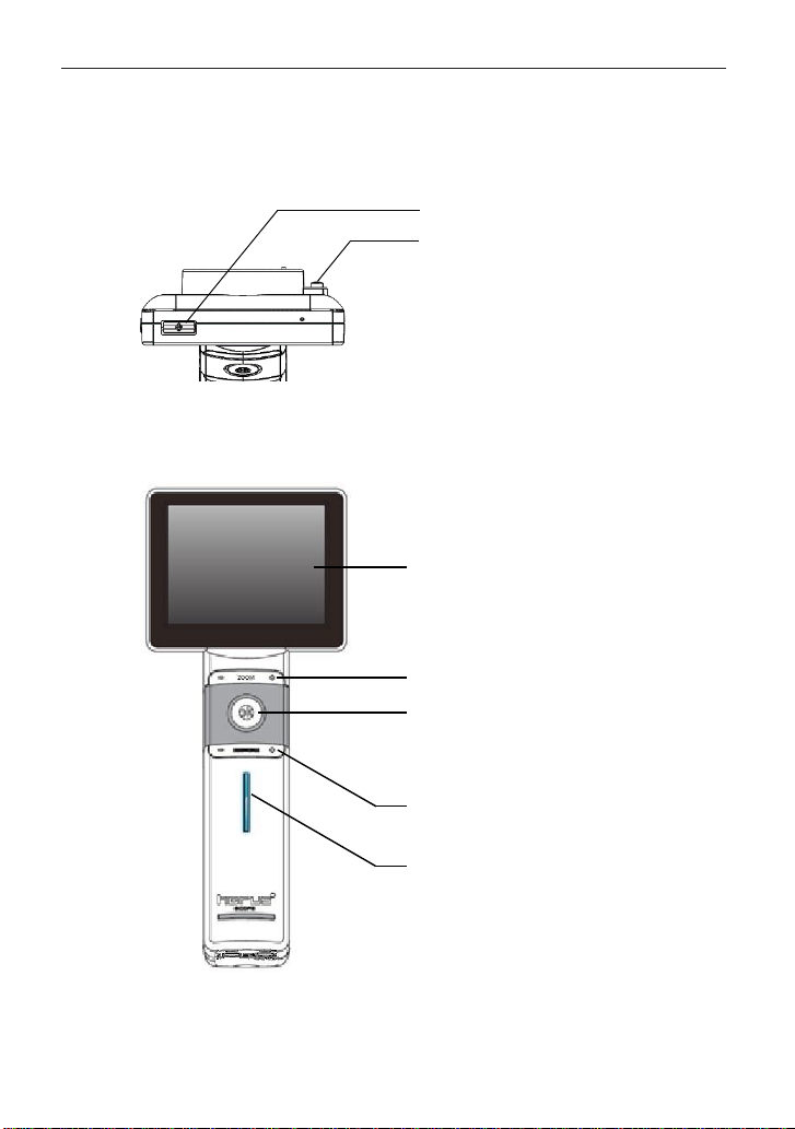
Control Unit (MiiS Horus Scope DSC 300P)
Top view ››
Front view ››
Power button
Lens locker
3.5” LCD touch panel
Zoom button: zoom in/out in live view
OK button:
•
Press down half or fully to take photo
•
Back to photo/video mode while reviewing
photos
Brightness decrease/increase in photo mode
Power indicator

13
Bottom view ››
Rear view ››
Mini USB
Pogo pad
Micro HDMI
Strap hook
Lens assembling mark
Cover glass and sensor
Lens connecting pins
Focus wheel
Battery cover
Memory card slot
Repair hole
Preparations
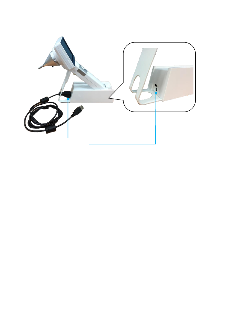
14
Charging Station
Mini USB connector

15
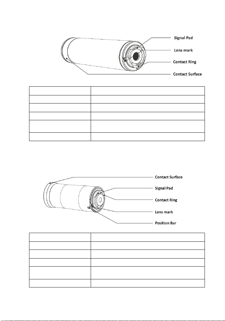
Introduction of scopes
MiiS Horus Scope DOC 300S/ DOC 100S (Digital Otoscope)
DOC 300S
Structure
Technical Description
Focus Range
7~20 mm (Typical)
Dimension
4.3 X 4.3 X 7 cm (Typical)
Weight
69 Grams (Typical)
Camera / video light source
Natural White Light Emitting Diode (LED)
DOC 100S
Structure
Technical Description
Focus Range
5~50 mm (Typical)
Dimension
4.3 X 4.3 X 7 cm (Typical)
Weight
69 Grams (Typical)
Camera / video light source
Natural White Light Emitting Diode (LED)
Specula (Disposal)
Contact Ring
Lens mark
Signal Pad
Position Bar
Preparations
16
MiiS Horus Scope DEC 100/ EEC 100 (Digital Eye Fundus Camera)
DEC 100
Structure
Technical Description
View Angle
40 Degree (Typical)
Diopter
-20 ~ +20D (Typical)
Dimension
4.3 X 4.3 X 12.9cm (Typical)
Weight
141 Grams (Typical)
Search Fundus Lighting
Two modes, natural white Light Emitting Diode (LED)
or infrared LED
Camera / video flash light
Natural White Light Emitting Diode (LED)
EEC 100
Structure
Technical Description
View Angle
25 Degree (Typical)
Diopter
-20 ~ +20D (Typical)
Dimension
4.3 X 4.3 X 10.7 cm (Typical)
Weight
111 Grams (Typical)
Search Fundus Lighting
Two modes, natural white Light Emitting Diode (LED)
or infrared LED
Camera / video flash light
Natural White Light Emitting Diode (LED)

17
①②
③
④
⑥⑤
⑦
MiiS Horus Scope DEA 100 (Digital Anterior Scope)
Structure
Technical Description
Working Distance
30 mm (Typical)
Dimension
4.3 X 4.3 X 4.5 cm (Typical)
Weight
91 Grams (Typical)
Camera / video light source
Natural White/Blue Light Emitting Diode (LED)
LED Switching Button
Switch to blue LED by the LED Switching Button to
take fluorescence image of cornea.
Illumination Light Source (ILS 100) for DEA 100 (Eye anterior illumination system)
Structure
①Forehead stopper
②Locking structure
③Battery groove
④Slit light control knob
⑤Auxiliary light position switch
⑥Filter wheel
⑦Auxiliary lighting
⑧Charge port
⑨Auxiliary light control knob
⑩Silt wheel
⑧
⑨⑩
LED Switching Button
Lens Cover
Position Bar
Lens mark
Signal Pad
Contact Ring
Preparations

18
NOTE
If you connect the USB cable of ILS 100 with a computer, please connect two USBs. If you use
the power adapter to charge, just use one USB.
Technical Description
Working
distanc
e
80mm
Illumination Angle
(deg
rees)
± 45 degree (± 5%
)
Slit
Length(
mm
)
10 mm (± 10
%)
Min Slit Width
(m
m
)
<0.2 mm
Max Slit Width
(m
m
)
Equal to slit length.
(1
0mm
)
Slit Width
S
e
lection
<0.2, 0.2, 0.5, 2, 5, φ10 mm
Filter
Transparent, Cobalt Blue, Red-free (Green)
Li
g
ht
Conform to Group II of ISO 15004-2:2007(
E)
We
i
g
ht
345g (typ. , include Forehead stopper)
Dimension
178mm x 160mm x 107mm
Power source
Rechargeable Li-ion Battery 3.7V/800mAh
(2.96Wh)
Operation time
90 minutes for continuously use
Power Indicator
Fully charged: Green
color
Charging: Orange color

19
MiiS Horus Scope DEA 200P (Digital Anterior Scope)
Structure
Technical Description
View Area
Wide End (H) 23.88*(V) 17.71 mm (typ. ±5%)
Tele End (H) 11.88*(V) 8.91 mm (typ. ±5%)
Working
distanc
e
80mm (Typical)
Illumination Angle
(deg
rees)
± 45 degree (± 5%
)
Slit Length
10mm (typ. ±10%)
Slit Width Selection
<0.2, 0.2, 0.5, 2.0, φ10 mm
Filter
Transparent, Cobalt blue, Red-free (Green)
Light
Conform to group II of ISO 15004-2:2007
Weight
580 Grams (Typical)
Preparations

20
MiiS Horus Scope DDC 200/ DDC 100 (Digital Dermatoscope)
DDC 200
Structure
Technical Description
View Angle
20 mm diameters (Diagonal) (Typical)
Dimension
5.5 X 5.5 X 3.45 cm (Typical)
Weight
65 Grams (Typical)
Camera / video light source
Natural White Light Emitting Diode (LED)
Polarization function
Polarized light changed by the icon on screen
DDC 100
Structure
Technical Description
View Angle
10 mm diameters (Diagonal) (Typical)
Dimension
4.5 X 4.5 X 5.6 cm (Typical)
Weight
114 Grams (Typical)
Camera / video light source
Natural White Light Emitting Diode (LED)
Polarization function
Polarized light changed by the switch on lens
Table of contents
Other MiiS Medical Equipment manuals


