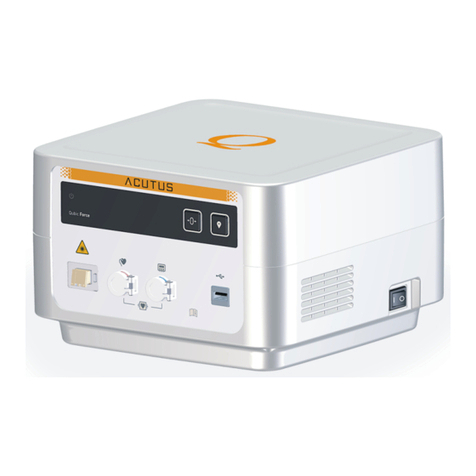
Acutus Medical AcQMap 900100 User manual

900100 AcQMap®
High Resolution Imaging and Mapping System
AcQTrack™ Conduction Pattern Software
SuperMap™ Hover Mapping Mode
Operator Manual
CAUTION: Federal law (USA) restricts this device to sale by or on the order of a physician


a
AcQMap High Resolution Imaging and Mapping System Operator Manual
Contents
Explanation of Symbols...........................................................................................01
Explanation of Icons................................................................................................03
CHAPTER 1 — Introduction ....................................................................................06
1.1. — AcQMap System Description...................................................................06
CHAPTER 2 — Warnings and Precautions ..............................................................08
CHAPTER 3 — Safety Essentials ............................................................................14
3.1. — Indication for Use.....................................................................................14
3.2. — Contraindications.....................................................................................14
3.3. — Potential Adverse Events .........................................................................14
CHAPTER 4 — AcQMap System Component Descriptions......................................15
CHAPTER 5 — AcQMap System Installation & Setup..............................................18
5.1. — AcQMap System Installation....................................................................18
CHAPTER 6 — AcQMap System Patient Preparation ..............................................24
6.1. — Patient Electrode Identification................................................................24
6.2. — Patient Electrode Placement....................................................................24
6.3. — Electrical Reference Sheath or Catheter Placement................................27
6.4. — Anatomic Reference Catheter Placement................................................27
6.5. — AcQMap Catheter ....................................................................................27
CHAPTER 7 — Navigating the User Interface .........................................................28
7.1. — Operating Modes .....................................................................................28
7.2. — Main Window Components .....................................................................28
7.3. — Patient Records and Notes Window........................................................29
7.4. — Common Controls....................................................................................33
7.5. — Using the Mouse......................................................................................34
7.6. — Live Signals Window – Non-contact and Contact Mapping....................37
7.7. — Acquisition Window .................................................................................39
7.8. — Maps Window ..........................................................................................45
7.9. — Configure 3-D Display..............................................................................47
7.10. — Electrode Highlighting............................................................................51
7.11. — Cut-Plane Tool .......................................................................................51
7.12. — 3D Settings – View Catheter Silhouette .................................................52
7.13. — 3D Settings – Add Catheter Shadows ...................................................52

bAcQMap High Resolution Imaging and Mapping System Operator Manual
CHAPTER 8 — Starting a Study ..............................................................................53
8.1. — Starting the AcQMap System Software...................................................53
8.2. — Starting a New Study ...............................................................................54
CHAPTER 9 — Setup for Non-contact Mapping......................................................60
9.1. — Checking Signals .....................................................................................61
9.2. — Acquisition Setup.....................................................................................71
9.3. — Configure Trace Display ...........................................................................77
CHAPTER 10 — Building a Surface Anatomy Using Ultrasound .............................80
10.1. — Step 1: Verify Settings............................................................................80
10.2. — Step 2: Configure and Enable Ultrasound .............................................81
10.3. — Step 3: Surface Build Menu ...................................................................83
10.4. — Step 4: Build a Surface Anatomy...........................................................83
10.5. — Pausing or Resuming an Anatomy Acquisition......................................88
10.6. — Exit the Anatomy Editor .........................................................................96
10.7. — Adding Definition to the Pulmonary Vein Structures..............................96
10.8. — Surface Processing of the Modified Anatomy .......................................99
10.9. — Automatic Identification of Added Structures........................................99
10.10. — Use a Surface Reconstruction in Acquisition Mode ............................99
10.11. — Resume an Existing Surface Reconstruction.....................................100
CHAPTER 11 — Acquiring Recordings..................................................................101
CHAPTER 12 — Reviewing Recordings ................................................................103
12.1. — Signal View and Filter Settings ............................................................104
12.2. — Full-Screen Multi-Channel Visualization ..............................................106
12.3. — Select a Time Window for Mapping.....................................................109
12.4. — Exclusion of Signal Traces for Mapping...............................................110
12.5. — VWave Removal and Zeroing in Atrial Fibrillation ................................113
12.6. — Export Data for Mapping......................................................................114
CHAPTER 13 — Mapping, Labels and Markers.....................................................115
13.1. — The Maps Screen.................................................................................116
13.2. — Creating Maps......................................................................................118
13.3. — Placing Labels......................................................................................124
13.4. — Placing Markers ...................................................................................125
13.5. — AcQTrack™ Conduction Pattern Software ..........................................128
13.6. — Complex Fractionated Atrial Electrograms (CFAE) Tool.......................131
13.7. — Composite Mapping ............................................................................132

c
AcQMap High Resolution Imaging and Mapping System Operator Manual
CHAPTER 14 — SuperMap ...................................................................................135
14.1. — Data Acquisition...................................................................................135
14.2. — Waveform Analysis ..............................................................................137
14.3. — Displaying a SuperMap .......................................................................141
14.4. — Displaying a Propagation History Map with an Amplitude Map ..........144
CHAPTER 15 — Expert Mode................................................................................146
15.1. — Common Controls................................................................................146
15.2. — AcQMap Set Up...................................................................................146
15.3. — Acquisition Window Expert Mode........................................................147
15.4. — Ultrasound Surface Anatomy in Expert Mode .....................................151
15.5. — Reviewing Recordings in Expert Mode................................................152
15.6. — Mapping, Labels and Markers in Expert Mode....................................156
15.7. — SuperMap in Expert Mode...................................................................158
CHAPTER 16 — Setup Contact mapping ..............................................................161
16.1. — Setup Contact Mapping Catheters and Detection Criteria..................161
16.2. — Select Catheter to Establish Localization and Field Scaling................168
16.3. — Field Scaling.........................................................................................169
CHAPTER 17 — Creating a Contact Anatomy .......................................................170
17.1. — Collecting Anatomy Points...................................................................170
17.2. — Editing an Anatomy..............................................................................172
17.3. — Add a New Structure............................................................................174
CHAPTER 18 — Contact Mapping.........................................................................175
18.1. — Configure Live Annotation Window .....................................................175
18.2. — Creating a Contact Electroanatomic Map............................................178
18.3. — Displaying Maps ..................................................................................182
18.4. — Reviewing Maps...................................................................................187
18.5. — Adding/Deleting a Map ........................................................................188
18.6. — Copying a Map.....................................................................................189
Table of contents
Other Acutus Medical Medical Equipment manuals
Popular Medical Equipment manuals by other brands

Getinge
Getinge Arjohuntleigh Nimbus 3 Professional Instructions for use

Mettler Electronics
Mettler Electronics Sonicator 730 Maintenance manual

Pressalit Care
Pressalit Care R1100 Mounting instruction

Denas MS
Denas MS DENAS-T operating manual

bort medical
bort medical ActiveColor quick guide

AccuVein
AccuVein AV400 user manual


