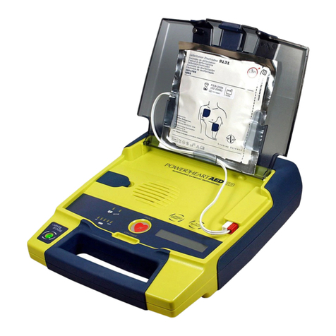
AUDICOR 200S User manual

12-Lead Solution Lead Placement
PREPARE THE PATIENT
ENTER PATIENT INFO
Patients over 40 with shortness of breath or other symptoms of heart failure (use AUDICOR Sensors to assess both heart sounds and ECG)
Patients over 18 with chest pain or other symptoms of Acute Ischemia (use AUDICOR Sensors to assess both heart sounds and ECG or conventional
ECG electrodes to assess only the ECG)
APPLY SENSORS & ELECTRODES
Turn on
AUDICOR console
and printer
1
Enter patient
number or name.
Touch OK to accept
3
Tap the Patient ID
window
2
Select appropriate
gender and age
range, verify date
and time, then
touch OK to continue
4
WHO SHOULD RECEIVE AN AUDICOR TEST?
*RA – Right clavicle
*LA – Left clavicle
RL – Right hip
LL – Left hip
V1 – Fourth intercostal space at right sternal border
V2 – Fourth intercostal space at left sternal border
*V3 – AUDICOR sensor Midway between V2 and V4.
*V4 – AUDICOR sensor at Fifth intercostal space left of midclavicular line.
V5 – Anterior axillary line at same level as V4
V6 – Midaxillary line at same level as V4
LEAD PLACEMENT
VERIFY TRACE & RECORD
Ensure clean Sound and ECG
traces before recording.
Touch Leads to cycle through
all limb and chest leads.
Keep patient and environment as
quiet as possible
Wait for strong, clear sound
signal from both sensors
After three clean sweeps, touch
Record
If data quality is unsatisfactory for
analysis, touch Cancel, make
adjustments as necessary (see
“Getting Good Sound Traces”)
and record again
Test results will be displayed and the
report printed automatically.
OR
Touch Print Copy within range of the
wireless printer
To later print a copy of a stored test. Touch
Recall Test in the Patient Information
screen, check the box next to the desired
report(s), then touch Print Checked.
LARA
V3 V4
AUDICOR®
Sensors
4-Lead Solution
Place V3 between fourth intercostal
space at left sternal border and V4
Apply Sensors to V3 & V4 with
moderate pressure and smooth
the entire pad to ensure secure,
complete seal.
With rocking motion, press the sound
connector securely on top of the
AUDICOR sensor. Rotate adapter to
ensure good contact.
Lift the green tab on top of the ECG Clip, place the tab electrode
between the metal “teeth” of the adapter or place the adapter on
the snap electrode and push down the green tab
1 2
3
OR
*Only leads V3, V4, RA and LA are
required for the 4-lead solution.
AUDICOR®200S Quick Reference Guide

Inovise Medical, Inc.
10565 SW Nimbus Avenue, #100 | Portland, OR 97223
TF: (USA) 877.466.8473 | T: 503.431.3800 | F: 503.431.3801 | Email: info@inovise.com
www.audicor.com
THE AUDICOR 12-LEAD REPORT
GETTING GOOD SOUND TRACES
Y
Z
[
X
\
nSummary of ECG and Heart Sound Findings
YSample Sound Complex - S1, S2 and, if present, the S3 and/or S4
ZMI Size/Location - If there is ECG evidence of cardiac injury, a 3D heart
graphic suggests the area at risk. Dotted lines indicate acute MI and dark
shading indicates MI of undetermined age.
[Strength of ECG Evidence for LVH - If detected, is displayed as horizontal
bar graph.
\10 second Heart Sound Trace
CHANGING DATE & TIME
From the Patient Information Screen, Touch
Settings, Select the Date Time tab, adjust to
correct date & time, touch OK
MAINTAINING AUDICOR®200
RESETTING THE CONSOLE
Touch Reset Button
to restore normal operation
CHANGING THE PAPER
• Load up to 30 sheets of paper
• Red grid faces the front of the printer with the white margin to
the left side
• Ensure all pages settle completely down into the feed slot.
FOR MORE INFORMATION refer to AUDICOR®200S User’s Guide
and Physician’s Guide. Both are available on line at www.audicor.com
!
CUSTOMER SUPPORT: 877.466.8473 / OPTION 1
© 2006 All rights reserved. Rx only
AUDICOR 200S simultaneously integrates heart sounds and ECG data to generate multiple parameters that
correlate to established hemodynamic measures:
Roos M, Toggweler S, Zuber M, Jamshidi P, Erne P. Acoustic cardiographic parameters and their relationship to invasive hemodynamic measurements in patients with LV systolic
dysfunction. Congestive Heart Failure. 2006;12(4 suppl 1):19-24.
UNDERSTANDING THE AUDICOR PARAMETERS
Lengthened LVST correlates with
improved EF
Time between mitral valve closure
(S1) to the aortic valve closure (S2)
LVST
(Left ventricular
systolic time)
S3 Strength >5 correlates with
LVEDP >15mmHg
Overall acoustic energy of third
heart sound. Range 0 – 10 units
S3
Strength
Shortened EMAT correlates with
increased contractility and short
electro-mechanical delays
CORRELATING HEMODYNAMIC
PARAMETERS
Duration from the onset of the QRS
complex to the closure of the mitral
valve
EMAT
(Electro-mechanical
activation time)
DEFINITIONPARAMETER
MAINTAINING AUDICOR®200
TRACE DESCRIPTION SUGGESTED RESPONSE
Good Signal Acceptable traces have clear, well-
defined first and second heart
sounds and relatively clean
baselines.
• Use a gauze pad to remove oils &
perspiration for dry, pink skin
• Shave as necessary
• Place limb electrodes on torso near
hips and clavicles.
• Check all electrodes for good
contact
• Ensure patient is still and relaxed
Sporadic Noise • Ensure patient is not talking and
surrounding environment is as
quiet as possible
• Do not touch or rub sensors and
wires when recording
• Ensure sensors are properly
positioned
Weak Signal • Ensure sensor firmly sealed to
patient.
• Shave chest hair
• Place sensor underneath breast
• Replace sensor as necessary
Leads Off Ensure firm connections:
• Adapters to Sensors
• Adapters to lead wires
• Patient cables to console.
Excessive Baseline Artifact
*The AUDICOR 4-lead report displays one ECG lead trace, sound trace,
heart sound snapshot and statement of heart sound findings.
P/N 40153B
Popular Medical Equipment manuals by other brands

Prestige medical
Prestige medical Classic Media 210047 operating instructions
ADC
ADC PG-PLUS PLL-734 manual
Axion
Axion STIM-PRO I-2000 manual
Omron
Omron Pocket Pain Pro PM400 instruction manual
Bowa
Bowa TissueSeal PLUS COMFORT Series operating manual
ultraMEDIC
ultraMEDIC ultraCHECK 200 Instructions for use
