5
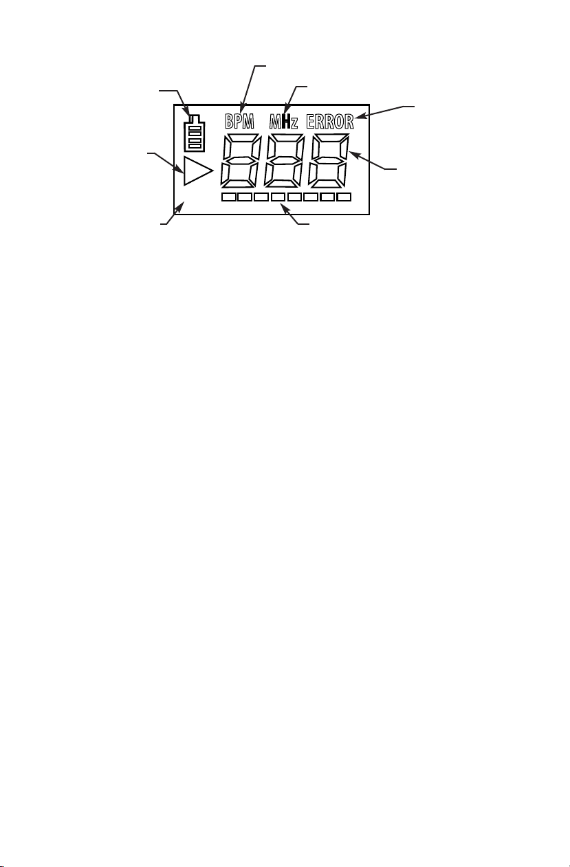
Signal Quality Indicator (Display units only)
An inadequate signal can produce erroneous rates from the heart rate calculation. The signal
level that is being obtained is shown on the Signal Quality Indicator bars. This indicator
provides a visual aid in obtaining a strong audio signal by showing the pulsatile nature of the
signal. A large difference between the highest and lowest signal bars that are lit confirms
that the quality of the signal is good and thus ensures the heart rate calculation is operating
at peak performance. The heart rate can be verified manually by counting the audible beats
for 20 seconds and multiplying by 3, or for 15 seconds and then multiplying by 4. Counting
for less than 15 seconds is not recommended due to a decrease in accuracy with the small
sample size.
Obstetrical
Fetal heart sounds are quite different from peripheral vascular blood flow sounds. Fetal sounds
are typically much lower in frequency and much higher in rate. For early term fetal detection,
start the probe at the pubic bone and slowly move along the midline – rocking the probe slowly
from side to side until a heartbeat is heard. For mid to late term fetal detection the best chance
of finding the heart sounds are to start on the fundus and move toward the navel and from one
side of the abdomen to the other, slowly rocking the probe until the heartbeat is heard. The fetal
heart reminds many people of a galloping horse and can vary in tone from a distant swishing
sound to a hard clopping.
Many times when attempting to detect the fetal heart, the maternal vascular sounds are heard
instead of (or in some cases, in addition to) the fetal sounds. These maternal sounds can come
from one of the major arteries, the placenta or the umbilical cord. The maternal vascular sounds
are typically higher in frequency at a lower rate. The heart rate calculation will display either the
maternal rate or the fetal rate, whichever portion of the signal is stronger.
If the fetal heart sounds cannot be located using the McKesson LUM ON®S RI S Doppler
procedure as described above, a second exam should be performed using another
commercially available fetal monitor as a repeated test.
5 and 8 MHz Vascular
Peripheral arterial sounds are typically higher in frequency. For the best sounds, angle the probe
approximately 45 degrees from the skin surface over the general location of the vessel. Slowly
move the probe side to side and vary the angle of the probe until the vascular sounds are heard.
Changing the angle of the probe has an effect on the frequency of the sound. The steeper the
probe angle is, the higher the frequency of the sound.
Peripheral venous sounds are not typically periodic and vary greatly depending on patient move-
ment and breathing. These sounds are more like the wind at the ocean and vary in pitch as the
patient moves or breathes.
4 MHz Vascular
The use of the 4 MHz vascular probe is the same as the 8 MHz as described above, except
tilting the probe 45 degrees is not necessary since the crystals are angled inside the probe cap.
This allows the user to simply place the probe flat on the peripheral vascular surface to scan for
the flow sounds by moving the flat probe face across the skin surface above the vessel.
Proper alignment of the 4 MHz vascular probe
with respect to the vessel