Second Sight Argus II User manual

900028-001 Rev D
Argus®II
Retinal Prosthesis System
Patient Manual
090000-002
Rx Only: Federal law restricts this device to sale
by or on the order of a physician.
HUMANITARIAN DEVICE: Authorized by Federal
(U.S.) law to provide electrical stimulation of the
retina to induce visual perception in blind patients
with severe to profound retinitis pigmentosa and
bare light or no light perception in both eyes. The
effectiveness of this device for this use has not
been demonstrated.


Argus®II
Retinal Prosthesis System
Patient Manual
Second Sight Medical Products, Inc.
12744 San Fernando Rd., Building 3
Sylmar, CA 91342, USA
Phone: +1 818 833 5000
Fax: +1 818 833 5067
E-mail: service@2-sight.com
Visit us at www.2-sight.com
Copyright © 2013
Second Sight Medical Products, Inc.
Argus, Second Sight and the Second Sight Logo
are registered trademarks of
Second Sight Medical Products, Inc.


Table of Contents
Chapter 1: Glossary............................................1
Chapter 2: Descriptive Information...................7
Indications for Use.............................................7
Device Description.............................................8
When the Device Should Not be Used
(Contraindications)...........................................28
General Warnings and Precautions.................29
Your Patient Identification Card.......................44
Risks and Probable Benefits............................47
Chapter 3: What to Expect Before, During and
After Surgery.....................................................59
Before Surgery ................................................59
The Day of Surgery..........................................59
After Surgery ...................................................61
Chapter 4: Using Your Device .........................68
Setup Instructions............................................68
Operating Instructions......................................72
Checking the Function of the Device...............79
Cleaning ..........................................................80
Maintenance....................................................81
Handling and Storage......................................81
Expected Failure Time and Mode and Its Effect
on You.............................................................84
How to Safely Dispose of the Device...............85
Chapter 5: Troubleshooting.............................89
Chapter 6: Additional information.................103
Clinical Studies..............................................103
Information about Retinitis Pigmentosa.........113
Warranty........................................................114

Chapter 7: User Assistance Information......118
Chapter 8: Symbols and Regulatory
Classifications................................................120
Symbols ........................................................120
Regulatory Classifications.............................122
Index................................................................ 126
Appendix A: Potential Effects of
Electromagnetic Interference (EMI).............. 132
Appendix B: Electromagnetic Environments
.........................................................................138

Chapter 1: Glossary Page 1
Chapter 1: Glossary
Term
Definition
Choroid{XE
"choroid" }
A thin layer of cells
between the retina and
the sclera that contains
pigments and blood
vessels that bring
oxygen and nutrients to
the retina
(See Figure 1)
Communication
Adapter (CA)
A device that is
connected to the Video
Processing Unit (VPU)
when the VPU is hooked
up to a computer in the
clinic
Conjunctiva{
XE
"conjunctiva" }
A thin layer of tissue that
covers the white part of
the eye and the inner
surface of the eyelids
(See Figure 1)

Chapter 1: Glossary Page 2
Term Definition
Cornea{XE
"cornea" }
The clear layer of tissue,
shaped like a dome, that
lies on top of the iris and
the pupil. The cornea is
the eye’s outer lens. It
gives the eye its major
focusing ability.
(See Figure 1)
Cyst
A closed sack of
abnormal tissue which
may contain air, fluids, or
semi-solid material
Diagnosis
The identification of
disease by its symptoms
and signs
Electrode Array
A rectangular grid of
electrodes used to
stimulate the retina
Electrical
Stimulation
A technique that uses
electrical currents to
activate nerve fibers

Chapter 1: Glossary Page 3
Term Definition
Electromagnetic
Interference{
XE
"electromagneti
c interference
(EMI)" }(EMI)
A field of energy
(electrical, magnetic, or
both) created by
electronic equipment.
This field of energy may
be strong enough to
interfere with the normal
operation of your Argus II
System.
Electrostatic
Discharge
(ESD{XE
"electrostatic
discharge
(ESD)" })
A momentary unwanted
flow of electrical current
that can cause damage
to electronic equipment
Incision The surgical cut created
in your eye by the doctor
so that the Argus II
Implant can be placed in
your eye

Chapter 1: Glossary Page 4
Term Definition
Iris{XE "iris" }
The iris is the round
structure in the eye that
gives someone his or her
eye color. For example,
blue-eyed people have a
blue iris while brown
eyed people have a
brown iris. The center of
the iris is an opening
called the pupil. The iris
controls the size of the
pupil when it reacts to
the amount of light that is
present.
(See Figure 1)
Radio
Frequency{XE
"Radio
Frequency
(RF)" }(RF)
Any electromagnetic
frequency within the
range used for wireless
communication
Retina{XE
"retina" }
A thin layer of nerve cells
at the back of the eyeball
which converts light into
nerve impulses that
travel to the brain
(See Figure 1)

Chapter 1: Glossary Page 5
Term Definition
Sclera{XE
"sclera" }
The white outer coating
of the eye made of tough
tissue which allows the
eye to keep its shape
and helps to protect the
delicate inner parts of the
eye (See Figure 1)
Therapy
Treatment of disease or
disorders
VPU{XE "VPU"
}(Video
Processing
Unit)
The part of the Argus II
System that processes
the information that is
sent to and from the
implant inside your eye
Chapter 1: Glossary Page 6
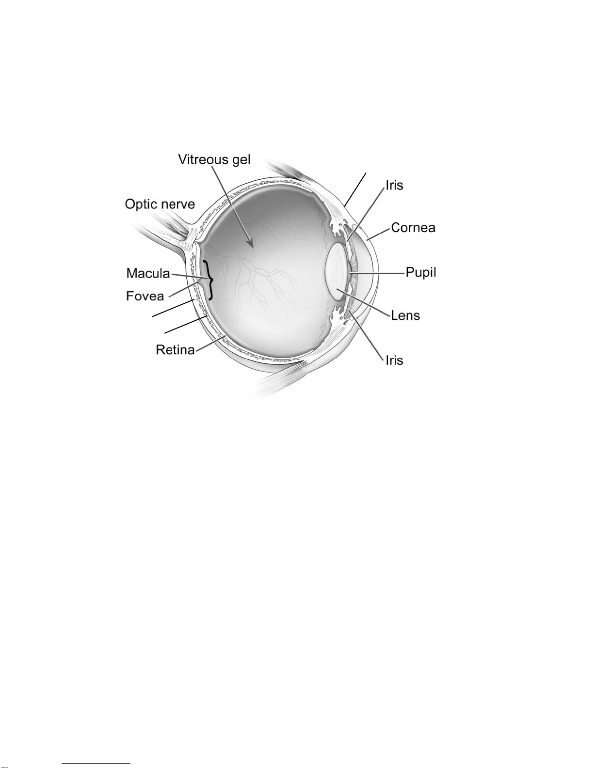
Figure 1: Parts of the Human Eye
Image courtesy of the National Eye Institute,
National Institutes of Health
Sclera
Choroid
Conjunctiva

Chapter 2: Descriptive Information Page 7
Chapter 2: Descriptive Information
Indications for Use{ XE "indications for
use" }
The Argus II Retinal Prosthesis System is
intended to provide electrical stimulation of the
retina to induce visual perception in blind patients.
You are eligible for the Argus II system if you have
severe to profound retinitis pigmentosa and you
meet the following criteria:
• You must be an adult, age 25 years or
older.
• You must have bare light or no light
perception in both eyes. If you do not have
any remaining light perception, your doctor
will test your eye to make sure it will
respond to electrical stimulation.
• You need to have been able to see objects,
shapes and lines in the past.
• In the eye that will be implanted, you either
need to have an artificial lens or no lens at
all. (If the eye that will be implanted still
has a natural lens, your doctor will remove
this lens during the implant surgery.)
• You must be willing and able to follow the
recommended schedule of clinical follow-

Chapter 2: Descriptive Information Page 8
up, device programming and visual
rehabilitation after you are implanted.
Your doctor will implant the Argus II Implant in only
one of your eyes, most likely the eye that has the
worse vision. Your doctor will discuss with you
which eye is best for the implant before your
implant surgery.
Device Description
The Argus II Retinal Prosthesis System consists of
the following main parts and accessories:
• Argus II Retinal Prosthesis (Implant)
• Argus II Video Processing Unit (VPU)
• Argus II Glasses (Glasses)
• Accessories:
• VPU Rechargeable Battery
• VPU Battery Charger
• VPU Pouch
• Travel Case
WARNING
Do not use any equipment with
your Argus II System other than
that supplied by Second Sight.

Chapter 2: Descriptive Information Page 9
If you use cables or batteries not supplied by
Second Sight, your Argus II system may be
more likely to experience interference from
other electronic devices. The use of non-
approved cables or batteries may also cause
the Argus II System to interfere with other
electronic equipment.
Refer to the Appendices A and B for more
information about interference with other electronic
equipment.
How Does the Argus II System Work?
You will have the Argus II Retinal Prosthesis
implanted in and around your eyeball. To turn on
and use the implant, you need to wear the glasses
and VPU.
When you are using the system, a miniature video
camera on the glasses captures images in real
time. The glasses send these images to the VPU.
The VPU converts these video images into
electrical signals and send them back to the
glasses. The coil on the glasses sends the signals
wirelessly to the implant. The implant then sends
out small pulses of electricity to the retina in your
eye. These pulses stimulate your retina. Your
retina sends the nerve signals along the optic
nerve to your brain. You perceive these pulses as
patterns of light. Over time, you may learn how to

Chapter 2: Descriptive Information Page 10
interpret these visual patterns as objects and
shapes.
Note: The implant is on only when you are wearing
the glasses and have the VPU turned on.
Otherwise, the implant is off.
The sections below describe each of the parts of
the Argus II System.
Argus II Retinal Prosthesis (Implant)
The implant consists of four parts: (1) the
electronics case (2) the implant coil, (3) the
electrode array, and (4) the scleral band.
Figure 2 shows the implant as it looks after it has
been implanted. Part of the implant sits on the
outside of your eye and part goes inside your eye.
The implant is not visible to other people.
The electronics case, the implant coil and the
scleral band sit on the outside of the eye. The
scleral band wraps around your eye and holds the
implant in place. A thin layer of tissue that covers
the white part of the eye also covers the parts of
the implant that sit on the outside of the eye.
A cable connects the electronics package to the
electrode array. This cable enters your eye
through an incision made during surgery. At the
end of cable is the electrode array. The electrode

Chapter 2: Descriptive Information Page 11
array is attached to the surface of your retina with
a retinal tack.
The electrode array provides electrical stimulation
to your retina. It has 60 electrodes arranged in a
rectangular grid. Fifty-five of these electrodes are
turned on at the time of implant. Up to 5 of the
remaining electrodes may be functional and could
be turned on to replace an electrode that is not
working.
Patient Contacting Materials of the Implant and
Tack
The implant and retinal tack are made of following
materials:
• Niobium
• Platinum
• Polyimide (plastic)
• Silicone Rubber
• Titanium

Chapter 2: Descriptive Information Page 12
Figure 2: Implant on a Right Eye
(looking at your eyeball)
Electronics Case
(outside the eye)
Implant Coil
(outside the eye)
Scleral Band
(outside the eye)
Electrode Array
(inside the eye)
Chapter 2: Descriptive Information Page 13
External Equipment
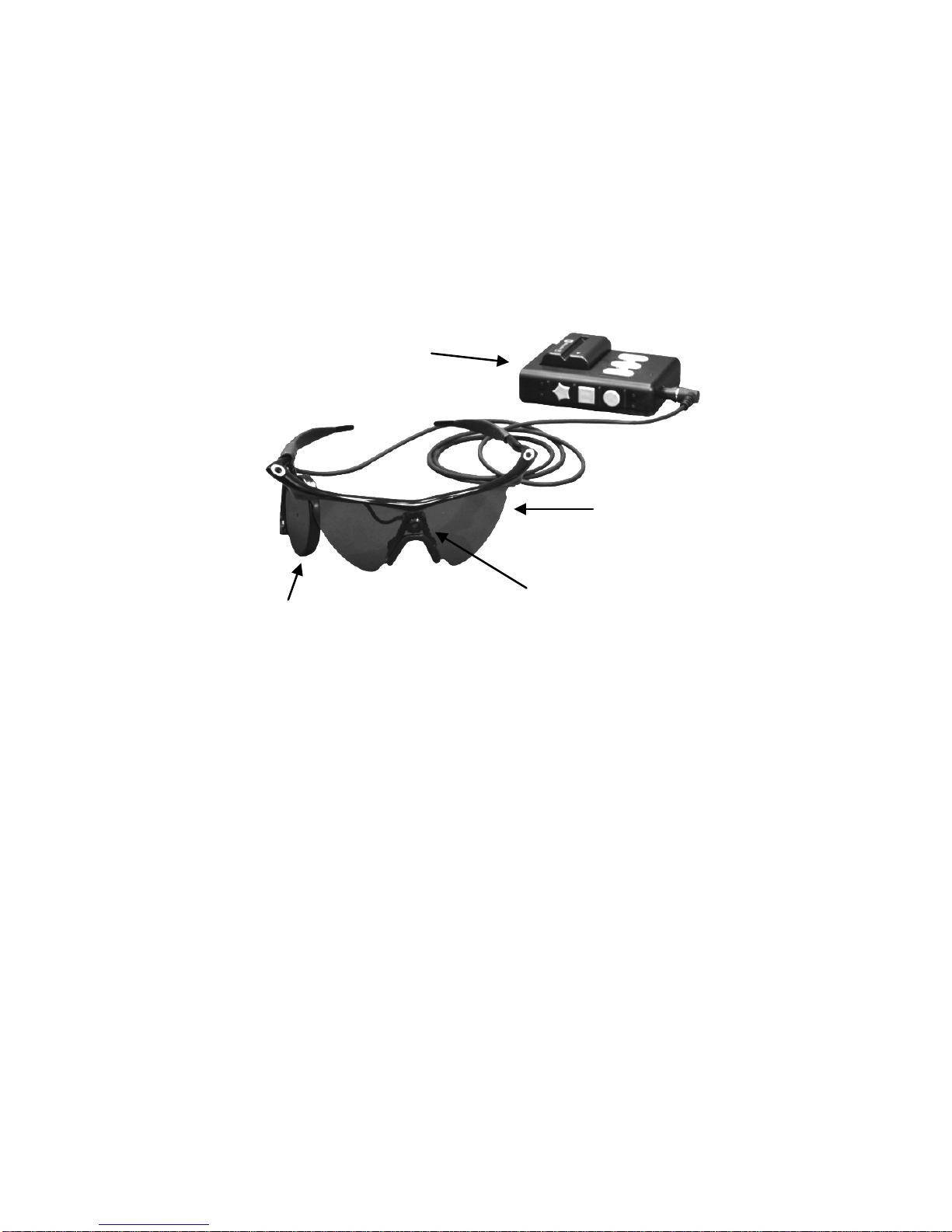
Figure 3 shows the VPU, glasses, and battery{XE
"external equipment" }.
Figure 3: External Equipment
Video Processing Unit (VPU){XE "VPU" }
The VPU allows you to turn stimulation on and off.
Using the buttons on the VPU, you can change the
stimulation program to suit your current
environment. The VPU buttons are large and have
distinct shapes so that you can easily identify them
by touch.
The VPU connects to the glasses using a cable.
The cable from the glasses plugs into the glasses
receptacle on the VPU to connect these two parts.
You must wear both the VPU and glasses for the
system to work.
Camera
Glasses
VPU
Glasses Coil

Chapter 2: Descriptive Information Page 14
The VPU keeps track of when you turn it on and
off, and it keeps a record of how well your implant
and VPU are functioning. The VPU also records
when there is break in the wireless link between
the implant and glasses. Your clinician can check
all of this information when you visit the clinic.
There is a “communication adaptor connector” on
the bottom of the VPU. Your clinician will use this
connector in the clinic to connect the VPU to a
computer. A metal door covers this connector. The
VPU with the battery weighs about half a pound
(0.23 kilograms). See Figure 4 for a diagram of the
VPU.
Table of contents
Other Second Sight Medical Equipment manuals


