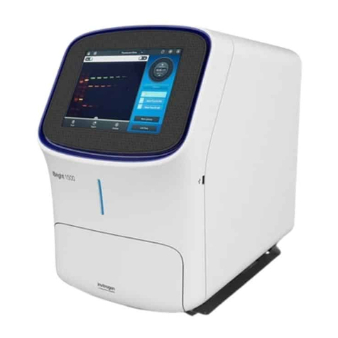
Thermo Scientific iBright CL1500 User manual

iBright™CL1500 and FL1500 Imaging Systems
Catalog Numbers A43678, A43679, A44114, A44115, A44240, and A44241
Pub. No. 100085108 Rev. A
WARNING! Read the Safety Data Sheets (SDSs) and follow the handling instructions. Wear appropriate protective eyewear, clothing, and
gloves. Safety Data Sheets (SDSs) are available from thermosher.com/support.
Set up after unpacking
Once unpacked and installed, power on the imager and follow the touchscreen prompts to set up a network conguration, enter instrument
information, and install the turntable.
Package should include: iBright™ Imaging System, Sample Stage/Turntable, White Transilluminator Screen, Sample Blot, Product Information
Sheet, Quick Reference Guide, and Safe Imager™ Viewing Glasses. Region-specic power cord shipped separately.
Note: For detailed information and safety symbols on this instrument, refer to the iBright™ Imaging Systems User Manual (Pub. no. MAN0018592).
Modes
Invitrogen™ iBright™ Imaging Systems allow users to image in a mode specic to a sample type. Each mode is optimized to produce a high quality
image for further analysis and data collection.
Chemi Blots
Mode to image chemiluminescent Western blots.
Fluorescent Blots (FL1500 model only)
Mode to image uorescent Western blots using a single channel or up to 4 channels using excitation wavelengths of 455-485 nm, 515-545 nm,
610-635 nm, 610-660 nm, and 750-768 nm with corresponding emission wavelengths of 510-555 nm, 565-615 nm, 675-720 nm, 710-730 nm, and
800-850 nm.
Nucleic Acid Gels
Mode to image DNA and RNA gels stained with uorescent nucleic acid stains such as Ethidium Bromide, SYBR™ Safe, SYBR™ Green (I or II),
SYBR™ Gold and similar products.
Protein Gels (Protein Visible and Protein Fluorescence)
Mode to image visible, stained protein gels (using white screen) and uorescent, stained, or labeled protein gels.
Note: Do not use the white screen for protein uorescence.
Note: Stained or labeled protein on nitrocellulose or PVDF membrane should be imaged using Universal mode.
Universal
Mode to image samples containing multiple signals, such as chemiluminescence, uorescence, and/or colorimetric stains. Image display is similar
to Fluorescent Blots and allows you to assign false color to any sample.
Gallery
Mode to retrieve, analyze, and manage images. Images are saved into a guest gallery or a registered user's gallery.
Get started
If the unit is off
If the unit is in sleep mode, touch the screen to activate the instrument.
Note: If the instrument does not respond to touch, ensure it is plugged in and turn on using the switch on the back of instrument. Continue to
step 1 below.
QUICK REFERENCE
For Research Use Only. Not for use in diagnostic procedures.

If the unit is on and in active mode
1. Touch to sign in to use or set up a user prole.
Note: If security mode is active, the system will default to the sign-in page. For those not logged on as a user (guest), acquired images are
saved to a guest gallery. Images cannot be transferred between user and guest galleries. Registered users can access images in guest gallery
by checking View guest's images under the lter options in Gallery. When logged in as a guest, images cannot be exported to a network
drive or cloud storage.
2. From the Welcome screen, choose the appropriate Mode from the drop-down menu.
3. Touch to open drawer.
4. Insert sample into the middle of the viewing area.
5. Touch to close drawer.
The system will auto zoom, auto focus, and auto rotate the sample for preview imaging.
Acquire an image
Image using Chemi Blot
1. Touch Smart Exposure. View the image preview and recommended exposure time.
2. Based on the image preview, use the controls on the right side of the screen to adjust the exposure time. The image preview will update in real
time. To adjust the time:
a. Use the + or – to adjust exposure time.
b. Finger swipe in the segmented dial to adjust exposure time.
c. Touch the center box to select a preset exposure time from a menu.
d. Enter a custom time by touching Custom.
3. Touch Capture to acquire image with the indicated exposure time.
Captured image appears on screen and automatically saves to the gallery.
• If an acceptable image, touch Export, Gallery or Analyze.
• If an unacceptable image, touch Trash to remove image, then return to step 2 to adjust exposure conditions.
Image using Fluorescent Blot (FL1500 model only)
1. Assign dyes from the list to up to 4 channels.
Note: Multiplexing orange and red dyes is not recommended.
2. Touch Smart Exposure. View the preview image and recommended exposure time.
Two images will display. The top image is the selected grayscale channel, and the boom image is the color composite of the overlaid, selected
channels.
3. Toggle eyeballs on or o to display in the composite overlaid image. Select the appropriate channel for editing (displayed in top image
location).
4. Proceed to step 2 in “Image using Chemi Blot“ on page 2 to adjust and capture the image.
Image using Nucleic Acid Gel
1. Touch Smart Exposure. View the preview image and recommended exposure time.
2. Proceed to step 2 in “Image using Chemi Blot“ on page 2 to adjust and capture the image.
3. (Optional) Band excision can be done by touching More Options4Band Excision and following the screen instructions.
Image using Protein Gel (Protein Visible)
1. Place the white screen directly on the transilluminator glass.
2. Place the sample directly on the white screen.
Note: We recommend the white screen for colorimetric stained protein gels).
3. Touch Smart Exposure. View the preview image and recommended exposure time.
4. Proceed to step 2 in “Image using Chemi Blot“ on page 2 to adjust and capture the image.
Note: Universal mode is recommended for colorimetric-stained protein on nitrocellulose or PVDF membrane.
Image using Protein Gel (Protein Fluorescent)
1. Place sample directly on transilluminator glass.
Note: Do no use white screen.
2. Touch Smart Exposure. View the preview image and recommended exposure time.
3. Proceed to step 2 in “Image using Chemi Blot“ on page 2 to adjust and capture the image.
2
iBright
™
CL1500 and FL1500 Imaging Systems Quick Reference

Image using Universal mode
1. Assign up to 4 channels from the list and select dyes/stains/substrate from the sub list to assign correct imaging conditions.
• FLUOR TRANS: Image uorescent samples, such as SYPRO™-stained uorescent gels, using the green transilluminator as excitation source.
• CHEMI: Image blots containing chemiluminescent samples.
• VISIBLE: Image membranes or opaque samples containing colorimetric signal, such as Thermo Scientic™ Pierce™ Reversible Membrane
Stain or Ponceau S stained membranes.
• PROTEIN GEL (Protein Visible): Image gels stained with colorimetric stains (e.g., Coomassie or silver-stained gels). Ensure the white screen
is placed under the sample.
• PROTEIN GEL (Protein Fluorescent): Image protein gels stained with uorescent stains/labels (e.g., No-Stain™ Protein Labeling Reagent
and SYPRO™ dyes). Ensure the sample is placed directly on the transilluminator glass.
• TPN: Designate channel as standard for total protein-based normalization (TPN). During analysis, software will automatically perform
TPN using this channel as the reference channel.
2. Touch Smart Exposure. View the preview image and recommended exposure time.
Two images will display. The top image is the grayscale image of the selected channel, and the boom image is the color composite of all the
channels selected for display.
3. Toggle eyeballs on or o to display channels in the composite image. Select the appropriate channel for editing (displayed in top image
location).
4. Proceed to step 2 in “Image using Chemi Blot“ on page 2 to adjust and capture the image.
Gallery
• View image les as thumbnails or a list.
• Select one, multiple, or all image les in the gallery. Select Actions to review the selected image le(s) and perform additional functions.
– Delete a selected image le from the tray using Trash. Deleted images are removed from the gallery.
– Export image les in the tray using Export.
– Analyze a selected image le in the tray using Analyze.
– View the image information for the selected image le using Image Information.
– Adjust the appearance of a selected image using Image Adjust.
– To generate a pdf report for a selected image le, touch Analyze4Next4More Options4Generate Report. Report contains images, image
information, and densitometry data in table format. Report can be printed to a network printer and/or exported to a USB or network drive.
Export
1. Choose appropriate export destination from the drop-down menu Destination.
• Connect
• USB port
• Network drive
• Printer
2. For exporting to a USB drive, network drive, or a network printer, choose the appropriate le type from the menu under File Type. More than
one le type can be selected at a time. Click Next.
• Publication (TIFF, JPEG, PNG): Images are exported as 24-bit RGB image les as they appear on the instrument with any image adjustments
or false coloring applied. Optimal format for the printing or presentation of images.
• Analysis (TIFF, G2I): Images are exported as 16-bit raw image les. The exported les represent the raw data with no image adjustments.
Optimal format for downstream analysis and quantitation of the image using appropriate external analysis software. G2i les can only be
analyzed using iBright™ Analysis Software.
3. Select the resolution and image size (only for images selected for publication).
4. Select Export or Next. If exporting multiple les, enter the folder name and select Export.
Analyze (on board)
• Automatically analyzes selected image le by identifying up to 4 analysis frames and the lanes and bands associated with each frame.
• Automatically saves analysis results and adjustments to the image le.
• See the user guide for detailed instructions.
• Additional cloud- and desktop-based analysis software is available. See “iBright™ Analysis Software“ on page 4.
iBright
™
CL1500 and FL1500 Imaging Systems Quick Reference
3

iBright™Analysis Software
For access to the Connect version of the analysis software, go to thermosher.com/cloud. Sign-in or register, then select iBright™ Analysis Software
under Protein Biology.
For desktop-based software (Windows™ or Apple™ OS, go to thermosher.com/ibrightanalysis. View the system requirements in the table below.
After completing the download, perform the installation process based on your conguration.
IMPORTANT! You must be logged into the system using an Administrator-type account.
Downloading a new installer:
1. Locate the installer at thermosher.com/ibrightanalysis.
2. Click Download.
3. Fill in the web form.
4. On the Thank You page, click the system-specic installer link to begin the download.
Note: For PC, download the installer appropriate for your system (e.g., 32-bit or 64-bit). To determine your system conguration, click the
Windows™ start buon, then enter "system information". Click on System Information to bring up your system conguration.
5. Perform the installation process based on your system conguration:
PC Mac
1. Open the installer file (iBrightAnalysisSoftware.exe) from the download
folder or location where web-browser downloads are saved.
2. Accept the terms and conditions (End User Licensing Agreement).
3. Click Install.
4. Launch the application from the shortcut created on your desktop.
1. Open the download folder and double-click on
iBrightAnalysisSoftware.zip to extract files.
2. Open the iBrightAnalysisSoftware.pkg.
Note: You will see a pop-up window if xcode command line tool is not
installed on your system. Accept the Apple™Xcode license and install
the software.
3. Accept the terms and conditions (End User Licensing Agreement).
4. Click Install. A prompt will ask your password for application
installation.
5. Launch the application from the Applications folder.
PC Mac
System configuration Windows™7, 8, 8.1, and 10 with 32-bit and 64-bit
operating system
OS X 10.10: Yosemite, OS X 10.11: El Capitan, macOS 10.12: Sierra,
macOS 10.13: High Sierra, macOS 10.14: Mojave
RAM 8 GB minimum 8 GB minimum
Note: Should you experience any issues with installation or have further questions, contact our technical support at
technicalsupport@thermosher.com.
Limited product warranty
Life Technologies Corporation and/or its aliate(s) warrant their products as set forth in the Life Technologies' General Terms and Conditions of
Sale at www.thermosher.com/us/en/home/global/terms-and-conditions.html. If you have any questions, please contact Life Technologies at
www.thermosher.com/support.
Life Technologies Holdings Pte Ltd | Block 33 | Marsiling Industrial Estate Road 3 | #07-06, Singapore 739256
For descriptions of symbols on product labels or product documents, go to thermofisher.com/symbols-definition.
The information in this guide is subject to change without notice.
DISCLAIMER: TO THE EXTENT ALLOWED BY LAW, THERMO FISHER SCIENTIFIC INC. AND/OR ITS AFFILIATE(S) WILL NOT BE LIABLE FOR SPECIAL, INCIDENTAL, INDIRECT,
PUNITIVE, MULTIPLE, OR CONSEQUENTIAL DAMAGES IN CONNECTION WITH OR ARISING FROM THIS DOCUMENT, INCLUDING YOUR USE OF IT.
Revision history: Pub. No. 100085108
Revision Date Description
A 08 May 2019 New quick reference for iBright™
CL1500/FL1500 Imaging Systems.
Important Licensing Information: These products may be covered by one or more Limited Use Label Licenses. By use of these products, you accept the terms and conditions of all
applicable Limited Use Label Licenses.
©2019 Thermo Fisher Scientific Inc. All rights reserved. All trademarks are the property of Thermo Fisher Scientific and its subsidiaries unless otherwise specified.
thermofisher.com/support | thermofisher.com/askaquestion
thermofisher.com
8 May 2019
Other manuals for iBright CL1500
1
This manual suits for next models
1
Table of contents
Other Thermo Scientific All In One Printer manuals

Thermo Scientific
Thermo Scientific iBright CL750 User manual

Thermo Scientific
Thermo Scientific Invitrogen FLoid User manual

Thermo Scientific
Thermo Scientific invitrogen EVOS FL User manual

Thermo Scientific
Thermo Scientific EVOS M5000 User manual

Thermo Scientific
Thermo Scientific iBright CL1500 User manual

Thermo Scientific
Thermo Scientific EVOS M5000 User manual

Thermo Scientific
Thermo Scientific EVOS M5000 User manual