Arrow International MAC User manual

Inspected Dimensions:
Folded Length: 7-1/2" (19 cm)
Folded Width: 5-1/2" (14 cm)
MAC™Multi-Lumen Central Venous Access Product 1
Dispositif d’accès veineux central à lumières multiples
MAC 9
MAC viellumigen zentraler Venenkatheter
15
Prodotto per accesso alla vena centrale a multilume MAC 21
Wielokanałowe urządzenie dostępowe do żył centralnych
MAC 27
Conjunto de Acesso Venoso Central de Lúmen Múltiplo
MAC 33
Многопросветное изделие для центрального венозного
доступа MAC 39
Pripomoček za centralni venski dostop z več svetlinami
MAC 45
Producto para acceso venoso central de luz múltiple MAC 50
MAC produkt för centralt ventillträde med erkanalig 56
MAC Çoklu Lümen Santral Venöz Erişim Ürünü 61


1
MAC™Multi-Lumen Central Venous Access Product
with ARROWg
+
ard®Antimicrobial Surface and Sharps Safety Features
Safety and Efcacy Considerations:
Do not use if package has been previously opened or damaged.
Warning: Read all package insert warnings, precautions, and
instructions prior to use. Failure to do so may result in severe
patient injury or death.
Do not alter the access device or any other kit/set component
during insertion, use, or removal.
Procedure must be performed by trained personnel well
versed in anatomical landmarks, safe technique, and potential
complications.
MAC™Multi-Lumen Access Catheters should be maintained
in closely monitored environments (eg., critical care unit,
operating room, recovery room), not in general nursing units.30
Precaution: When using Central Venous Catheterization
Product with Contamination Guard for use only with
MAC™ Multi-Lumen Central Venous Access Device (MAC™
companion product), clinicians must be aware of the potential
complication of Cardiac Tamponade (see complications
warning included in all Arrow®Central Venous Catheter
Products) (refer to Fig. 1).
Fig. 1
ARROWg+
ard® Antimicrobial Surface: The Arrow®
antimicrobial access device consists of our polyurethane access
device, plus our ARROWg+
ard Blue®exterior antimicrobial
surface treatment of chlorhexidine acetate and silver sulfadiazine.
The nominal amount of chlorhexidine, silver, and sulfadiazine
applied to the external surface of the MAC™Multi-Lumen Central
Venous Access Device is 208 µg/cm, 31 µg/cm and 73 µg/cm,
respectively.
To demonstrate effectiveness of the ARROWg+
ard®antimicrobial
surface treatment, data was submitted to FDA concerning the
Arrow®14 Fr. hemodialysis catheter, a device with identical
external dimensions to the MAC™Multi-Lumen Access Device.
Sample results of chlorhexidine acetate, silver and sulfadiazine
from a hemodialysis catheter containing identical external
dimensions are 208 µg/cm, 40 µg/cm, and 85 µg/cm respectively.
Antimicrobial activity associated with ARROWg+
ard®on
catheters and/or access devices has been demonstrated in the
following ways:
14 Fr. Catheter In Vitro Results:
Antimicrobial activity associated with the ARROWg+
ard
Blue®hemodialysis catheter has been demonstrated in vitro
using a modied Kirby-Bauer technique utilizing the vertical
catheter segment placement method, in the following ways:
• ARROWg+
ard Blue®hemodialysis catheters produced zones
of inhibition greater than 9 mm in diameter after 24 hours
against:31
Candida albicans
Staphylococcus aureus (methicillin resistant)*
Staphylococcus epidermidis
Streptococcus pyogenes
Klebsiella pneumoniae
Xanthomonas maltophilia
Escherichia coli (ß-lactamase producer)
Pseudomonas aeruginosa
Enterobacter faecalis
Enterobacter cloacea
Enterobacter aerogenes
Acinetobacter baumannii
• ARROWg+
ard Blue®hemodialysis catheters retained
antimicrobial activity (zones of inhibition greater than 5 mm
in diameter) after 7 days against:31
Staphylococcus aureus (methicillin resistant)*
Staphylococcus epidermidis
Streptococcus pyogenes
Klebsiella pneumoniae
Xanthomonas maltophilia
Escherichia coli (ß-lactamase producer)
Enterobacter faecalis
Enterobacter cloacea
* Note: This is not the prevalent strain in catheter-related
infections.
• Marked decreases in antimicrobial activity against all
organisms are apparent at Day 7 of in vitro analysis.
Clinical Efcacy:
Antimicrobial activity data associated with the ARROWg+
ard
Blue®catheter have not been collected with the MAC™Multi-
Lumen Central Venous Access Device.
The following clinical study was conducted on the original
formulation 7 Fr. and 12 Fr. ARROWg+
ard Blue®central
venous catheters.
• A prospective, randomized, controlled clinical trial of 237
large-bore and central venous catheter insertions in 115
patients demonstrated that catheter-related bloodstream
infections rates were 1.14/1000 catheter days for
ARROWg+
ard Blue®catheters versus 3.95/1000 catheter
days for nonimpregnated catheters (p=0.31).9
The following clinical study was conducted on the original
formulation 7 Fr. triple-lumen ARROWg+
ard Blue®catheter.

2
• A prospective, randomized, controlled clinical trial of 403
central venous catheter insertions in 158 adult patients in
a medical-surgical ICU showed that ARROWg+ard Blue®
catheters were 50% less likely to be colonized at removal
than the control catheters (13.5 compared to 24.1 colonized
catheters per 100 catheters, p=0.005) and were 80% less
likely to produce a bloodstream infection (1.0 compared
to 4.7 infections per 100 catheters; 1.6 compared to 7.6
infections per 1000 catheter days, p=0.03).28
• No adverse effects were seen from the antimicrobial catheter,
and none of the isolates obtained from infected catheters in
either group showed in vitro resistance to chlorhexidine or
silver sulfadiazine.28
• Complete data was obtained for 403 central venous catheters
(195 control catheters and 208 antimicrobial catheters) in
158 patients. Control catheters removed from patients who
were receiving systemic antibiotic therapy occasionally
showed low-level surface activity that was unrelated to the
length of time the catheter had been in place (mean zone of
inhibition ± SD, 1.7 ± 2.8 mm); in contrast, antimicrobial
catheters uniformly showed residual surface activity (mean
zone of inhibition, 5.4 ± 2.2 mm; P < 0.002), which declined
after prolonged periods in situ. Antimicrobial activity was
seen with antimicrobial catheters that had been in place for
as long as 15 days.28
The following clinical study was conducted on the original
formulation 7 Fr. triple-lumen ARROWg+
ard Blue®catheter.
• The ARROWg+
ard Blue®catheter has demonstrated a
signicant decrease in the rate of bacterial colonization
along the catheter in limited animal studies.16
• An independent review of 11 randomized clinical trials on
the ARROWg+
ard Blue®antimicrobial catheters (MEDLINE
search from January 1966 to January 1998) concluded that
central venous catheters impregnated with a combination of
chlorhexidine acetate and silver sulfadiazine are effective
in reducing the incidence of both catheter colonization and
catheter-related bloodstream infections in patients at high
risk for catheter-related infections.43
If the total amount of silver sulfadiazine and chlorhexidine
contained in the antimicrobial surface was released from the
catheter as a single dose, the blood levels of silver, sulfadiazine,
and chlorhexidine that would be found would be less than the
blood levels found after clinical usage of these compounds in
established safe dosages as administered via mucous membranes
and skin.12
The potential exposure of patients to the two agents, silver
sulfadiazine and chlorhexidine, on the antimicrobial surface is
signicantly less than that encountered when these compounds are
used on burn wounds, on cutaneous wounds, or as mucosal irrigants.12
The worldwide reported incident rate due to hypersensitivity
reactions is 0.00023% with a conrmed incident rate of
0.000077%.10
Indications for Use:
The MAC™Multi-Lumen Central Venous Access Device
with ARROWg+
ard Blue®permits venous access and catheter
introduction to the central circulation. It may be inserted into
the jugular, subclavian, or femoral veins. The ARROWg+
ard®
technology is intended to help provide protection against catheter-
related infections. Clinical data have not been collected that
demonstrate the use of the ARROWg+
ard® antimicrobial surface
in decreasing catheter-related infections for this device. It is not
intended to be used as a treatment for existing infections, nor is it
indicated for long-term use.
Contraindications:
The ARROWg+
ard Blue®antimicrobial catheter is contraindicated
for patients with known hypersensitivity to chlorhexidine acetate,
silver sulfadiazine, and/or sulfa drugs.
Hypersensitivity reactions are a concern with antimicrobial
catheters, in that they can be very serious and even life-
threatening. The ARROWg+
ard Blue®antimicrobial catheter was
introduced worldwide in 1990, and six years elapsed before the
rst hypersensitivity reaction was reported in Japan in May 1996.10
To date (August 2003) the ARROWg+
ard Blue®reported incident
rate has been extremely low, at 1 per 503,081 exposures, and
the skin test conrmed rate is even lower, at 1 per 1,446,360
exposures. The vast majority of these episodes (17) have been
endemic to individuals of Japanese extraction living in Japan.
The literature indicates that individuals of Japanese extraction
are known to have had similar hypersensitive reactions following
topical chlorhexidine administration.14,19,25,26,34,35,39,42 Three (3)
incidents have been reported in the UK, two (2) in the USA,
and one (1) in New Zealand. If adverse reactions occur after
catheter placement, remove catheter immediately.
Special Patient Populations:
Controlled studies of the antimicrobial catheter have not been
conducted in pregnant women,32 pediatric or neonatal patients
and patients with known sulfonamide hypersensitivity, erythema
multiforme, Stevens-Johnson syndrome,12 and glucose-6-
phosphate dehydrogenase deciency. The benets of the use of
this catheter should be weighed against any possible risk.
Warnings and Precautions:*
1. Warning: Sterile, Single use: Do not reuse, reprocess
or resterilize. Reuse of device creates a potential risk of
serious injury and/or infection which may lead to death.
2. Warning: Chlorhexidine-containing compounds have
been used as topical disinfectants since the mid-1970’s.
An effective antimicrobial agent, chlorhexidine found
use in many antiseptic skin creams, mouth rinses,
and disinfectants used to prepare the skin for surgical
procedures. In addition, chlorhexidine has been
incorporated into cosmetic products where it reportedly
functions as a cosmetic biocide. In the early 1990’s, the
FDA cleared three types of medical devices containing
chlorhexidine: intravenous catheters, topical antimicrobial
skin dressings, and an implanted surgical mesh.13
Hypersensitivity reactions are a concern with antimicrobial
catheters, in that they can be very serious and even life-
threatening. The ARROWg+
ard Blue®antimicrobial
catheter was introduced worldwide in 1990, and six years
elapsed before the rst hypersensitivity reaction was
reported in Japan in May 1996.10
To date (August 2003) the ARROWg+
ard Blue®reported
incident rate has been extremely low, at 1 per 503,081
exposures, and the skin test conrmed rate is even lower,
at 1 per 1,446,360 exposures. The vast majority of these
episodes (17) have been endemic to individuals of Japanese
extraction living in Japan. The literature indicates that
individuals of Japanese extraction are known to have
had similar hypersensitive reactions following topical
chlorhexidine administration.14,19,25,26,34,35,39,42 Three (3)
incidents have been reported in the UK, two (2) in the USA,
and one (1) in New Zealand. If adverse reactions occur
after catheter placement, remove catheter immediately.
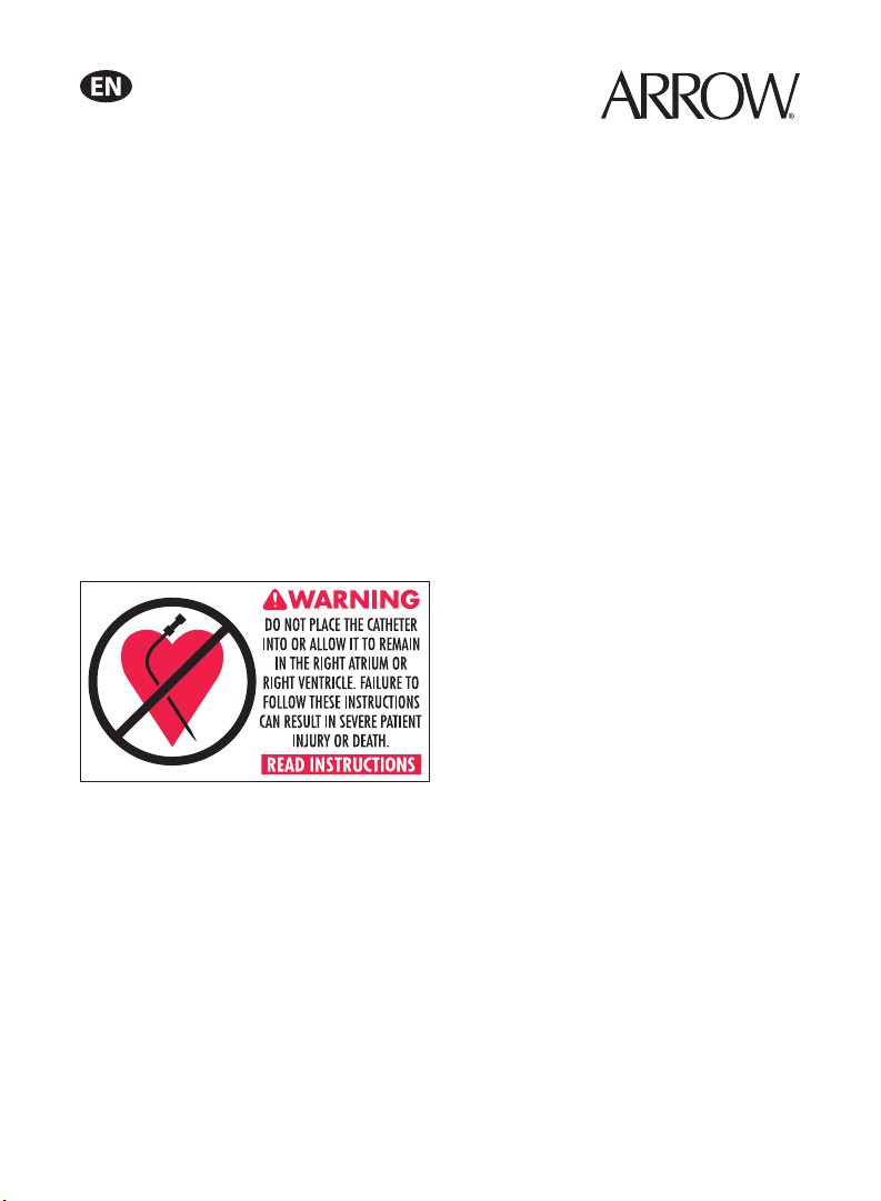
3. Warning: Practitioners must be aware of complications
associated with percutaneous access device introduction
including vessel wall perforation,38 pleural and mediastinal
injuries,1,27 air embolism,7,18,23,30 sheath embolism, thoracic
duct laceration,4 bacteremia, septicemia, thrombosis,5
inadvertent arterial puncture,8 nerve damage, hematoma,
hemorrhage,6 dysrhythmias, and occlusion.

3
4. Precaution: Promptly remove any intravascular catheter
that is no longer essential.17 Evaluate the need for large
bore venous access against the patient’s current therapy
requirements. Should this device be used for continued
venous access, maintain distal lumen sideport patency with
KVO (Keep Vein Open) I.V. rate as per hospital protocol.
5. Warning: Do not apply excessive force in removing guide
wire, dilator or access device. If withdrawal cannot be
easily accomplished, a chest x-ray should be obtained and
further consultation requested.
6. Warning: The practitioner must be aware of potential air
embolism associated with leaving open needles, sheaths, or
catheters in venous puncture sites or as a consequence of
inadvertent disconnects. To lessen the risk of disconnects,
only securely tightened Luer-Lock connections should
be used with this device. Follow hospital protocol for all
sheath and side port maintenance.
7. Warning: Hemostasis valve must be occluded at all times
to minimize the risk of air embolism or hemorrhage. If
catheter introduction is delayed, or catheter is removed,
temporarily cover valve opening with sterile-gloved
nger until catheter or obturator is inserted. Use Arrow®
obturator, either included with this product or sold
separately, as dummy catheter with hemostasis valve
assembly. This will ensure that leakage does not occur and
inner seal is protected from contamination.30
8. Warning: Passage of the guide wire into the right heart
can cause dysrhythmias, right bundle branch block,11 and
a perforation of the vessel wall, atrial or ventricular.
9. Warning: Practitioners must be aware of the potential for
entrapment of guide wire by any implanted device in the
circulatory system (ie. vena cava lters, stents). Review
patient’s history before catheterization procedure to assess
for possible implants. Care should be taken regarding the
length of spring-wire guide inserted.3It is recommended
that if patient has a circulatory system implant, catheter
procedure be done under direct visualization to minimize
the risk of guidewire entrapment.
10. Warning: This kit is designed to reduce the risk of
accidental needle and sharps related sticks. Care must
still be taken to minimize the risk of sharps injury.
Clinicians must adhere to state/federal OSHA standards
for blood borne pathogens when starting, discontinuing,
or maintaining a central venous catheter to minimize the
risk of exposure.
11. Warning: Due to the risk of exposure to HIV (Human
Immunodeciency Virus) or other blood borne pathogens,
health care workers should routinely use universal blood
and body-uid precautions in the care of all patients.
12. Precaution: For blood sampling, temporarily shut off
remaining port(s) through which solutions are being
infused.
13. Precaution: Do not suture directly to the outside diameter
of access device to minimize the risk of cutting or
damaging the catheter or impeding device ow.
14. Precaution: Indwelling access devices should be routinely
inspected for desired ow rate, security of dressing, correct
position and for proper Luer-Lock connection.
15. Precaution: Maintain the insertion site with regular
meticulous redressing using aseptic technique.
16. Precaution: Alcohol and acetone can weaken the structure
of polyurethane materials. Check ingredients of prep
sprays and swabs for acetone and alcohol content.
Acetone: Do not use acetone on access device surface.
Acetone may be applied to skin but must be allowed to dry
completely prior to applying dressing.
Alcohol: Do not use alcohol to soak access device surface
or to restore patency. Care should be taken when instilling
drugs containing high concentration of alcohol. Always
allow alcohol to dry completely prior to applying dressing.
17. Precaution: Some disinfectants used at the access device
insertion site contain solvents, which can attack the access
device material. Assure insertion site is dry before dressing.
18. Precaution: Properly dispose of sharps in sharps container
in accordance with state/federal OSHA standards for
blood borne pathogens and/or institutional policy.
A Suggested Procedure:
Use sterile technique.
1. Precaution: Place patient in slight Trendelenburg position
as tolerated to reduce the risk of air embolism. If femoral
approach is used, place patient in supine position.
2. Prep area of anticipated venipuncture.
3. Drape puncture site as required.
4. Perform skin wheal with desired needle (25 Ga. or 22 Ga.
needle). In kits where provided, the SharpsAway II™Locking
Disposal Cup is used for the disposal of needles (15 Ga. - 30 Ga.).
• Using one-handed technique, rmly push needles into
disposal cup holes (refer to Fig. 2).
Fig. 2
• Once placed into the disposal cup, needles will be
automatically secured in place so that they cannot be reused.
• Discard the entire cup, at completion of the procedure, into
an approved sharps container
Precaution: Do not attempt to remove needles that have
been placed into cup. These needles are secured in place.
Damage may occur to the needles if they are forced out of
the disposal cup.
5. Insert tip of desired catheter through Tuohy-Borst adapter end
of catheter contamination shield. Advance catheter through
tubing and hub at other end (refer to Fig. 3).
Tuohy-Borst adapter
Catheter tip
Catheter
contamination shield Catheter
Fig. 3
6. Slide entire catheter contamination shield to proximal end of
catheter.
7. If ow-directed catheter is used, inate and deate balloon
with syringe to ensure integrity. Precaution: Do not exceed
balloon catheter manufacturer’s recommended volume.
Place catheter and catheter contamination shield on sterile
eld awaiting nal placement.
8. Insert entire length of dilator through hemostasis valve into
access device, pressing hub of dilator rmly into hub of
hemostasis valve assembly. Place assembly on sterile eld
awaiting nal placement.

4
9. Insert introducer needle with attached Arrow®Raulerson
Syringe into vein and aspirate. (If larger introducer needle is
used, vessel may be pre-located with 22 Ga. locater needle and
syringe.) Remove locater needle.
Alternate Technique:
Catheter/needle may be used in the standard manner as
alternative to introducer needle. If catheter/needle is used,
Arrow®Raulerson Syringe will function as a standard syringe,
but will not pass spring-wire guide. If no free ow of venous
blood is observed after needle is removed, attach syringe
to the catheter and aspirate until good venous blood ow is
established. Precaution: The color of the blood aspirated is
not always a reliable indicator of venous access.21 Do not
reinsert needle into introducer catheter.
10. Because of the potential for inadvertent arterial placement, one
of the following techniques should be utilized to verify venous
access. Insert the uid primed blunt tip transduction probe into
the rear of the plunger and through the valves of the Arrow®
Raulerson Syringe. Observe for central venous placement via
a wave form obtained by a calibrated pressure transducer.
Remove transduction probe (refer to Fig. 4).
20
0
Fig. 4
Alternate Technique:
If hemodynamic monitoring equipment is not available to permit
transducing a central venous wave form, check for pulsatile
ow by either using the transduction probe to open the syringe
valving system or by disconnecting the syringe from the needle.
Pulsatile ow is usually an indicator of inadvertent arterial
puncture. Precaution: The color of the blood aspirated is not
always a reliable indicator of venous access.21
11. Using the two-piece Arrow Advancer™, advance spring-wire
guide through syringe into vein. Warning: Aspiration with
spring-wire guide in place will cause introduction of air
into syringe. Precaution: To minimize the risk of leakage of
blood from syringe cap, do not reinfuse blood with spring-
wire guide in place.
Two-Piece Arrow Advancer™ Instructions:
• Using your thumb, straighten the “J” by retracting the spring-
wire guide into the Arrow Advancer™(refer to Figs. 5, 6).
Fig. 5
Fig. 6
When the tip is straightened, the spring-wire guide is ready
for insertion. Centimeter marks are referenced from “J” end.
One band indicates 10 cm, two bands 20 cm, and three bands
30 cm.
Introducing the Spring-Wire Guide:
• Place the tip of the Arrow Advancer™– with “J” retracted – into
the hole
in the rear of the Arrow® Raulerson Syringe plunger
(refer to Fig. 7).
Fig. 7
• Advance spring-wire guide into Arrow®Raulerson Syringe
approximately 10 cm until it passes through syringe valves
(refer to Fig. 8).
Fig. 8
• Raise your thumb and pull the Arrow Advancer™
approximately 4 cm to 8 cm away from the syringe. Lower
thumb onto the Arrow Advancer™and while maintaining a
rm grip on the spring-wire guide, push the assembly into
the syringe barrel to further advance the spring-wire guide.
Continue until spring-wire guide reaches desired depth (refer
to Fig. 9).

5
Fig. 9
Alternate Technique:
If a simple straightening tube is preferred, the straightening
tube portion of the Arrow Advancer™can be disconnected
from the unit and used separately.
Separate the Arrow Advancer™tip or straightening tube from the
blue Arrow Advancer™unit. If the “J” – tip portion of the spring-
wire guide is used, prepare for insertion by sliding the plastic
tube over the “J” to straighten.The spring-wire guide should then
be advanced in the routine fashion to the desired depth.
12. Advance the guide wire until triple band mark reaches rear of
syringe plunger. Advancement of “J” tip may require a gentle
rotating motion. Warning: Do not cut spring-wire guide to
alter length. Do not withdraw spring-wire guide against
needle bevel to minimize the risk of possible severing or
damaging of spring-wire guide.
13. Hold spring-wire guide in place and remove introducer needle
and Arrow®Raulerson Syringe (or catheter). Precaution:
Maintain rm grip on spring-wire guide at all times. Use
centimeter markings on spring-wire guide to adjust indwelling
length according to desired depth of indwelling access device
placement.
14. Enlarge cutaneous puncture site with cutting edge of scalpel
positioned away from the spring-wire guide. Precaution: Do
not cut guide wire. In kits where provided, retract scalpel
in the protected position (refer to Fig. 10). Use tissue dilator
to enlarge puncture site as required. Warning: Do not leave
tissue dilator in place as an indwelling catheter to minimize
the risk of possible vessel wall perforation.
Fig. 10
15. Thread tapered tip of dilator/access device assembly over
spring-wire guide. Grasping near skin, advance assembly with
slight twisting motion to a depth sufcient to enter vessel.
Dilator may be partially withdrawn to facilitate advancement
of access device through tortuous vessel. Precaution: Do not
withdraw dilator until the access device is well within the
vessel to minimize the risk of damaging tip.
16. Advance access device assembly off dilator into vessel, again
grasping near skin and using slight twisting motion.
17. To check for proper access device placement within the vessel,
attach syringe to distal side port for aspiration. Hold access
device assembly in place and withdraw spring-wire guide and
dilator sufciently to allow venous blood ow to be aspirated
into distal side port. Precaution: Maintain rm grip on
spring-wire guide at all times.
18. Holding access device assembly in place, remove guide
wire and dilator as a unit. Place sterile-gloved nger over
hemostasis valve. Warning: To minimize the risk of
possible vessel wall perforation, do not leave tissue dilator
in place as an indwelling catheter. Warning: Although the
incidence of spring-wire guide failure is extremely low,
practitioner should be aware of the potential for breakage
if undue force is applied to the wire. Flush and connect distal
side port to appropriate line as necessary.
Conrm and monitor proximal port by aspirating until free
ow of venous blood is observed. Connect all extension lines to
appropriate Luer-Lock line(s) as required. Unused port(s) may
be “locked” through injection cap(s) using standard hospital
protocol. Pinch/slide clamps are provided on extension lines
to occlude ow through each lumen during line and injection
cap changes. Precaution: To minimize the risk of damage to
extension lines from excessive pressure, each clamp must
be opened prior to infusing through that lumen.
19. Feed catheter through access device assembly into vessel.
Advance catheter to desired position. Warning: Hemostasis
valve must be occluded at all times to minimize the risk
of air embolism or hemorrhage. If catheter introduction
is delayed, temporarily cover valve opening with sterile-
gloved nger until obturator is inserted. Use Arrow
obturator, either included with this product or sold
separately, as dummy catheter with hemostasis valve
assembly. This will ensure that leakage does not occur and
inner seal is protected from contamination.30
20. Hold access device in place and reposition catheter
contamination shield so that distal hub is approximately ve
inches (12.7 cm) from hemostasis valve (refer to Fig. 11).
5 in. approx.
Fig. 11
21. Hold rear hub (Tuohy-Borst adapter end) of catheter
contamination shield in place. Disengage distal hub from inner
feed tube by moving forward. Advance distal hub forward
toward hemostasis valve assembly. Hold assembly in place
(refer to Fig. 12).
Press distal hub over
cap seal (twist to lock)
Tuohy-Borst adapter
hold in place
Advance toward
Hemostasis valve
Fig. 12

6
22. Press distal hub of catheter contamination shield over
assembly cap. Twist to lock (refer to Fig. 13).
Fig. 13
• Orient slot in hub with locking pin on assembly cap.
• Slide hub forward over cap and twist.
23. Grasp insertion catheter through front portion of catheter
contamination shield and hold in place while repositioning
Tuohy-Borst adapter end as desired (refer to Fig. 14).
Precaution: Do not reposition Tuohy-Borst adapter end on
insertion catheter once moved to this nal position.
Reposition seal
end as desired
Grasp catheter here
Fig. 14
24. Tighten Tuohy-Borst adapter by pressing down on cap
and simultaneously turning clockwise to secure hub to
catheter. Gently pull insertion catheter to verify securement.
Precaution: Do not overtighten Tuohy-Borst adapter
to minimize the risk of lumen constriction or insertion
catheter damage.
25. Tuohy-Borst adapter end of catheter contamination shield
should be secured with sterile tape to inhibit insertion catheter
movement (refer to Fig. 15). Precaution: Do not apply tape
to the transparent sheathing on the shield to minimize the
risk of tearing material.
Catheter
Tuohy-Borst adapter
Sterile tape
Fig. 15
26. Use triangular juncture hub with side wings as primary suture
site. In kits where provided, the catheter clamp and fastener
should be utilized as a secondary suture site as necessary.
Precaution: Do not staple directly to the outside diameter
of the catheter to minimize the risk of cutting or damaging
the catheter or impeding catheter ow. Dress insertion site
according to hospital protocol.
Staple Anchoring Device Instructions:
(Where Provided)
• Position thumb and index nger of dominant hand on
indented surface of staple anchoring device.
• Pass staple point through eye of catheter suture hub (refer
to Fig. 16).
Fig. 16
• Tent skin and position with hub eye between staple opening.
Precaution: Do not place staple over catheter body or
extension lines except at indicated anchoring location to
minimize the risk of damage to catheter.
• Firmly squeeze anchoring device together to close staple and
secure catheter to skin (refer to Fig. 17).
Fig. 17
• Repeat procedure through other suture eyes, if applicable.
Discard anchoring device upon completion.
27. Dress puncture site per hospital protocol. Precaution:
Maintain the insertion site with regular, meticulous
redressing using aseptic technique.
28. Record the insertion procedure on the patient’s chart.
Catheter Removal Procedure:
1. Precaution: Place the patient in a supine position.
2. Remove dressing, if applicable. Precaution: To minimize
the risk of cutting the access device, do not use scissors to
remove dressing.
3. Twist distal hub of catheter contamination shield to allow
removal from locking pin on hemostasis valve assembly.
Withdraw catheter from valve. Warning: Hemostasis valve
must be occluded at all times to minimize the risk of air
embolism or hemorrhage. Temporarily cover valve opening
with sterile-gloved nger until catheter or obturator is inserted.
Access Device Removal Procedure:
1. Precaution: Promptly remove any intravascular catheter
that is no longer essential.17 Evaluate the need for large
bore venous access against the patient’s current therapy
requirements. Should this device be used for continued
venous access, maintain distal lumen sideport patency with
KVO (Keep Vein Open) I.V. rate as per hospital protocol.
2. Precaution: Place the patient in a supine position.
3. Remove dressing, if applicable. Precaution: To minimize
the risk of cutting the access device, do not use scissors to
remove the dressing.

7
4. Using staple remover, remove staple(s), where applicable,
or remove sutures from primary suture site. Precaution: Be
careful not to cut the access device.
5. Withdraw device from hemostasis valve. Cover hemostasis
valve with sterile-gloved nger. Warning: Hemostasis valve
must be occluded at all times to minimize the risk of air
embolism or hemorrhage.
6. Warning: Exposure of central vein to atmospheric
pressure may result in entry of air into central venous
system. Using staple remover, where applicable, remove
staples(s) from catheter clamp, and primary suture site. Be
careful not to cut catheter. Remove catheter slowly, pulling
parallel to skin. As catheter exits site, apply pressure with
a dressing impermeable to air, e.g. VASELINE®† gauze.
Because residual catheter track remains an air entry point until
completely sealed, the occlusive dressing should remain in
place for at least 24-72 hours dependent upon amount of time
catheter was indwelling.23,35,37,40
7. Upon removal of the access device, inspect it to make sure that
the entire length has been withdrawn.
8. Document removal procedure.
References:
1. Albertson TE, Fisher CJ, Vera Z. Accidental mediastinal entry
via left internal jugular vein cannulation. Intensive Care Med.
1985;11:154-157.
2. Allgeyer DO, Herr GP, Szeftel A, Shapiro SM, et al.
Evaluation of a Sharps Safety-Enhanced Device for Skin
Fixation of Central Venous Catheters. Anesthesiology.
93(3A);2000:A-593.
3. Andrews RT, Bova DA, Venbrux AC. How much guidewire is
too much? Direct measurement of the distance from subclavian
and internal jugular vein access sites to the superior vena cava-
atrial junction during central venous catheter placement. Crit
Care Med. Jan. 2000;28:138-142.
4. Ariditis J, Giala M, Anagnostidou A. Accidental puncture of
the right lymphatic duct during pulmonary artery ization. Acta
Anaesthesiol Scand. 1988;32:67-68.
5. Benumof JL. Thrombosis after pulmonary-artery
catheterization via the internal jugular vein. NEJM.
1982;306:1487. Letter.
6. Benya RV. Fibrin sheath formation surrounding a pulmonary
artery catheter sheath: eversion of the sleeve during catheter
removal. Crit Care Med. 1990;18:345. Letter.
7. Bristow A, Batjer H, Chow V, Rosenstein J. Air embolism
via a pulmonary artery catheter introducer. Anesthesiology.
1985;63:340-341. Letter.
8. Brzowski BK, Mills JL, Beckett WC. Iatrogenic subclavian
artery pseudoaneurysms: case reports. J Trauma. 1990;30:616-
618.
9. Collin GR, Decreasing Catheter Colonization Through the
Use of an Antiseptic-Impregnated Catheter. Chest. June
1999;115:1632-1640.
10. Data on le, Arrow International, Inc.
11. Eissa NT, Kvetan V. Guide wire as a cause of complete heart
block in patients with preexisting left bundle branch block.
Anesthesiology. 1990;73:772-774.
12. Farber T. ARROWg
+
ard®antiseptic surface toxicology review.
Monograph. Published by Arrow International, Inc. April,
1992.
13. FDA Public Health Notice. Potential hypersensitivity reactions
to chlorhexidine-impregnated medical devices. Food and Drug
Administration Web site. Available at: http://www.fda.gov/
cdrh/chlorhex.html. Accessed October 26, 1999.
14. Fukui A, Ohsumi A, Takaori M. A case of anaphylactic shock
induced by chlorhexidine gluconate. J Japan Society Clin
Anesthesia. 1989;9:356-360.
15. Greene ES, Berry AJ, Jagger J, Hanley E, et al. Multicenter
Study of Contaminated Percutaneous Injuries in Anesthesia
Personnel. Anesthesiology. 1998;89:1362-72.
16. Greenfeld JI, Sampath L, Popilskis SJ, et al. Decreased
Bacterial Adherence and biolm formation on chlorhexidine
and silver sulfadiazine-impregnated central venous catheters
implanted in swine. Crit Care Med. 1995;23:894-900.
17. Guidelines for the Prevention of Intravascular Catheter
Related Infections. CDC. 2002;51(RR10);1-26.
18. Hartung EJ, Ender J, Sgouropoulou S, Bierl R, Engelhardt W,
Engemann R. Severe air embolism caused by a pulmonary
artery introducer sheath. Anesthesiology. 1994;80:1402.
Letter.
19. Harukuni I, Ishizawa Y, Nishikawa T, Takeshima R, Dohi
S, Naito H. Anaphylactic shock with ventricular brillation
induced by chlorhexidine. Japanese J Anesthesiology.
1992;41:455-459.
20. Hightower D, March J, Ausband S, Brown LH. Comparison
of Staples vs Suturing for Securing Central Venous Catheters.
Acad. Emerg. Med. 1996;3:1103-5.
21. Jobes DR, Schwartz AJ, Greenhow DE, Stephenson LW,
Ellison N. Safer jugular vein cannulation: recognition of
arterial punctures and preferential use of the external jugular
route. Anesthesiology. 1983;59:353-355.
22. Kanegaye JT, Vance CW, Chan L, Schonfeld N. Comparison
of skin stapling devices and standard sutures for pediatric scalp
lacerations: A randomized study of cost and time benets. The
Journal of Pediatrics.1996;130:808-13.
23. Kashuk JL, Penn I. Air embolism after central venous
catheterization. Surg Gynecol Obstet. September
1984;159:249-252.
24. Kondo K, O’Reily LP, Chiota J. Air embolism associated with
an introducer for pulmonary arterial catheters. Anesth Analg.
1984;63:871-872.
25. Kubo H, Akiyama Y, Honda K, Nakajo N. Anaphylaxis
following oral irrigation with chlorhexidine gluconate. J
Japanese Dental Society Anesthesiology. 1985;13:659-663.
26. Layton GT, Stanworth DR, Amos HE. The incidence of IgE
and IgG antibodies to chlorhexidine. Clin Experimental
Allergy. 1989;19:307-314.
27. Macksood MJ, Setter M. Hydrothorax and hydromediastinum
after use of an indwelling percutaneous catheter introducer.
Crit Care Med. 1983;11:957-958.
28. Maki DG, Wheeler SJ, Stolz SM, Mermel LA. Clinical trial of
a novel antiseptic-coated central venous catheter. Abstract of
paper presented at 31st ICAAC Clinical Infections, Chicago,
October 1, 1991.
29. Maki DG, Wheeler SJ, Stolz SM, Mermel LA. Prevention of
central venous catheter-related bloodstream infection by use
of an antiseptic-impregnated catheter. Ann Int Med. August 15,
1997;127:257-266.
30. Mihm FG, Rosenthal MH. Pulmonary artery catheterization.
In: Benumof JL, ed. Clinical Procedures in Anesthesia and
Intensive Care. Philadelphia, PA: JB Lippincott; 1992:419.
31. Modak SM, Sampath L. Development and evaluation of a
new polyurethane central venous antiseptic catheter: reducing
central venous catheter infections. Infections in Medicine. June
1992:23-29.
32. Modak SM. (Written communication, June 1991).
33. Occupational Exposure to Bloodborne Pathogens; Needlestick
and Other Sharps Injuries. 66 Federal Register 5317 (2001)
(Codied at 29 CFR~1910.1030).

8
34. Okano M, Nomura, Hata S, et al. Anaphylactic symptoms due
to chlorhexidine gluconate. Arch Dermatol. 1989;125:50-52.
35. Okano M, Nomura M, Okada N, Sato K, Tashiro M. Four cases
presenting anaphylactic reactions due to topical application of
Hibitane®. Skin Research. 1983;25:587-592.
36. Paskin DL, Hoffman WS, Tuddenham WJ. A new
complication of subclavian vein catheterization. Ann Surg.
March 1974;179:266-268.
37. Phifer TJ, Bridges M, Conrad SA. The residual central venous
catheter track – an occult source of lethal air embolism: case
report. J Trauma. 1991;31:1558-1560.
38. Roy RC. Possible hazards from catheter sheath introducers.
Crit Care Med. 1984;12:616. Letter.
39. Takeda K, Inoue K, Matsuya T, et al. An allergic shock
possibly induced by the chlorhexidine; report of a case. J
Osaka Univ Dent Soc. 1985;30:221-225.
40. Thielen JB, Nyquist J. Subclavian catheter removal. J
Intravenous Nurs. March/April 1991;14:114-118.
41. Tomkins DP, Van Der Walt JH. Needleless and Sharp-Free
Anaesthesia. Anaesthesia and Intensive Care. 1996;24:164-7.
42. Tsuneto S, Watanabe S, Koyama K, Nakayama K, Saito H,
Saito K. Anaphylactic shock induced by chlorhexidine mixed
in the vial of lidocaine. J Japan Society Clin Anesthesia.
1987;7:272-277.
43. Veenstra DL, Saint S, Saha S, et al. Efcacy of Antiseptic –
impregnated cantral venous catheters in preventing catheter-
related bloodstream infection, A meta – analysis. JAMA.
1999;281:261-267.
Arrow International, Inc. recommends that the user be acquainted
with the reference literature.
*If you have any questions or would like additional reference
information, please contact Arrow International, Inc.
† A registered trademark of Unilever Supply Chain, Inc.
Rx only.

9
Dispositif d’accès veineux central à lumières multiples MAC
à revêtement antimicrobien ARROWg+
ard et protection
contre les instruments tranchants et pointus
Efcacité et sécurité :
N’utilisez pas si l’emballage a été endommagé ou est déjà
ouvert.
Avertissement : Avant l’utilisation, lisez tous les
avertissements ainsi que toutes les mises en garde et les
instructions de la notice accompagnant le produit. Le non-
respect du mode d’emploi risquerait de blesser grièvement le
malade ou d’entraîner sa mort.
N’altérez pas le dispositif d’accès ou tout autre composant de ce
kit/jeu durant l’insertion, l’utilisation ou le retrait du matériel.
La procédure doit être effectuée par un personnel qualié
appliquant une technique sans danger, et faisant preuve d’une
excellente connaissance des points de repères anatomiques et
des complications potentielles.
Les cathéters d’accès multi-lumière MAC doivent être conservés
dans un environnement soigneusement surveillé (tel que salle de
réanimation, salle d’opération, salle de récupération) et non
dans des salles de soins inrmiers généraux.30
Précaution : Lorsqu’ils utilisent le produit de cathétérisme
veineux central avec bouclier anticontamination pour usage
exclusif avec le dispositif d’accès veineux central à lumières
multiples MAC (le produit connexe MAC), les cliniciens doivent
être conscients du potentiel de tamponnade cardiaque (cf. les
avertissements relatifs aux complications accompagnant tous
les produits de cathétérisme veineux central Arrow) (cf. Fig. 1).
Surface antimicrobienne ARROWg+
ard : Le dispositif
d’accès antimicrobien Arrow consiste en un dispositif d’accès
en polyuréthane complété par un traitement extérieur de surface
antimicrobien standard ARROWg
+
ard Blue à base d’acétate de
chlorhexidine et de sulfadiazine d’argent. Les produits suivants sont
appliqués en quantité minimale à la surface externe du dispositif
d’accès veineux central à lumières multiples MAC : chlorhexidine
(208 µg/cm) ; argent (31 µg/cm) et sulfadiazine (73 µg/cm).
En vue de démontrer l’efcacité du traitement de surface
antimicrobien
ARROWg
+
ard
, nous avons présenté des données
à la FDA concernant le cathéter d’hémodialyse 14 Fr. Arrow, un
dispositif dont les dimensions externes sont identiques à celles du
dispositif d’accès veineux à lumières multiples MAC. Les résultats
des tests réalisés sur un cathéter d’hémodialyse aux dimensions
externes identiques sont les suivants : acétate de chlorhexidine,
208 µg/cm ; argent, 40 µg/cm et sulfadiazine, 85 µg/cm. L’activité
antimicrobienne associée aux cathéters et/ou aux dispositifs
d’accès
ARROWg
+
ard
a été démontrée des façons suivantes :
Résultats In Vitro de cathéters de 14 Fr. :
L’activité antimicrobienne associée au cathéter
ARROWg
+
ard
Blue a été établie in vitro en utilisant une variante de la technique
de Kirby-Bauer (un segment du cathéter a été placé en position
verticale). Les résultats suivants ont été obtenus :
• Après 24 heures, les cathéters d’hémodialyse
ARROWg
+
ard
Blue ont produit des zones d’inhibition d’un diamètre
supérieur à 9 mm contre les micro-organismes suivants :31
Candida albicans
Staphylococcus aureus (résistant à la méthicilline)*
Staphylococcus epidermidis
Streptococcus pyogenes
Klebsiella pneumoniae
Xanthomonas maltophilia
Escherichia coli (producteur de ß-lactamase)
Pseudomonas aeruginosa
Enterobacter faecalis
Enterobacter cloacea
Enterobacter aerogenes
Acinetobacter baumannii
• Après 7 jours, les cathéters d’hémodialyse
ARROWg
+
ard
Blue ont maintenu leur activité antimicrobienne (en
produisant des zones d’inhibition d’un diamètre supérieur à
5 mm) contre les micro-organismes suivants :31
Staphylococcus aureus (résistant à la méthicilline)*
Staphylococcus epidermidis
Streptococcus pyogenes
Klebsiella pneumoniae
Xanthomonas maltophilia
Escherichia coli (producteur de ß-lactamase)
Enterobacter faecalis
Enterobacter cloacea
*Note : Cette souche n’est pas prédominante dans les
infections associées aux cathéters.
• Une diminution de l’activité antimicrobienne vis à vis de
tous les organisme est apparente au Jour 7 de l’analyse
in vitro.
Efcacité clinique :
Les données de l’activité antimicrobienne associée au cathéter
ARROWg
+
ard
Blue n’ont pas été collectées pour le dispositif
d’accès veineux central à lumières multiples MAC.
L’étude clinique suivante a été réalisée sur la version originale
des cathéters veineux centraux
ARROWg
+
ard
Blue 7 Fr. et 12 Fr.
• Un essai clinique prospectif randomisé contrôlé portant
sur l’insertion de 237 cathéters veineux centraux de gros
calibre chez 115 patients a révélé un taux d’infection du
débit sanguin liée au cathéter de 1,14/1 000 jours x cathéters
pour le cathéter
ARROWg
+
ard
Blue et de 3,95/1 000 jours x
cathéters pour les cathéters non imprégnés (p=0,31).9
L’étude clinique suivante a été réalisée sur la version originale
du cathéter à trois lumières
ARROWg
+
ard
Blue 7 Fr.
• Un essai clinique prospectif randomisé contrôlé portant
sur l’insertion de 403 cathéters veineux centraux chez
158 patients adultes dans un service médico-chirurgical de
soins intensifs a révélé que, comparé aux cathéters témoins,
le cathéter
ARROWg
+
ard
Blue présente un risque 50 % plus
faible d’être colonisé au moment du retrait (respectivement
13,5 et 24,1 cathéters colonisés par 100 cathéters, p=0,005)
et une probabilité 80 % inférieure de produire une infection
du débit sanguin (respectivement 1,0 et 4,7 infections
par 100 cathéters ; 1,6 et 7,6 infections par 1000 jours x
cathéters, p=0,03).28

10
• Aucun effet indésirable n’a été observé avec le cathéter
antimicrobien et aucun des isolats obtenus des cathéters
infectés des deux groupes n’a révélé in vitro de résistance à
la chlorhexidine ou à la sulfadiazine d’argent.28
• Des données complètes ont été obtenues pour 403 cathéters
veineux centraux (195 cathéters témoins et 208 cathéters
antimicrobiens) chez 158 patients. Les cathéters témoins
retirés des patients qui recevaient un traitement antibiotique
systémique ont montré à l’occasion une activité de surface
de faible niveau sans aucun rapport avec la durée de séjour
du cathéter (zone moyenne d’inhibition écart type, 1,7
± 2,8 mm) ; par contre, les cathéters antimicrobiens ont
montré uniformément une activité de surface résiduelle
(zone moyenne d’inhibition, 5,4 ± 2,2 mm ; P < 0,002)
qui a décliné après des périodes prolongées in situ.
L’activité antimicrobienne a été constatée avec les cathéters
antimicrobiens qui sont restés en place pendant une période
pouvant atteindre 15 jours.28
L’étude clinique suivante a été réalisée sur la version originale du
cathéter à trois lumières
ARROWg
+
ard
Blue 7 Fr.
• Des études limitées sur l’animal ont démontré une
diminution signicative du taux de colonisation bactérienne
le long du cathéter
ARROWg
+
ard
Blue.16
• L’analyse indépendante de 11 essais cliniques randomisés
de cathéters antimicrobiens
ARROWg
+
ard
Blue (recherche
MEDLINE de janvier 1966 à janvier 1998) conclut que les
cathéters veineux centraux imprégnés d’une combinaison
d’acétate de chlorhexidine et de sulfadiazine d’argent sont
efcaces pour diminuer à la fois le risque de colonisation
du cathéter et celui d’une infection du débit sanguin chez le
patient à haut risque d’infection liée au cathéter.43
Si la quantité totale de sulfadiazine d’argent et de chlorhexidine
contenue sur la surface antimicrobienne était relâchée en une
dose unique, les taux sanguins d’argent, de sulfadiazine et de
chlorhexidine seraient inférieurs à ceux qui sont mesurés après
usage clinique de ces substances à des posologies reconnues sans
danger lors de leur administration à travers les muqueuses ou la
peau.12
Le danger potentiel d’exposition des patients à ces deux agents,
la sulfadiazine d’argent et la chlorhexidine, sur la surface
antimicrobienne, est considérablement inférieur au danger
rencontré avec ces substances lors de soins pour des brûlures ou
des blessures cutanées, ou lors d’irrigations des muqueuses.12
Le taux mondial rapporté d’incidents dus à des réactions
d’hypersensibilité est de 0,00023 % avec un taux conrmé
d’incidents de 0,000077 %.10
Indications d’emploi :
Le dispositif d’accès veineux central à lumières multiples MAC
avec
ARROWg
+
ard
Blue permet un accès veineux et l’introduction
de cathéters dans la circulation centrale. Il peut être introduit par
la veine jugulaire, sous-clavière ou fémorale. La technologie
ARROWg
+
ard
a pour fonction d’aider à la prévention des infections
associées au cathétérisme. Il n’a pas été collecté de données
cliniques portant sur l’utilisation de la surface antimicrobienne
ARROWg
+
ard
pour diminuer les infections liées au cathéter pour
ce dispositif. Elle ne doit pas être utilisée pour traiter des infections
existantes et n’est pas indiquée pour un usage à long terme.
Contre-indications :
Le cathéter antimicrobien
ARROWg
+
ard
Blue est contre-indiqué
chez les patients présentant une hypersensibilité connue à l’acétate
de chlorhexidine, à la sulfadiazine d’argent ou aux sulfamides.
Les réactions d’hypersensibilité sont à craindre avec les cathéters
antimicrobiens car elles peuvent être très graves, voire susceptibles
d’engager le pronostic vital. Le premier cathéter antimicrobien
ARROWg
+
ard
Blue a été introduit à l’échelle internationale en
1990, et six ans se sont écoulés avant que la première réaction
d’hypersensibilité ait été signalée au Japon en mai 1996.10
À l’heure actuelle (Août 2003), le taux d’incidents associés au
cathéter
ARROWg
+
ard
Blue a été extrêmement bas, à 1 sur
503,081 expositions et le taux validé de tests cutanés est encore
plus bas, à 1 sur 1,446,360 expositions. La grande majorité de
ces épisodes (17) était endémique de sujets d’origine japonaise
vivant au Japon. La littérature indique que des personnes d’origine
japonaise ont eu de semblables réactions d’hypersensibilité à la
suite d’une administration locale de chlorhexidine.14,19,25,26,34,35,39,42
Trois (3) incidents ont été signalés au Royaume-Uni, deux (2) aux
États-Unis et un (1) en Nouvelle-Zélande. Si une réaction
indésirable survient après la mise en place du cathéter, le
retirer immédiatement.
Groupes de patients particuliers :
Aucune étude contrôlée du cathéter antimicrobien n’a été réalisée
chez la femme enceinte,32 le patient pédiatrique, le nouveau-né ou
le patient avec hypersensibilité avérée aux sulfamides, érythème
multiforme, syndrome de Stevens-Johnson12 ou décience
glucose-6-phosphate déshydrogénase. Les avantages découlant de
l’utilisation de ce cathéter doivent être évalués en fonction des
risques potentiels.
Avertissements et précautions : *
1. Avertissement : Stérile, à usage unique : Ne pas réutiliser,
retraiter ou restériliser. La réutilisation du produit crée
un risque potentiel de blessure et/ou d’infection grave
pouvant causer le décès.
2. Avertissement : Des composés contenant de la
chlorhexidine sont utilisés en tant que désinfectants locaux
depuis le milieu des années 1970. Agent antimicrobien
efcace, la chlorhexidine est très utilisée dans les crèmes
antiseptiques pour la peau, les eaux dentifrices et les
désinfectants utilisés pour préparer la peau en vue
d’interventions chirurgicales. La chlorhexidine est en
outre incorporée dans des produits cosmétiques à titre de
bactéricide cosmétique. Au début des années 1990, la FDA
a approuvé trois types de dispositifs médicaux contenant
de la chlorhexidine : des cathéters intraveineux, des
pansements cutanés antimicrobiens locaux et des treillis
chirurgicaux implantables.13
Les réactions d’hypersensibilité sont à craindre avec les
cathéters antimicrobiens car elles peuvent être très graves,
voire susceptibles d’engager le pronostic vital. Le premier
cathéter antimicrobien ARROWg
+
ard Blue a été introduit
à l’échelle internationale en 1990, et six ans se sont écoulés
avant que la première réaction d’hypersensibilité ait été
signalée au Japon en mai 1996.10
À l’heure actuelle (Août 2003), le taux d’incidents associés
au cathéter ARROWg
+
ard Blue a été extrêmement bas, à 1
sur 503,081 expositions et le taux validé de tests cutanés est
encore plus bas, à 1 sur 1,446,360 expositions. La grande
majorité de ces épisodes (17) était endémique de sujets
d’origine japonaise vivant au Japon. La littérature indique
que des personnes d’origine japonaise ont eu de semblables
réactions d’hypersensibilité à la suite d’une administration
locale de chlorhexidine.14,19,25,26,34,35,39,42 Trois (3) incidents
ont été signalés au Royaume-Uni, deux (2) aux États-Unis
et un (1) en Nouvelle-Zélande. Si une réaction indésirable
survient après la mise en place du cathéter, le retirer
immédiatement.

11
3. Avertissement : Les praticiens doivent être conscients
des complications associées à l’introduction des
dispositifs d’accès par voie percutanée, y compris la
perforation de parois vasculaires,38 des lésions pleurales et
médiastinales,1,27 des embolies gazeuses,7,18,23,30 des embolies
dues aux gaines, la lacération du canal thoracique,4
la bactériémie, la septicémie, des thromboses,5des
ponctions artérielles inadvertantes,8 des lésions nerveuses,
la formation d’hématomes, des hémorragies,6des
dysrythmies et occlusion.
4. Précaution : Retirer promptement tout cathéter
intravasculaire qui n’est plus essentiel.17 Évaluer le besoin
d’établir un accès veineux au moyen d’un dispositif de
gros calibre dans le cadre des nécessités thérapeutiques
actuelles du patient. Si ce dispositif est utilisé pour établir
un accès veineux continu, maintenir la perméabilité de
l’orice latéral de la lumière distale en utilisant un débit
KVO (maintien veine ouverte) conforme au protocole
hospitalier.
5. Avertissement : N’appliquez pas une force excessive en
retirant le l-guide, le dilatateur ou la gaine. Si le retrait
n’est pas aisé, il faut effectuer une radiographie du thorax
et demander une consultation supplémentaire.
6. Avertissement : Le praticien doit être conscient des risques
d’embolie gazeuse associés au fait de laisser des aiguilles,
des gaines ou des cathéters ouverts sur des sites de ponction
veineuse ou à la suite d’une déconnexion accidentelle. Pour
diminuer les risques de déconnexion, il ne faut utiliser
avec cet appareil que des connexions de type Luer-Lock
très bien serrées. Suivez le protocole hospitalier pour
l’entretien de toutes les gaines et de tous les ports latéraux.
7. Avertissement : La valve hémostatique doit toujours
être bouchée pour réduire au minimum le risque
d’embolie gazeuse ou d’hémorragie. Si l’introduction
du cathéter est retardée, ou bien si le cathéter est retiré,
couvrez temporairement l’ouverture de la valve d’un
doigt recouvert d’un gant stérile jusqu’à l’insertion
du cathéter ou d’un obturateur. Utilisez un obturateur
Arrow, éventuellement inclus avec ce produit ou acheté
séparément, comme cathéter factice avec l’ensemble
de valve hémostatique. Ceci garantira l’absence de
fuites et protégera le joint d’étanchéité intérieur de la
contamination.30
8. Avertissement : Le passage du l de guidage dans le cœur
droit peut causer des dysrythmies, un bloc de branche
droite,11 et une perforation de la paroi vasculaire, dans
l’oreillette ou le ventricule.
9. Avertissement : Le praticien doit être conscient du
potentiel d’emprisonnement du l-guide par tout dispositif
implanté dans l’appareil circulatoire. (par exemple les
ltres pour occlusion de la veine cave, les stents) Passez
en revue les antécédents du patient avant de commencer
la procédure de cathétérisme pour déterminer la présence
ou non d’implants. Il faut être prudent en ce qui concerne
la longueur du l-guide insérée.3Si l’appareil circulatoire
du patient abrite un implant, il est recommandé que la
procédure de cathétérisme soit réalisée sous visualisation
directe an de réduire le plus possible le risque
d’emprisonnement du l-guide.
10. Avertissement : Ce kit est conçu pour réduire le risque
de piqûres accidentelles causées par des aiguilles et des
objets pointus et tranchants. Il vous incombe tout de
même de prendre les mesures nécessaires pour réduire
le plus possible le risque de blessures causées par de tels
objets pointus et tranchants. Tout praticien doit respecter
les normes de l’OSHA relatives aux agents pathogènes à
diffusion hématogène lors de la mise en place, du retrait ou
du maintien d’un cathéter veineux central an de limiter
le plus possible le risque d’exposition.
11. Avertissement : En raison du risque d’exposition au virus
de l’immunodécience humaine (VIH) ou à d’autres
agents pathogènes à diffusion hématogène, le personnel
médical doit prendre toutes les mesures de protection
indispensables lors de la manipulation de sang ou d’autres
uides corporels, et ceci avec tous les patients.
12. Précaution : Pour faire une prise de sang, fermez
temporairement le ou les orices à travers lesquels les
solutions sont perfusées.
13. Précaution : Pour réduire au minimum les risques
de coupure, de détérioration ou de blocage du débit,
n’effectuez aucune suture directement sur le diamètre
extérieur du dispositif d’accès.
14. Précaution : En présence de dispositifs d’accès à demeure,
il faut contrôler régulièrement le débit désiré, la sûreté du
pansement, la bonne position du dispositif d’accès et les
raccords Luer-Lock.
15. Précaution : Prenez soin du site d’insertion en changeant
les pansements régulièrement et méticuleusement sous
asepsie.
16. Précaution : L’alcool et l’acétone peuvent affaiblir la
structure des matériaux en polyuréthane. Vériez que
les pulvérisateurs et les tampons d’ouate utilisés pour la
préparation préopératoire ne contiennent ni acétone, ni
alcool.
Acétone : N’utilisez pas d’acétone sur la surface du
dispositif d’accès. Il est possible d’appliquer de l’acétone
sur la peau, mais il faut la laisser sécher complètement
avant d’appliquer un pansement.
Alcool : Ne faites pas tremper le dispositif d’accès dans
de l’alcool et n’utilisez pas d’alcool pour désobstruer
le dispositif. La plus grande prudence s’impose en cas
d’administration de médicaments à forte concentration
en alcool. Attendez toujours le séchage complet de l’alcool
avant d’appliquer un pansement.
17. Précaution : Certains désinfectants utilisés sur le site
d’insertion du dispositif d’accès contiennent des solvants
qui peuvent attaquer le matériau du dispositif d’accès.
Assurez-vous que le site d’insertion est sec avant
d’appliquer le pansement.
18. Précaution : Jetez les objets pointus et tranchants
comme il se doit dans des conteneurs réservés à cet effet,
conformément aux normes de l’OSHA relatives aux agents
pathogènes à diffusion hématogène et/ou conformément
aux règlements de votre établissement médical.
Procédure suggérée :
Utilisez la technique stérile.
1.
Précaution : Pour diminuer le risque d’embolie gazeuse,
mettre le patient légèrement en position Trendelenburg selon
sa tolérance. En cas d’accès par voie fémorale, coucher le
patient sur le dos.
2. Nettoyez la zone prévue pour la ponction veineuse.
3. Recouvrez le champ opératoire selon les besoins.
4. Faites une saillie dans la peau avec une aiguille appropriée
(aiguille de 25 ou 22 Ga.). Dans certains kits, le conteneur
d’élimination à verrouillage SharpsAway II sert à éliminer les
aiguilles (de 15 à 30 Ga.).
• Pousser fermement les aiguilles d’une seule main dans les
compartiments du conteneur d’élimination (consulter la
Fig. 2).
• Une fois en place dans le conteneur d’élimination, les
aiguilles seront automatiquement immobilisées de façon à
ne pas pouvoir être réutilisées.

12
• À la conclusion de l’intervention, jeter tout le conteneur dans
un conteneur pour objets piquants/tranchants autorisé.
Précaution : Ne pas essayer de retirer les aiguilles qui ont
été placées dans le conteneur. Ces aiguilles sont immobilisées
en place. Les forcer hors du conteneur d’élimination les
endommagerait.
5. Insérez la pointe du cathéter désiré dans l’extrémité de
l’adaptateur Tuohy-Borst du bouclier de protection du
cathéter. Faites avancer le cathéter le long de la tubulure et à
travers la garde à l’autre extrémité (cf. Fig. 3).
6. Faites glisser tout le bouclier de protection du cathéter contre
la contamination jusqu’à l’extrémité proximale du cathéter.
7. Si un cathéter ottant est utilisé, gonez et dégonez
le ballonnet avec une seringue pour assurer l’intégrité.
Précaution : Ne dépassez pas le volume recommandé par
le fabricant du cathéter à ballonnet.
Placez le cathéter et le
bouclier de protection du cathéter contre la contamination sur
un champ stérile en attendant le placement nal.
8. Insérez toute la longueur du dilatateur à travers la valve
hémostatique jusqu’à l’intérieur du dispositif d’accès en
enfonçant la garde du dilatateur fermement dans la garde de
l’ensemble de valve hémostatique. Placez l’ensemble sur un
champ stérile en attendant le placement nal.
9. Enfoncez l’aiguille d’introduction xée à la seringue
Arrow Raulerson dans la veine et aspirer. (Si une aiguille
d’introduction plus grosse est utilisée, il est possible de
localiser le vaisseau au préalable à l’aide d’une aiguille de
positionnement N° 22 et d’une seringue.) Retirez l’aiguille de
positionnement.
Autre technique :
Le montage aiguille/cathéter peut être utilisé de façon
standard à la place d’une aiguille d’introduction. Si l’on
utilise le montage aiguille/cathéter, la seringue Arrow
Raulerson fonctionnera alors comme une seringue standard
mais ne dépassera pas le l de guidage metallisé spiralé. En
cas d’absence d’un reux de sang veineux après le retrait de
l’aiguille, attachez la seringue au cathéter et aspirer jusqu’à
l’apparition d’un reux veineux satisfaisant.
Précaution :
La couleur du sang aspiré n’est pas toujours un indicateur
able de l’accès veineux.21 Ne réinsérez pas l’aiguille dans le
cathéter introducteur.
10. En raison du danger d’accéder par mégarde à une artère, il faut
vérier à l’aide de l’une des techniques suivantes que l’accès
est bien veineux. Insérez la sonde de transduction amorcée par
son extrémité émoussée dans l’arrière du piston et à travers
les valves de la seringue Arrow Raulerson. La preuve d’avoir
accédé au système veineux central peut être observée par la
forme d’onde obtenue par le transducteur de pression étalonné.
Retirez la sonde de transduction (cf. Fig. 4).
Autre technique :
Si un matériel de surveillance hémodynamique n’est pas
disponible pour permettre la transduction d’une forme d’onde
veineuse centrale, vériez l’écoulement pulsé en utilisant
la sonde de transduction pour ouvrir le système de valve
de la seringue ou en déconnectant la seringue de l’aiguille.
L’écoulement pulsé est en général un indicateur de ponction
artérielle accidentelle.
Précaution : La couleur du sang aspiré
n’est pas toujours un indicateur able de l’accès veineux.21
11. Faites progresser le l de guidage metallisé spiralé dans la
veine à travers la seringue en utilisant le dispositif Arrow
Advancer en deux parties.
Avertissement : L’aspiration
effectuée en présence du l de guidage metallisé spiralé va
provoquer un appel d’air dans la seringue. Précaution :
Pour éviter une fuite de sang au niveau du capuchon de la
seringue, ne réintroduisez pas le sang en présence du l de
guidage metallisé spiralé.
Arrow Advancer en deux parties
Instructions :
• A l’aide du pouce, redressez la pointe en “J” en retirant
le l de guidage metallisé spiralé dans le dispositif Arrow
Advancer (cf. Fig. 5, 6).
Le l de guidage metallisé spiralé est prêt à être inséré lorsque
son extrémité est droite. Les points de référence en centimètres
sont mesurés à partir de l’extrémité en “J”. Une bande indique
10 cm, deux bandes, 20 cm et trois bandes, 30 cm.
Insertion du l de guidage metallisé spiralé :
• Placez l’extrémité du dispositif Arrow Advancer –
l’extrémité en “J” étant retirée – dans le trou situé à l’arrière
du piston de la seringue Arrow Raulerson (cf. Fig. 7).
• Faites avancer le l de guidage metallisé spiralé à l’intérieur
de la seringue Arrow Raulerson sur environ 10 cm jusqu’à ce
qu’il traverse les valves de la seringue (cf. Fig. 8).
• Relevez le pouce et éloignez le dispositif Arrow Advancer
de la seringue de 4 à 8 cm environ. Rabaissez le pouce sur
le dispositif Arrow Advancer et tout en agrippant fermement
le l de guidage metallisé spiralé, poussez ce montage dans
le corps de la seringue pour faire progresser le l de guidage
metallisé spiralé. Avancez celui-ci jusqu’à la profondeur
désirée (cf. Fig. 9).
Autre technique :
Si l’on préfère un tube de redressement, celui du dispositif
Arrow Advancer peut être détaché de l’unité et utilisé
séparément.
Détachez l’extrémité ou le tube de redressement Arrow
Advancer de l’unité Arrow Advancer bleue. Si l’on utilise le
l de guidage metallisé spiralé avec la pointe en “J”, préparez-
le pour l’insertion en le faisant glisser dans le manchon en
plastique pour le redresser. Le l de guidage metallisé spiralé
doit ensuite être avancé jusqu’à la profondeur voulue avec la
technique habituelle.
12. Faites progresser le l de guidage metallisé spiralé jusqu’à ce
que la bande triple atteigne l’arrière du piston de la seringue.
Un léger mouvement de rotation peut faciliter la progression
de la pointe en “J”.
Avertissement : Ne coupez pas le l de
guidage metallisé spiralé pour en altérer sa longueur. Pour
réduire au minimum le risque d’endommager ou de casser
le l de guidage metallisé spiralé, ne pas le retirer contre le
biseau de l’aiguille.
13. Maintenez le l de guidage metallisé spiralé en place et
retirez l’aiguille d’insertion et la seringue Arrow Raulerson
(ou le cathéter).
Précaution : Conservez en permanence une
prise ferme sur le l de guidage metallisé spiralé.
Utilisez
les repères en centimètres du l de guidage pour ajuster
la longueur introduite selon la profondeur désirée pour le
placement du dispositif d’accès.
14. Agrandissez le site de la ponction cutanée en tenant le
tranchant du bistouri éloigné du guide.
Précaution : Ne
coupez pas le guide. Dans les kits où il est fourni, rétractez
le bistouri jusque dans la position où il se trouve protégé (cf.
Fig. 10).
Utilisez un dilatateur vasculaire pour agrandir le site
de ponction suivant les besoins.
Avertissement : Ne laissez pas
en place l’écarteur de tissus en tant que cathéter à demeure
an de minimiser le risque de perforer la paroi vasculaire.
15. Enlez la pointe conique de l’ensemble dilatateur/dispositif
d’accès au-dessus du l de guidage metallisé spiralé. En le
saisissant à proximité de la peau, faites avancer l’ensemble
avec un léger mouvement de torsion jusqu’à une profondeur
sufsante pour pénétrer dans le vaisseau. Le dilatateur peut
être retiré partiellement pour faciliter l’avancement du
dispositif d’accès à travers un vaisseau tortueux.
Précaution :
Pour réduire au minimum le risque de détérioration de
l’extrémité, ne retirez le dilatateur que lorsque le dispositif
d’accès a bien pénétré dans le vaisseau.

13
16. Faites avancer l’ensemble de dispositif d’accès en dehors du
dilatateur et à l’intérieur du vaisseau, en saisissant à nouveau
près de la peau et en utilisant un léger mouvement de torsion.
17. Pour conrmer le placement correct du dispositif d’accès
à l’intérieur du vaisseau, raccordez la seringue au port
latéral distal en vue de l’aspiration. Maintenez l’ensemble
de dispositif d’accès en place et retirez sufsamment le l
de guidage metallisé spiralé et le dilatateur pour permettre
l’aspiration de sang veineux dans le port latéral distal.
Précaution : Conservez en permanence une prise ferme sur
le l de guidage metallisé spiralé.
18. Tout en maintenant l’ensemble de dispositif d’accès en place,
retirez en bloc le l de guidage et le dilatateur. Placez un
doigt recouvert d’un gant stérile sur la valve hémostatique.
Avertissement : An de réduire au minimum le risque
potentiel de perforation de la paroi vasculaire, n’utilisez
pas le dilatateur de tissus en guise de cathéter à demeure.
Avertissement : Bien que l’incidence de défaillance du l
de guidage metallisé spiralé soit extrêmement faible, le
praticien doit être au courant du risque de cassure si une
force excessive est appliquée sur le l.
Rincez et connectez le
port latéral distal à la conduite appropriée suivant les besoins.
Conrmez et contrôlez le port proximal en aspirant jusqu’à ce
que l’écoulement libre de sang veineux puisse être observé.
Raccorder tous les tubes de rallonge au(x) tube(s) à Luer-Lock
approprié(s) selon les besoins. Le port ou les ports inutilisés
peuvent être bouchés avec une solution de remplissage
introduite par un capuchon ou des capuchons d’injection en
suivant le protocole standard de l’hôpital. Les tubes de rallonge
comportent des clamps coulissants qui permettent d’occlure
individuellement les lumières pendant les changements de
tubes et de bouchons d’injection.
Précaution : Pour réduire
au minimum le risque d’endommagement de rallonge en
raison d’une pression excessive, il faut ouvrir chaque clamp
avant la perfusion à travers cette lumière.
19. Faites passer le cathéter à travers l’ensemble de dispositif
d’accès jusqu’à l’intérieur du vaisseau. Faites avancer le
cathéter jusqu’à la position désirée.
Avertissement : La valve
hémostatique doit être bouchée en permanence pour réduire
au minimum le risque d’embolie gazeuse ou d’hémorragie.
Si l’introduction du cathéter est retardée, couvrir
temporairement l’ouverture de la valve avec un doigt muni
d’un gant stérile jusqu’à l’insertion de l’obturateur. Utilisez
un obturateur Arrow, éventuellement inclus avec ce produit
ou acheté séparément, comme cathéter factice avec l’ensemble
de valve hémostatique. Ceci garantira l’absence de fuites et
protégera le joint d’étanchéité intérieur de la contamination.30
20. Maintenez le dispositif d’accès en place et changez la position
du bouclier de protection du cathéter contre la contamination
de façon à ce que la garde distale soit à environ 13 cm de
l’ensemble de valve hémostatique (cf. Fig. 11).
21. Maintenez la garde arrière (extrémité adaptateur Tuohy-Borst)
du bouclier de protection du cathéter contre la contamination
en place. Libérez la garde distale du tube d’alimentation
interne en le déplaçant vers l’avant. Faites avancer la garde
distale vers l’ensemble de valve hémostatique. Maintenez
l’ensemble en place (cf. Fig. 12).
22. Enfoncez la garde distale du bouclier sur le capuchon de
l’ensemble. Tournez pour verrouiller (cf. Fig. 13).
• Orientez la fente de la garde de façon à ce que la goupille
de verrouillage soit placée dans le capuchon de l’ensemble.
• Faites glisser le capuchon vers l’avant, au-dessus du
capuchon, et tournez.
23. Saisissez le cathéter d’insertion par l’avant du bouclier
et maintenez-le en place tout en changeant la position de
l’adaptateur Tuohy-Borst suivant les besoins (cf. Fig. 14).
Précaution : Ne changez pas la position de l’adaptateur
Tuohy-Borst sur le cathéter d’insertion lorsque ce dernier
est dans sa position nale.
24. Serrez l’adaptateur Tuohy-Borst en appuyant sur le capuchon
tout en le tournant dans le sens des aiguilles d’une montre an de
raccorder la garde au cathéter. Tirez doucement sur le cathéter
d’insertion pour vérier le bon raccordement. Précaution :
An de réduire au minimum les risques de constriction de
la lumière ou de détérioration du cathéter d’insertion, ne
serrez pas excessivement l’adaptateur Tuohy-Borst.
25. Appliquez un ruban adhésif stérile sur l’extrémité adaptateur
Tuohy-Borst du bouclier de protection pour empêcher tout
mouvement du cathéter (cf. Fig. 15).
Précaution : An de
réduire au minimum les risques de déchirure du matériau,
n’appliquez pas de ruban adhésif stérile sur la gaine
transparente du bouclier.
26. Utilisez une garde d’articulation triangulaire avec ailes latérales
incorporés comme site primaire pour la suture. Dans les
kits où ils sont fournis, il faut utiliser le clamp et l’attache du
cathéter comme site de suture secondaire suivant les besoins.
Précaution : Ne pas agrafer directement sur le diamètre
extérieur du cathéter pour éviter de le couper ou de
l’endommager, ou de bloquer l’écoulement dans le cathéter.
Pansez le site d’insertion conformément au protocole hospitalier.
Instructions pour le dispositif d’ancrage
par agrafes : (s’il a été fourni)
• Positionnez le pouce et l’index de votre main dominante sur
la surface alvéolaire du dispositif d’ancrage par agrafes.
• Faites passer la pointe de l’agrafe à travers la fente de la
garde de suture du cathéter (cf. Fig. 16).
• Soulevez la peau et positionnez-la avec la fente de la garde
au centre de l’agrafe.
Précaution : Ne placez pas l’agrafe
sur le corps du cathéter ou les tubulures de rallonge, sauf
à l’emplacement d’ancrage indiqué, an de réduire le plus
possible le risque d’endommager le cathéter.
• Pressez fermement le dispositif d’ancrage an de fermer
l’agrafe et d’assujettir le cathéter à la peau (cf. Fig. 17).
• Recommencer la procédure à travers les autres fentes de
suture, le cas échéant. Jetez le dispositif d’ancrage une fois
la procédure terminée.
27. Pansez le site de la ponction en suivant le protocole hospitalier.
Précaution : Prenez soin du site d’insertion en changeant les
pansements régulièrement et méticuleusement sous asepsie.
28. Inscrivez la procédure d’insertion sur la pancarte du patient.
Procédure pour le retrait du cathéter :
1.
Précaution : Couchez le patient sur le dos.
2. Retirez le pansement le cas échéant.
Précaution : Pour
réduire au minimum le risque de coupure du dispositif
d’accès, n’utilisez pas de ciseaux pour retirer le pansement.
3. Tournez la garde distale du bouclier de protection du cathéter
contre la contamination pour permettre le retrait de la
goupille de verrouillage de l’ensemble valve hémostatique.
Retirez le cathéter de la valve.
Avertissement : La valve
hémostatique doit toujours être bouchée pour réduire au
minimum le risque d’embolie gazeuse ou d’hémorragie.
Couvrez temporairement l’ouverture de la valve avec un doigt
recouvert d’un gant stérile jusqu’à l’insertion du cathéter ou
d’un obturateur.

14
Procédure de retrait du dispositif d’accès :
1.
Précaution : Retirer promptement tout cathéter
intravasculaire qui n’est plus essentiel.17 Évaluer le besoin
d’établir un accès veineux au moyen d’un dispositif de gros
calibre dans le cadre des nécessités thérapeutiques actuelles
du patient. Si ce dispositif est utilisé pour établir un accès
veineux continu, maintenir la perméabilité de l’orice latéral
de la lumière distale en utilisant un débit KVO (maintien
veine ouverte) conforme au protocole hospitalier.
2.
Précaution : Couchez le patient sur le dos.
3. Retirez le pansement le cas échéant.
Précaution : Pour
réduire au minimum le risque de coupure du dispositif
d’accès, n’utilisez pas de ciseaux pour retirer le pansement.
4. Le cas échéant, retirezlaoulesagrafesaumoyen de la dégrafeuse,
ou retirez les sutures du site de suture principal. Précaution :
Faites très attention de ne pas couper le dispositif d’accès.
5. Retirez le dispositif de la valve hémostatique. Couvrez la
valve hémostatique d’un doigt recouvert d’un gant stérile.
Avertissement : La valve hémostatique doit être bouchée en
permanence pour réduire au minimum le risque d’embolie
gazeuse ou d’hémorragie.
6.
Avertissement : L’exposition de la veine centrale à la
pression atmosphérique peut provoquer un appel d’air dans
le système veineux central.
À l’aide d’une dégrafeuse, s’il y
a lieu, retirez les agrafes du clamp du cathéter et du site de
suture primaire. Faites attention de ne pas couper le cathéter.
Retirez le cathéter lentement, en le tirant parallèlement
à la peau. Pendant le retrait du cathéter du site d’insertion,
comprimez avec un pansement étanche à l’air, par exemple
de la gaze imprégnée de VASELINE. Le canal résiduel laissé
par le cathéter étant un point d’entrée pour l’air jusqu’à
occlusion complète, le pansement occlusif doit rester en place
entre 24 et 72 heures au minimum, selon la durée du séjour
du cathéter.23,35,37,40
7. Après son retrait, vériez que le dispositif d’accès a été retiré
au complet.
8. Documentez le retrait.
Arrow International, Inc. recommande que l’utilisateur se
familiarise avec la documentation de référence.
*Si vous avez des questions ou si vous désirez des ouvrages de
référence supplémentaires, veuillez entrer en contact avec Arrow
International, Inc.

15
Hinweise zur Sicherheit und
Leistungsfähigkeit:
Nicht verwenden, wenn die Packung offen oder beschädigt ist.
Warnung: Vor Gebrauch alle Warnungen, Vorsichtsmaßnahmen
und Anleitungen in der Packungsbeilage lesen. Wenn dies nicht
getan wird, kann es zu schweren Verletzungen oder zum Tod des
Patienten kommen.
An der Einführvorrichtung sowie anderen Komponenten
der Ausstattung/Garnitur dürfen während der Einführung,
Verwendung oder Entfernung keinerlei Änderungen
vorgenommen werden.
Das Verfahren muß von geschultem Personal, das über die
anatomischen Verhältnisse, eine sichere Technik und potentielle
Komplikationen informiert ist, durchgeführt werden.
Mehrlumige MAC-Katheter sollten nur verwendet werden,
wenn Überwachungsmöglichkeiten bestehen, also z.B. auf
der Intensivstation, im Operationssaal oder Aufwachraum,
jedoch nicht auf Abteilungen mit allgemeiner Pege.30
Vorsichtsmaßnahme: Bei Verwendung des Einführbestecks
für zentralen Venenkatheter mit Kontaminationsschutz
zum ausschließlichen Gebrauch mit der viellumigen MAC-
Einführvorrichtung für zentralen Venenkatheter (MAC-
Begleitprodukt) müssen Ärzte über die mögliche Komplikation
einer Herztamponade informiert sein (vgl. die allen zentralen
Venenkathetern von Arrow beiliegende Warnung zu
Komplikationen) (Siehe Abbildung 1).
Antimikrobielle ARROWg+
ard-Oberäche: Bei der
antimikrobiellen Einführvorrichtung von Arrow handelt es sich
um unsere Polyurethan-Einführvorrichtung, deren Oberäche
zur Erzielung einer antimikrobiellen Wirkung mit unserer
ARROWg+
ard Blue-Beschichtung aus Chlorhexidinazetat und
Silber-Sulfadiazin versehen wurde. Die nominale Menge an
Chlorhexidin, Silber und Sulfadiazin, mit der die Außenäche der
viellumigen MAC-Einführvorrichtung für zentralen Venenkatheter
beschichtet wurde, beträgt 208 µg/cm, 31 µg/cm bzw. 73 µg/cm.
Zum Nachweis der Wirksamkeit der antimikrobiellen
ARROWg+
ard-Oberächenbeschichtung wurden Daten bei der
FDA eingereicht, die am 14 Fr.-Hämodialysekatheter von Arrow
ermittelt wurden. Diese Vorrichtung weist die gleichen äußeren
Abmessungen auf wie die veillumigen MAC-Einführvorrichtung.
Die Ergebnisse von Chlorhexidinazetat-, Silber- und
Sulfadiazinproben eines Hämodialysekatheters mit den gleichen
äußeren Abmessungen betrugen 208 µg/cm, 40 µg/cm bzw.
85 µg/cm. Eine antimikrobielle Aktivität von ARROWg+
ard
auf Kathetern und/oder Einführvorrichtungen konnte wie folgt
demonstriert werden:
In-vitro-Ergebnisse mit 14-Fr.-Kathetern:
Eine antimikrobielle Aktivität des ARROWg+
ard Blue-
Hämodialysekatheters wurde in vitro unter Verwendung einer
modizierten Kirby-Bauer-Methode (wobei das Kathetersegment
vertikal platziert wurde) wie folgt nachgewiesen:
• An ARROWg+
ard Blue-Hämodialysekathetern bildeten sich
nach 24 Stunden Hemmzonen mit einem Durchmesser von
mehr als 9 mm aus gegen:31
Candida albicans
Methicillin-resistenter* Staphylococcus aureus
Staphylococcus epidermidis
Streptococcus pyogenes
Klebsiella pneumoniae
Xanthomonas maltophilia
Escherichia coli (ß-Laktamase produzierend)
Pseudomonas aeruginosa
Enterobacter faecalis
Enterobacter cloacae
Enterobacter aerogenes
Acinetobacter baumannii
• Nach 7 Tagen wiesen ARROWg+
ard Blue-
Hämodialysekatheter noch eine antimikrobielle Aktivität
(Hemmzonen mit einem Durchmesser von mehr als 5 mm)
auf gegen:31
Methicillin-resistenter* Staphylococcus aureus
Staphylococcus epidermidis
Streptococcus pyogenes
Klebsiella pneumoniae
Xanthomonas maltophilia
Escherichia coli (ß-Laktamase produzierend)
Enterobacter faecalis
Enterobacter cloacae
*Hinweis: Es handelt sich hierbei nicht um den
vorherrschenden Stamm bei katheterbedingten Infektionen.
• Eine deutliche Abnahme der antimikrobiellen Aktivität
gegen alle Keime trat in vitro nach 7 Tagen auf.
Klinische Wirkung:
Die Daten zur antimikrobiellen Aktivität durch den
ARROWg+
ard Blue-Katheter wurden nicht anhand der
viellumigen MAC-Einführvorrichtung für zentralen
Venenkatheter gesammelt.
Die folgende klinische Studie wurde an ARROWg+
ard Blue
zentralen Venenkathetern der Größen 7 Fr. und 12 Fr. mit
Originalzusammensetzung durchgeführt:
• In einer prospektiven, randomisierten, kontrollierten
klinischen Studie, bei der 237 großlumige zentrale
Venenkatheter bei 115 Patienten gelegt wurden, traten
katheterbedingte Bakteriämien bei Verwendung des
ARROWg+
ard Blue-Katheters an 1,14/1000 Kathetertagen
auf. In der Kontrollgruppe fanden sich dagegen Bakteriämien
an 3,95/1000 Kathetertagen (p=0,31).9
Die folgende klinische Studie wurde an dreilumigen
ARROWg+
ard Blue-Kathetern der Größe 7 Fr. mit
Originalzusammensetzung durchgeführt:
• In einer prospektiven, randomisierten, kontrollierten,
klinischen Studie, bei der 403 zentrale Venenkatheter
bei 158 erwachsenen Patienten auf einer internistisch-
chirurgischen Intensivstation gelegt wurden, konnte gezeigt
werden, dass eine Kolonisierung des ARROWg+
ard Blue-
Katheters zum Zeitpunkt der Entfernung um 50% weniger
wahrscheinlich als eine Kolonisierung des Kontrollkatheters
MAC viellumigen zentraler Venenkatheter
mit antimikrobieller ARROW
g+
ard Oberächenbeschichtung und Verletzungsschutz

16
war (13,5 versus 24,1 Katheter mit Kolonisierung pro
100 Katheter, p=0,005). Die Wahrscheinlichkeit einer
Bakteriämie war bei Verwendung des ARROWg+
ard
Blue-Katheters um 80% geringer als bei Verwendung
des Kontrollkatheters (1,0 versus 4,7 Infektionen
pro 100 Katheter; 1,6 versus 7,6 Infektionen pro
1000 Kathetertage, p=0,03).28
• Nebenwirkungen des antimikrobiellen Katheters wurden
nicht beobachtet. Keines der Isolate von inzierten
Kathetern beider Gruppen zeigte eine In-vitro-Resistenz
gegenüber Chlorhexidin oder Silber-Sulfadiazin.28
• Detaillierte Daten wurden von 403 zentralen Venenkathetern
(195 Kontrollkatheter und 208 antimikrobielle Katheter),
die bei 158 Patienten verwendet wurden, erhalten.
Kontrollkatheter, die von Patienten, die mit systemischen
Antibiotika behandelt wurden, entfernt wurden, zeigten
gelegentlich eine geringe Oberächenaktivität (mittlere
Hemmzone: 1,7 ± 2,8 mm SD), die unabhängig von der
Dauer der Katheterimplantation war. Im Gegensatz dazu
zeigten alle antimikrobiellen Katheter eine residuelle
Oberächenaktivität (mittlere Hemmzone: 5,4 ± 2,2 mm;
P<0,002), die nach längerer Zeit in situ abnahm.
Antimikrobielle Katheter zeigten noch nach 15-tägiger
Anwendung am Patienten eine antimikrobielle Wirkung.28
Die folgende klinische Studie wurde an dreilumigenARROWg+
ard
Blue-Kathetern der Größe 7 Fr. mit Originalzusammensetzung
durchgeführt:
• In begrenzten Tierversuchen wurde eine signikante
Abnahme der bakteriellen Besiedlung an der Oberäche des
ARROWg+
ard Blue-Katheters gezeigt.16
• In einem unabhängigen Überblick über 11 randomisierte,
klinische Studien mit dem antimikrobiellen ARROWg+
ard
Blue-Katheter (MEDLINE-Suche nach Artikeln, die
zwischen Januar 1966 und Januar 1998 publiziert wurden)
wurde der Schluss gezogen, dass die Häugkeit einer
Katheterkolonisierung und einer katheterbedingten
Bakteriämie durch die Verwendung zentraler Venenkatheter
mit einer Oberächenbeschichtung aus Chlorhexidinazetat
und Silber-Sulfadiazin bei Patienten mit einem hohen Risiko
für katheterbedingte Infektionen reduziert wird.43
Wenn die Gesamtmenge von Silber-Sulfadiazin und Chlorhexidin,
die in der antimikrobiellen Oberächenbeschichtung enthalten
ist, vom Katheter als Einzeldosis freigesetzt würde, wären die
Silber-, Sulfadiazin- und Chlorhexidinblutspiegel geringer als
die Blutspiegel nach klinischer Anwendung festgelegter, sicherer
Dosen dieser Komponenten auf Haut oder Schleimhäuten.12
Der potentielle Kontakt des Patienten mit Silber-Sulfadiazin und
Chlorhexidin an der antimikrobiellen Oberäche ist wesentlich
geringer als der Kontakt mit diesen Substanzen, wenn diese zur
Behandlung von Brandwunden oder Hautabschürfungen bzw. zur
Spülung von Schleimhäuten eingesetzt werden.12
Die weltweit berichtete Vorfallrate aufgrund von
Überempndlichkeitsreaktionen beträgt 0,00023% mit einer
bestätigten Vorfallrate von 0,000077%.10
Indikationen:
Die viellumigen MAC-Einführvorrichtung für zentralen
Venenkatheter mitARROWg+
ard Blue ermöglicht venösen Zugang
und die Einführung von Kathetern in den zentralen Kreislauf.
Sie kann in die V. jugularis, V. subclavia oder V. femoralis
eingeführt werden. Die ARROWg+
ard-Technologie soll Schutz
gegen katheterbedingte Infektionen bieten. Klinische Daten über
eine Reduzierung katheterbedingter Infektionen durch Katheter
mit antimikrobieller ARROWg+
ard-Oberächenbeschichtung
existieren zur Zeit noch nicht. Sie ist weder zur Behandlung
existierender Infektionen noch zum Gebrauch über längere Zeit
gedacht.
Kontraindikationen:
Der antimikrobielle ARROWg+
ard Blue Katheter ist bei Patienten
mit bekannter Überempndlichkeit gegen Chlorhexidinazetat,
Silber-Sulfadiazin und/oder Sulfon-amide kontraindiziert.
Überempndlichkeitsreaktionen können bei antimikrobiellen
Kathetern problematisch sein, da es sich um sehr ernste, selbst
lebensbedrohende Ereignisse handeln kann. Der antimikrobielle
ARROWg+
ard Blue Katheter wurde im Jahr 1990 weltweit
eingeführt und es vergingen sechs Jahre, bis die erste
Überempndlichkeitsreaktion gemeldet wurde (im Mai 1996 aus
Japan).10
Bis heute (August 2003) ist die Anzahl der zum ARROWg+
ard
Blue gemeldeten Vorfälle extrem niedrig (1 Vorfall pro 503,081
Anwendungen), und die über Hauttests bestätigte Rate ist sogar
noch geringer (1 Vorfall pro 1,446,360 Anwendungen). Die
überwiegende Mehrzahl dieser Episoden (17) trat endemisch bei
Personen japanischer Abstammung, die in Japan leben, auf. In
der Literatur nden sich Berichte, die ähnliche anaphylaktische
Reaktionen auf topische Chlorhexidin-Verabreichung bei
Patienten japanischer Abstammung beschreiben.14,19,25,26,34,35,39,42
Aus Großbritannien sind drei (3) Vorfälle, aus den USA zwei
(2) und aus Neuseeland ein (1) Vorfall bekannt. Den Katheter
unverzüglich entfernen, falls nach der Platzierung des
Katheters Nebenwirkungen auftreten.
Besondere Patientengruppen:
Bei schwangeren Frauen,32 Kindern und Neugeborenen,
Patienten mit bekannter Sulfonamid-Überempndlichkeit,
Erythema multiforme, Stevens-Johnson-Syndrom12 und
Glucose-6-Phosphat-Dehydrogenase-Mangel wurden bisher
keine kontrollierten Studien durchgeführt. Die Vorteile einer
Verwendung dieses Katheters sollten gegen die möglichen Risiken
abgewogen werden.
Warnungen und Vorsichtsmaßnahmen:*
1. Warnung: Steril, für den Einmalgebrauch: Nicht
wiederverwenden, wiederaufbereiten oder erneut
sterilisieren. Eine Wiederverwendung der Vorrichtung
birgt das potenzielle Risiko einer schweren Verletzung
und/oder Infektion, die zum Tod führen kann.
2. Warnung: Chlorhexidinhaltige Mittel werden bereits
seit den 70er Jahren als topische Desinfektionsmittel
verwendet. Als wirksames antimikrobielles Mittel ndet
Chlorhexidin Anwendung in zahlreichen antiseptischen
Hautcremes, Mundspülungen und Desinfektionsmitteln
zur Vorbereitung der Haut bei chirurgischen Eingriffen.
Chlorhexidin wird außerdem in Kosmetikprodukten
verwendet, wo es laut Herstellerangaben als kosmetisches
Biozid wirkt. In den frühen 90er Jahren gab die FDA
drei Arten von medizinischen Geräten frei, welche
Chlorhexidin enthalten: intravenöse Katheter, topische
antimikrobielle Hautpaster und ein Netzimplantat.13
Überempndlichkeitsreaktionen können bei
antimikrobiellen Kathetern problematisch sein, da es sich
um sehr ernste, selbst lebensbedrohende Ereignisse handeln
kann. Der antimikrobielle ARROWg+
ard Blue Katheter
wurde im Jahr 1990 weltweit eingeführt und es vergingen
sechs Jahre, bis die erste Überempndlichkeitsreaktion
gemeldet wurde (im Mai 1996 aus Japan). 10
Bis heute (August 2003) ist die Anzahl der zum
ARROWg+
ard Blue gemeldeten Vorfälle extrem
niedrig (1 Vorfall pro 503,081 Anwendungen), und die
über Hauttests bestätigte Rate ist sogar noch geringer
(1 Vorfall pro 1,446,360 Anwendungen). Die überwiegende
Mehrzahl dieser Episoden (17) trat endemisch bei
Personen japanischer Abstammung, die in Japan leben,
auf. In der Literatur nden sich Berichte, die ähnliche

17
anaphylaktische Reaktionen auf topische Chlorhexidin-
Verabreichung bei Patienten japanischer Abstammung
beschreiben.14,19,25,26,34,35,39,42 Aus Großbritannien sind drei
(3) Vorfälle, aus den USA zwei (2) und aus Neuseeland ein
(1) Vorfall bekannt. Den Katheter unverzüglich entfernen,
falls nach der Platzierung des Katheters Nebenwirkungen
auftreten.
3. Warnung: Ärzte müssen über folgende Komplikationen,
die beim Legen von perkutanen Einführvorrichtungen
auftreten können, informiert sein: Gefäßwandperforation,38
Verletzungen der Pleura bzw. des Mediastinums,1,27
Luftembolie,7,18,23,30 embolische Verschleppung der
Schleuse, Lazeration des Ductus thoracicus,4 Bakteriämie,
Septikämie, Thrombose,5unbeabsichtigte arterielle
Punktion,8 Verletzung von Nerven, Bildung von
Hämatomen, Blutungen,6 Dysrhythmien und Verschluss.
4. Vorsichtsmaßnahme: Einen nicht mehr benötigten
intravasalen Katheter umgehend entfernen.17 Die
Notwendigkeit eines venösen Zugangs mit großlumigen
Vorrichtungen sorgfältig gegen die aktuellen
therapeutischen Bedürfnisse des Patienten abwägen. Wenn
diese Vorrichtung für kontinuierlichen venösen Zugang
verwendet wird, muss die Offenheit des Seitenanschlusses
am distalen Lumen durch Einstellen einer zum Offenhalten
der Vene erforderlichen Infusionsrate sichergestellt
werden (Klinikprotokoll beachten).
5. Warnung: Keine übermäßige Kraft beim Entfernen von
Führungsdrähten, Dilatatoren oder Einführvorrichtungen
anwenden. Falls das Entfernen nicht ohne Schwierigkeiten
möglich ist, sollte ein Thoraxröntgen gemacht und das
weitere Vorgehen besprochen werden.
6. Warnung: Ärzte müssen über die potentielle Gefahr
einer Luftembolie informiert sein, die auftreten kann,
wenn Kanülen, Schleusen oder Katheter in venösen
Punktionsstellen offen liegen gelassen werden oder wenn
es zu unbeabsichtigten Diskonnektionen kommt. Um
das Risiko von Diskonnektionen zu verringern, sollten
nur fest angezogene Luer-Lock-Verbindungen mit dieser
Vorrichtung verwendet werden. Folgen Sie den in Ihrer
Klinik geltenden Richtlinien zur Pege von Schleusen und
Seitenanschlüssen.
7. Warnung: Das Hämostaseventil muß immer verschlossen
sein, um das Risiko einer Luftembolie oder Blutung auf
ein Minimum herabzusetzen. Falls sich die Einführung
des Katheters verzögert oder der Katheter entfernt
wird, sollte die Ventilöffnung mit einem Finger in einem
sterilen Handschuh vorübergehend bedeckt werden,
bis der Katheter oder Obturator eingeführt wird. Dabei
sollte ein Arrow-Obturator, der entweder dem Produkt
beigelegt oder separat erhältlich ist, als Blindkatheter
mit der Hämostaseventil-Baugruppe verwendet werden.
Dadurch wird das Auftreten einer Leckage verhindert und
sichergestellt, daß der innere Verschluß vor Kontamination
geschützt ist.30
8. Warnung: Bei Einführen des Führungsdrahtes in die
rechte Herzhälfte kann es zu Rhythmusstörungen,
Rechtsschenkelblock11 sowie Perforationen der
Gefäßwand, des Atriums oder des Ventrikels kommen.
9. Warnung: Ärzte müssen darüber informiert sein, dass
sich Führungsdrähte an Implantaten im Blutkreislauf
(d.h. Vena-Cava-Filtern, Stents) verfangen können. Vor
dem Kathetereingriff die Krankengeschichte des Patienten
auf das Vorhandensein von etwaigen Implantaten
überprüfen. Hinsichtlich der eingeführten Länge des
Federführungsdrahtes ist Vorsicht angebracht.3 Es wird
empfohlen, den Kathetereingriff bei Patienten mit einem
Implantat im Blutkreislauf unter direkter Sichtkontrolle
durchzuführen, um das Risiko eines Verfangens des
Führungsdrahtes auf ein Minimum herabzusetzen.
10. Warnung: Diese Garnitur dient dazu, das Risiko von
Verletzungen durch Kanülen und scharfe Gegenstände
zu verringern. Trotzdem muss darauf geachtet werden,
das Risiko von Verletzungen durch scharfe Gegenstände
auf ein Minimum zu reduzieren. Das Klinikpersonal muss
beim Legen, Entfernen und bei der Pege eines zentralen
Venenkatheters die staatlichen bzw. bundesstaatlichen
Arbeitsschutznormen bezüglich durch Blut übertragbarer
Keime befolgen, um das Risiko eines Kontaktes mit
Keimen auf ein Minimum zu reduzieren.
11. Warnung: Da das Risiko einer Infektion mit dem
humanen Immundezienzvirus (HIV) und anderen
durch Blut übertragbaren Keimen besteht, sollte das
Personal bei der Pege aller Patienten immer allgemeine
Vorsichtsmaßnahmen im Umgang mit Blut und
Körperüssigkeiten treffen.
12. Vorsichtsmaßnahme: Zur Entnahme von Blutproben
müssen alle übrigen Anschlüsse, durch die Lösungen
infundiert werden, vorübergehend verschlossen werden.
13. Vorsichtsmaßnahme: Nähte nicht an der Außenseite
der Einführvorrichtung anlegen, um das Risiko von
Einstichen oder Beschädigungen der Einführvorrichtung
auf ein Minimum herabzusetzen und den Durchuß nicht
zu behindern.
14. Vorsichtsmaßnahme: Verweil-Einführvorrichtungen
sollten routinemäßig auf die gewünschte Flußrate,
sicheren Verband, korrekte Lage und sichere Luer-Lock-
Verbindungen untersucht werden.
15. Vorsichtsmaßnahme: Punktionsstelle regelmäßig aseptisch
verbinden.
16. Vorsichtsmaßnahme: Alkohol und Azeton können die
Struktur von Polyurethan schwächen. Bestandteile von
Sprays zur Vorbereitung der Haut für chirurgische
Eingriffe und Tupfern auf Azeton und Alkohol überprüfen.
Azeton: Azeton nicht auf die Oberäche der
Einführvorrichtung aufbringen. Azeton kann auf der Haut
verwendet werden, muß aber vollständig getrocknet sein,
bevor ein Verband ngelegt wird.
Alkohol: Einführvorrichtungen dürfen nicht in
Alkohol eingeweicht werden. Alkohol darf nicht
zur Wiederherstellung der Durchgängigkeit einer
Einführvorrichtung verwendet werden. Bei der
Verabreichung von Medikamenten, die hochkonzentrierten
Alkohol enthalten, sollte besonders vorsichtig vorgegangen
werden. Alkohol immer vollständig trocknen lassen, bevor
ein Verband angelegt wird.
17. Vorsichtsmaßnahme: Manche an der Einführungsstelle
der Einführvorrichtung verwendete Desinfektionsmittel
enthalten Lösemittel, die das Material des
Einführvorrichtung angreifen können. Vergewissern Sie
sich, daß die Einführungsstelle trocken ist, bevor der
Verband angelegt wird.
18. Vorsichtsmaßnahme: Zur Entsorgung scharfer
Gegenstände müssen entsprechende Behälter verwendet
werden, in Übereinstimmung mit staatlichen bzw.
bundesstaatlichen Arbeitsschutznormen bezüglich durch
Blut übertragbarer Keime und/oder nach den Richtlinien
des jeweiligen Krankenhauses.
Vorgeschlagenes Vorgehen:
Eine sterile Technik verwenden.
1.
Vorsichtsmaßnahme: Bringen Sie den Patienten in eine
leichte Trendelenburg-Lage, sofern diese toleriert wird, um
das Risiko einer Luftembolie zu mindern. Beim Zugang über
die V. femoralis sollte der Patient auf dem Rücken liegen.
2. Vorgesehene Venenpunktionsstelle vorbereiten.

18
3. Punktionsstelle wie erforderlich abdecken.
4. Lokalanästhesie mit einer 25-Ga.- bzw. 22-Ga.-Kanüle
durchführen. Soweit zur Garnitur gehörig, der Entsorgungsnapf
SharpsAway II mit Sperrfunktion wird zur Entsorgung von
Kanülen (15 Ga. - 30 Ga.) verwendet.
• Mit einer Hand die Kanüle fest in eine der Öffnungen des
Entsorgungsnapfes drücken (siehe Abb. 2).
• Sobald eine Kanüle in den Entsorgungsnapf eingeführt wird,
wird sie automatisch festgehalten, so dass sie nicht wieder
verwendet werden kann.
• Nach Beendigung des Verfahrens den gesamten Napf in
einem zugelassenen Behälter für scharfe und spitze Abfälle
entsorgen.
Vorsicht: Nicht versuchen, eine in den Napf eingeführte
Kanüle wieder zu entfernen. Die Kanülen werden
festgehalten. Beim gewaltsamen Herausziehen einer Kanüle
aus dem Entsorgungsnapf kann diese beschädigt werden.
5. Spitze des vorgesehenen Katheters durch das Tuohy-Borst-
Adapterende des Katheter-Kontaminationsschutzes einführen.
Katheter durch den Schlauch und das Ansatzstück am anderen
Ende vorschieben (siehe Abb. 3).
6. Den ganzen Katheter-Kontaminationsschutz zum proximalen
Katheterende schieben.
7. Falls ein Einschwemmkatheter verwendet wird, Ballon
mit einer Spritze füllen und wieder entleeren, um seine
Unversehrtheit sicherzustellen.
Vorsichtsmaßnahme: Das
vom Hersteller empfohlene Volumen des Ballonkatheters
darf nicht überschritten werden.
Katheter und Katheter-
Kontaminationsschutz vor der endgültigen Plazierung auf ein
steriles Tuch legen.
8. Dilatator in seiner ganzen Länge durch das
Hämostaseventil in die Einführvorrichtung einführen,
wobei das Dilatatoransatzstück fest in das Ansatzstück
des Hämostaseventils gedrückt wird. Vor Plazierung der
Einführvorrichtung Baugruppe auf ein steriles Tuch legen.
9. Einführungskanüle mit befestigter Arrow-Raulerson-Spritze
in die Vene einführen und aufziehen. (Wenn eine stärkere
Einführungskanüle verwendet wird, kann das Gefäß mit einer
22-Ga.-Pilotnadel auf einer Spritze vorpunktiert werden.)
Pilotnadel entfernen.
Alternative Methode:
Ein Katheter mit Kanüle kann anstelle der Einführungskanüle
wie gewöhnlich verwendet werden. In diesem Falle
funktioniert die Arrow-Raulerson-Spritze wie eine
herkömmliche Injektionsspritze; der Federführungsdraht
kann jedoch nicht durchgeführt werden. Wenn nach Entfernen
der Kanüle kein venöses Blut herausießt, Spritze auf den
Katheter setzen und aspirieren, bis venöses Blut sichtbar
wird.
Vorsichtsmaßnahme: Die Farbe des aspirierten
Blutes ist nicht immer ein zuverlässiges Anzeichen
für venösen Zugang.21 Kanüle nicht nochmals in den
Einführungskatheter einführen.
10. Da die Möglichkeit einer versehentlichen Plazierung in
einer Arterie besteht, sollte eine der folgenden Methoden
zur Verizierung des venösen Zugangs verwendet werden.
Führen Sie das mit Flüssigkeit gefüllte, stumpfe Ende
der Druckübertragungs-Sonde durch die Ventile in den
hinteren Teil des Kolbens der Arrow-Raulerson-Spritze ein.
Kontrollieren Sie die Plazierung in der Vene anhand der
Druckkurve eines geeichten Druckumwandlers. Entfernen Sie
die Druckübertragungssonde (siehe Abb. 4).
Alternative Methode:
Wenn keine hämodynamischen Vorrichtungen zur
Aufzeichnung einer Venen-Welle zur Verfügung stehen,
sollte festgestellt werden, ob der Blutuß pulsiert,
indem eine Druckübertragungssonde zur Öffnung des
Spritzenventilsystems benutzt oder die Kanüle von der
Spritze abgenommen wird. Pulsierender Blutuß zeigt
gewöhnlich eine versehentliche Punktion einer Arterie an.
Vorsichtsmaßnahme: Die Farbe des aspirierten Blutes
ist nicht immer ein zuverlässiges Anzeichen für venösen
Zugang.21
11. Federführungsdraht unter Verwendung des zweiteiligen
Arrow-Advancers durch die Spritze in die Vene vorschieben.
Warnung: Wenn bei liegendem Federführungsdraht
aspiriert wird, kommt es zum Lufteintritt in die Spritze.
Vorsichtsmaßnahme: Nachdem der Federführungsdraht
eingeführt wurde, darf Blut nicht wieder infundiert
werden, um das Risiko eines Durchsickerns von Blut vom
Spritzenstopfen auf ein Minimum herabzusetzen.
Zweiteiliger Arrow-Advancer
Anleitungen:
• Mit dem Daumen das J-förmige Ende des
Federführungsdrahtes gerade biegen, wobei der
Führungsdraht in den Arrow-Advancer zurückgezogen wird
(siehe Abb. 5, 6).
Wenn die Spitze geradegebogen ist, kann der
Federführungsdraht eingeführt werden. Die Zentimeter-
Markierungen beziehen sich auf den Abstand vom J-förmigen
Ende. Ein Streifen zeigt 10 cm, zwei Streifen zeigen 20 cm und
drei Streifen 30 cm an.
Einführen des Federführungsdrahtes:
• Spitze des Arrow-Advancers mit dem zurückgezogenen
J-förmigen Ende in das Loch auf der Rückseite des Arrow-
Raulerson-Spritzenkolbens einführen (siehe Abb. 7).
• Federführungsdraht etwa 10 cm weit in die Arrow-
Raulerson-Spritze einführen, bis er die Spritzenventile
passiert (siehe Abb. 8).
• Daumen anheben und den Arrow-Advancer ungefähr 4 bis
8 cm weit von der Spritze wegziehen. Daumen wieder auf
den Arrow-Advancer setzen, Federführungsdraht festhalten
und die Baugruppe in den Spritzenzylinder drücken, um den
Federführungsdraht weiter vorzuschieben. So fortfahren, bis
der Federführungsdraht die gewünschte Position erreicht
(siehe Abb. 9).
Alternative Methode:
Wenn ein einfacher Begradiger gewünscht wird, kann dieser
von der Arrow-Advancer-Einheit abgenommen und separat
benutzt werden.
Arrow-Advancer-Spitze oder Begradiger von der blauen
Arrow-Advancer-Einheit entfernen. Falls das J-förmige Ende
des Federführungsdrahtes verwendet wird, bereiten Sie diesen
zur Einführung vor, indem Sie das Plastikröhrchen über das
J schieben, um es geradezubiegen. Der Federführungsdraht
kann nun auf übliche Weise bis zur gewünschten Stelle
eingeführt werden.
12. Führungsdraht so weit vorschieben, bis die dreifache
Markierung den hinteren Teil des Spritzenkolbens erreicht
hat. Das Vorschieben des J-förmigen Endes kann eine leichte
Drehbewegung erfordern.
Warnung: Federführungsdraht
nicht kürzen. Den Federführungsdraht nicht gegen den
Kanülenschliff herausziehen, um das Risiko eines möglichen
Abscherens oder einer Beschädigung des Führungsdrahtes
auf ein Minimum herabzusetzen.
Other manuals for MAC
1
Table of contents
Languages:
Other Arrow International Medical Equipment manuals
Popular Medical Equipment manuals by other brands

Univentor
Univentor 400 quick start guide

Steris
Steris Quick Catch Instructions for use

MGE UPS Systems
MGE UPS Systems SA 2 Operating and maintenance manual

Lowenstein Medical
Lowenstein Medical JOYCEone Instructions for use

Drucker Diagnostics
Drucker Diagnostics DASH COAG Operator's manual

COOK Medical
COOK Medical FLOW J manual