BestScope International Limited
chair, violent shocks and collision can cause damage.
a. Single hand carry way: with one hand holding the body, the microscope will be hold by thumb
buttoned filed from inside.
b. Two hands lift way: one hand turn back the microscope slightly by holding body handle, the
another hand holding the microscope’s forepart.
4. Operation And Use
Bright field operation process instruction
△When inspecting to confirm polarizing direction, please finding a cleavage black mica from
mineral slice and place it in center of field, then rotate stage again. Stop rotating when black
mica’s color become very dark, the direction of cleavage seam just present the polarizing
direction.
△In positive cross polarized light, if interference colours of mineral slice is first class grey ,after
insert gypsum (λ) in optical path, please change to second class blue-green if homology radius
is parallel; if synonyms radius is parallel, change first class grey to first class orange color.
△In positive cross polarized light, if interference colours of mineral slice is first class purplish
red ,after insert mica(λ/4) in optical path, please change to second class blue; if synonyms
radius is parallel, lower to first class orange color.
△Analyzer change-over handle can convert optical path, switching observation from mono
polarized light to positive cross polarized light.
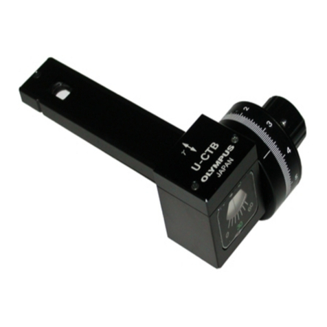
△Midbody, analyzer, compensator and Bertrand lens bearing.
1.Compensator socket: can plug in gypsum (λ), Mica (λ/4) or Quartz wedge.
Turn power switch, rotating screw to adjust
brightness of Field.
Place specimen on stage, moving to the light path
until to observe position, gently press clamps,
moving to slice’s both end then fixed.
Rotate nosepiece, put objective10X in light path;
rotate Coaxial Coarse And Fine Focusing handle
wheel to focusing until legible image.(adjust
tension ring to adjust coaxial coarse and fine
focusing circle’s tension.
Polarizing direction is reseted in horizontal level.
(can through complete cleavage black mica
sheet to define polarizing direction.
When polarizer and analyzer located in dial 0°, is
orthogonal each other and FOV will be dark(no
sample),if not, should check polarizing position.
According to mineral slice interference colors, plug
in slider.
1. Switch on the power, adjust brightness.
2. Place Mineral Specimen.
3. Put 10X objective into optical path and
focus.
4. Define polarizing direction.
5.Check polarizer and analyzer is
orthogonal.
6. Use compensator slider.
7. Observe with Bertrand lens
Conoscopic observation should use high power
eyepiece, push in Bertrand lens when in
orthogonal polarization, to determine mineral’s
optical sign and optic axial angle.


