CellSolutions 30 User manual

CellSolutions 30
Operator’s Manual
Document Number: CS-OP-30US
Rev. 07
Issue Date: July 24, 2020

CellSolutions 30 Operator’s Manual CS-OP-30US Rev. 07 ii
Copyright 2015 - CellSolutions, LLC. All rights reserved. No part of this manual or the described software
may be copied, reproduced, translated or reduced to any electronic medium or machine-readable form without
the prior written consent of CellSolutions, LLC., except that you may make one copy of the program and
related files for back-up purposes.
Although this manual was prepared with every precaution to ensure accuracy, CellSolutions assume no
liability for errors or omission, nor for danger resulting from application or use of this information.
C
P
CELLSOLUTIONS GmbH
Halbinselstr. 37
88142 Wasserburg, Germany
M
CellSolutions, LLC.
1100 Revolution Mill Drive
Suite 1
Greensboro, NC 27405, USA

CellSolutions 30 Operator’s Manual CS-OP-30US Rev. 07 iii
Table of Contents
Preface
v
Information about this Manual
v
General Information
v
Warranty and Contact Information
v
Section 1.0 Introduction
1-1
1.1 Intended Use
1-1
1.2 Requirements
1-1
1.3 Hazards and Warnings
1-1
Section 2.0 Specifications and Installation
2-1
2.1 Equipment Specifications
2-1
2.2 Recommended Installation Space
2-2
2.3 Installation and Setup
2-3
2.4 Power the Unit
2-3
2.5 Aligning Unit for Operation
2-4
2.6 Transport, Storage, Disposal
2-4
Section 3.0 Material Requirements
3-1
3.1 Reagents
3-1
3.2 Re-usable Materials
3-1
3.3 Consumable Materials
3-1
Section 4.0 Operation Overview
4-1
4.1 Specimen Identification
4-1
4.2 Slide Presentation and Barcoding
4-1
4.3 Specimen Volume Detection
4-1
4.4 Specimen Dilution
4-1
4.5 Specimen Mixing and Transfer
4-2
4.6 Specimen Application to Slide
4-2
4.7 Loading Slide into Staining Rack
4-2
4.8 Specimen Drying
4-2
Section 5.0 Sample Preparation
5-1
5.1 Sample Collection
5-1
5.2 Sample Identification and Tracking
5-1
5.3 Sample Transfer
5-1
5.4 Centrifugation
5-1
5.5 Decanting
5-2

CellSolutions 30 Operator’s Manual CS-OP-30US Rev. 07 iv
5.6 Vortexing
5-5
Section 6.0 Operating Procedure
6-1
6.1 Software Operator Interface
6-1
6.2 Operating Modes
6-1
6.3System Initialization
6-3
6.4 Startup Checks
6-3
6.5 Processing Samples
6-9
6.6System Utilities
6-12
6.7 System Shutdown
6-15
Section 7.0 Maintenance
7-1
7.1 Daily Maintenance
7-1
7.2 Weekly Maintenance
7-1
7.3 Semi-Annual Maintenance
7-2
Section 8.0 Troubleshooting
8-1
Appendix A Glossary of Terms
A-1
Appendix B Glossary of Symbols
B-1
Appendix C Microscope Slide Printer
C-1
Index

CellSolutions 30 Operator’s Manual CS-OP-30US Rev. 07 v
PREFACE
Information About This Manual
This manual provides information on the installation, operation and
maintenance of the CellSolutions 30 System and its software.
Throughout the manual the following three notices are used to highlight
important information:
WARNING: INDICATES THE POSSIBILITY OF SEVERE
PERSONAL INJURY OR LOSS OF LIFE IF INSTRUCTIONS ARE
NOT FOLLOWED.
Caution: Indicates the possibility of severe equipment damage if
instructions are not followed.
Note: Indicates useful information.
General Information
This device is intended for the preparation of thin-layer cell
presentations on microscope slides for subsequent staining and
evaluation. All users of the device should be appropriately trained on
the uses of the device and have an understanding of the overall slide
preparation and screening process.
WARRANTY INFORMATION
The CellSolutions 30 has a one-year warranty from the date of sale. For
technical support or repair information contact your designated local
representative or CellSolutions.
CellSolutions
1100 Revolution Mill Drive
Greensboro, NC 27405, USA
+1-336-510-1120

Introduction
CellSolutions 30 Operator’s Manual CS-OP-30US Rev. 07 1-1
1.0 INTRODUCTION
1.1 Intended Use
The CellSolutions 30 automates certain steps in the process of preparing a microscope slide with
a thin layer of cells for microscopic visual evaluation. The unit takes as input, preserved cell
samples that have already been concentrated by centrifugation. The system then outputs
optimized samples of approximately the same cellularity onto microscope slides that are ready for
staining.
1.2 Requirements
The device is designed to use the specific reagents and consumable materials identified in this
manual (i.e. reagents, disposable tubes, automated pipette tips, stain shield labels). Use of other
reagents and materials may damage the device and cause incorrect results as well as render the
warranty invalid.
The samples should be collected by experienced professionals using an acceptable cervical
sampling device that allows detachment or thorough rinsing of the brush or spatula head in the
preservative vials. The vials used for collection are BestPrep®General Cytology Preservative
Vials (C-101-500).
1.3 Hazards and Warnings
1.3.1 Chemical Hazards
The fluids processed by the device are biological samples that may contain infectious
material.
WARNING: SPECIMENS MAY CONTAIN INFECTIOUS MATERIAL.
WEAR PROTECTIVE CLOTHING AND AVOID CONTACT WITH
SPECIMEN.
WARNING: IF A SPILL OCCURS, WIPE CLEAN THE AFFECTED
AREA USING APPROPRIATE CLEANING MATERIAL FOR THE TYPE
OF SPILL. POTENTIAL BIOHAZARD CLEAN UP MAY USE A TOWEL
LIGHTLY DAMPENED WITH A 10% BLEACH SOLUTION.
1.3.2 Mechanical Hazards
The CellSolutions 30 is controlled by a computer in communication with sensors and
motors that when properly operated should prevent any accidental harm to the operator.
The operator should take reasonable care not to interfere with moving parts of the system
while in operation.

Introduction
CellSolutions 30 Operator’s Manual CS-OP-30US Rev. 07 1-2
1.3.3 Electrical Hazards
The CellSolutions 30 has 2 items that are separately plugged into an alternating current
supply. The 2 items are a computer and the CellSolutions 30 Processing Platform. Each
item operates on 100 to 240 volts and 50 to 60 Hz. Usual electrical precautions should
be observed.

Specifications and Installation
CellSolutions 30 Operator’s Manual CS-OP-30US Rev. 07 2-1
2.0 SPECIFICATIONS AND INSTALLATION
2.1 Equipment Specifications
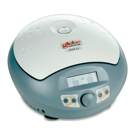
The system comes with a CellSolutions 30 processing platform, a computer and a Smart
Card reader. A separate centrifuge and vortex mixer that is not provided with the system
is needed to perform the overall process. The centrifuge and vortex mixer listed below
are suggested units, however, others may be used as long as they can achieve the
required G-forces and mixing requirements of the process. The physical dimensions and
specifications for each unit are as follows:
2.1.1 CellSolutions 30:
Dimensions: Width: 430 mm (17 inches)
Depth: 580 mm (23 inches)
Height: 740 mm (29 inches)
Power: Configuration 1: 120VAC, 60Hz
•CellSolutions 30 platform –6.2 amps
•Computer –0.5 amps
Configuration 2: 240VAC, 50Hz
•CellSolutions 30 platform –3.1 amps
•Computer –1 amp
(Note: Operation at 100VAC to 240VAC is
acceptable.)
Weight: 41 kg (90 lbs)
Operating Temperature: 5Cto 35C (41to 95F)
Relative Humidity: 30 to 80% RH, non-condensing
Throughput: 30 slides per hour (may vary based on sample size)
Barcode: Code 128, DataMatrix, PDF417 (Other formats available
–
Contact Authorized Representative)
Remote Access: Remote Troubleshooting Support (Contact
Authorized Representative for availability)
Computer: System runs on a computer with a Windows 7 or later
operating system that is connected with a USB cable.

Specifications and Installation
CellSolutions 30 Operator’s Manual CS-OP-30US Rev. 07 2-2
2.1.2 Centrifuge:
A centrifuge is required but not provided. The following centrifuge is suggested
as being one that is compatible with the CellSolutions 30 system:
Manufacturer: Drucker
Model: Horizon 24 Flex with six-bucket rotor
Dimensions: Width: 380 mm (15 inches)
Depth: 430 mm (17 inches)
Height: 230 mm (9 inches)
Power: Configuration 1: 120VAC, 60 Hz, 1 amp
Configuration 2: 240VAC, 50 Hz, 0.5 amps
Weight: 17 kg (37 lbs)
Capacity: 24 tubes (6 position rotor with a 4-tube
rack in each position)
2.1.3 Vortexer:
A vortex mixer is required but not provided.
A standard laboratory vortex mixer with comparable specifications to the unit
noted below is acceptable.
Manufacturer: Thermolyne
Model: Maxi Mix II, No. M37615
Dimensions: Width: 130 mm (5 inches)
Depth: 200 mm (8 inches)
Height: 150 mm (6 inches)
Weight: 3 kg (6 lbs)
2.2 Recommended Installation Space
In addition to the bench top space required to hold the CellSolutions 30 platform, space is
also needed for the computer and for handling tubes, racks, and slides.
Recommended Bench Space for CellSolutions 30:
Width: 1200 mm (48 inches)
Depth: 750 mm (30 inches)
Height: Approximately 800 mm (32 inches)
Recommended Bench Space for centrifuge, vortex mixer, and handling:
Width: 1200 mm (48 inches)
Depth: 750 mm (30 inches)
Height: Not critical.

Specifications and Installation
CellSolutions 30 Operator’s Manual CS-OP-30US Rev. 07 2-3
The above dimensions are recommended values. Each installation site’s space will vary
based on space constraints and usage volumes.
2.3 Installation and Setup
The CellSolutions 30 should be placed on a sturdy and stable table that does not tilt or
flex.
The unit can be placed with the back toward a wall so long as there is at least 50 mm (2
inches) of space between the unit’s back and the wall. This space provides ventilation for
unit cooling.
Once the unit is in its final place on the table, the 4 machine feet should be adjusted to
level the machine. The feet should be adjusted until the bubble in the level attached to
the rotary table is centered. All 4 feet must be adjusted so they are touching the table
and the unit does not tilt back and forth on two feet.
Note: It is critical that the machine be completely level so that the cell suspension
deposited on the slide does not run off the slide or pool toward one side of the deposit
area. If the solution pools to one side, that side will have a higher cell concentration than
the rest of the slide.
Note: Any time the machine is moved, the level should be re-checked and adjusted if
necessary.
The tubing to the pump should be placed in a reservoir bottle or container. The container
should be filled with a 50% ethanol solution.
The tubing that is connected to the priming station standpipe should be routed to a
discharge collection container or to a drain.
During operation, the unit ejects used pipette tips into the detachable tip chute toward the
back left of the instrument. The detachable tip chute is held in place with a threaded
thumb screw so the chute can be easily removed for cleaning with a dilute bleach
solution. A small metal pin is on the top of the tip chute for attaching a tip disposal bag
such as a Whitney Products Safe-Keeper Container (Item BH2005). Other suitable leak
proof tip disposal containers may also be used and are the option of the user.
2.4 Powering the Unit
The CellSolutions 30 processing platform and the computer have separate power cords.
Each of these components can be powered with 100 to 240 VAC and 50 to 60 Hz.
Check that the available power is correct before plugging the components into the wall
socket.
The computer is connected to the processing platform with a USB cable. The cable
should be connected to the USB connection marked on the computer and the square
USB connection under the air inlet on the back of the machine.
The Smart Card reader is plugged into a USB port on the computer.

Specifications and Installation
CellSolutions 30 Operator’s Manual CS-OP-30US Rev. 07 2-4
After all the connections noted above are made, the computer and the processing
platform can be turned on in any order. Once the computer is booted up, the
CellSolutions 30 software can be started by double clicking the icon on the computer
desktop.
2.5 Aligning Unit for Operation
After shipping or moving the CellSolutions 30 the mechanical alignment of the system
may have slightly changed. All of the System Checks listed in the Utilities Menu should
be performed during installation and after moving the unit. The System Checks verify
proper alignment and can be used to make some mechanical adjustments (i.e. leveling
the unit).
The unit has an initialization file that specifies motor alignment values (i.e. location of tip
rack or sample tubes) and calibration information (i.e. fluid pump calibration offsets). If
the System Checks indicate that adjustments are needed to the initialization file, trained
maintenance personnel should be contacted.
2.6 Transport, Storage, Disposal
Prior to removing the unit from service for decommissioning, storage or transport, the unit
must be cleaned/decontaminated. This is done by wiping all external surface of the unit
that may have come in contact with biological samples. The surfaces should be wiped
with a towel that is lightly dampened with a 10% bleach solution. Do not spray cleaning
solution directly on the unit.
The system should have fluids purged from the pump and tubing prior to removing unit
from service. This can be accomplished by removing the pump inlet tube from the diluent
reservoir and using the Prime Fluid Lines option in the Utilities Menu (see section 6.5) to
pump liquids out of the pump and tubing. At least 5 ml of air should be pumped through
the system.
If the equipment is to be permanently removed at the end of its service life cycle, it should
be handled as Waste Electrical and Electronic Equipment (WEEE). The equipment,
including accessories, does not belong in your regular waste. For disposal of the
equipment in the European Economic Area (EEA) or other areas with specified WEEE
regulations, contact your CellSolutions Representative for disposal guidance or dispose
of in accordance with your local regulations. The unit must first be cleaned and
decontaminated as noted above.

Material Requirements
CellSolutions 30 Operator’s Manual CS-OP-30US Rev. 07 3-1
3.0 MATERIAL REQUIREMENTS
3.1 Reagents
The device uses 2 different fluids:
•50% ethanol solution
•GluCyte™Cell Adherent: Part Number: GC 100
The amount of diluent used varies per sample with typical amounts between 100ul to 1000ul per
sample. The amount of GluCyte™used is about 200ul per sample.
3.2 Re-usable Materials
The sample racks provided with the machine hold 20 sample tubes along with 20
disposable tubes that are used during processing.
3.3 Consumable Materials
The device uses one of each of the items below for each sample when running in Single
Slide Mode. These items are either supplied in the Kit GCK 500-A that includes
GluCyte™, or purchased separately where mandated.
•BestPrep®General Cytology Preservative Kits (No. C-101-500) This item is not
supplied in the Kit GCK 500-A but is needed for the procedure. Please order this
item separately using Catalog No. C-101-500.
•Glass Slides (No. GCK D4) Supplied in Kit GCK 500-A
•Disposable Centrifuge Tubes: (No. GCK D1) Supplied in Kit GCK 500-A
•Disposable Tubes: 13x75mm, 5ml, round bottom tubes (No. 55.475) Supplied in
Kit GCK 500-A
•Automated Pipette Tips: (No. GCK D3) Supplied in Kit GCK 500-A (One Pipette
Tip is used for each sample plus one additional Pipette Tip is used at the
beginning of each run.)
When running in Dual-Slide Mode or Triple-Slide Mode, two or three Glass Slides are
used for each sample.
The system also uses a specialized ribbon for printing on the glass slides. One roll of
ribbon will print approximately 8000 slides. The ribbon is not supplied as part of the Kit
and should be purchased separately using Catalog No. GCK D7.

Operation Overview
CellSolutions 30 Operator’s Manual CS-OP-30US Rev. 07 4-1
4.0 OPERATION OVERVIEW
The objective of the CellSolutions 30 is to produce barcoded slides that are ready to be stained.
The slides prepared will have a thin layer of cells adhering in a defined area of the slide. The cell
deposition area has a controlled cellularity (number of cells per square millimeter) that is readily
suitable for evaluation either manually, using a microscope or a suitable microscopic imaging
system.
When running in Normal Single-Slide Mode the operation can be broken down into the steps
listed below.
4.1 Specimen Identification
A barcode scanner is used to read labels on the side of the sample tubes. The system
uses a mirror that allows the scanner to only read one tube’s barcode at a time. The
barcodes on adjacent tubes are not visible to the scanner so a mix-up of samples is not
possible.
4.2 Slide Presentation and Barcoding
The system feeds microscope slides from the bottom of a stack of slides onto a
processing platform. After the barcode on the tube is read a matching barcode is printed
on the microscope slide.
4.3 Specimen Volume Detection
To achieve a relatively consistent cellularity on the final slide, the device must first get an
approximation of how many cells there are in the original specimen tube. The device
uses an air pressure sensor in the pipetting system that can detect when a pipette tip
contacts a liquid level surface. Once the elevation of the pelletized sample inside the
tube is found, the unit can determine the volume of the pellet.
4.4 Specimen Dilution
The machine dilutes the sample in two different tubes (primary and secondary tubes) and
with two different fluids (diluent and GluCyte™) to achieve desired cellular concentration
prior to dispensing to the microscope slide. The amount of dilution is based on the
number of cells in the original sample. The system uses the volume of the pellet to
approximate the number of cells.
Each sample is diluted differently based on the number of cells in that sample. Normally,
the cell pellet is first diluted with 50% ethanol and then the diluted sample is further
diluted by mixing with GluCyte™. If there is an extremely small sample, the first dilution
step is skipped and the sample is just diluted with GluCyte™.

Operation Overview
CellSolutions 30 Operator’s Manual CS-OP-30US Rev. 07 4-2
4.5 Specimen Mixing and Transfer
The device uses a disposable pipette tip and a pipette pump to both mix and transfer
samples. The mixing is done by aspirating and dispensing the fluid multiple times to
ensure the cell suspension is a homogeneous mixture within the diluent or GluCyte™.
4.6 Specimen Application to Slide
The device aspirates a specific volume of the mixed cell and GluCyte™suspension and
transfers it to the slide. Once the solution is dispensed to the slide it is inscribed into a
defined pattern by bringing the pipette tip near the surface and spreading it into a
rectangular pattern. The robot arm ejects the pipette tip into a collection container after
the cell mixture is inscribed on the slide.
4.7 Loading Slide into Staining Rack
The rack used to collect slides after the sample has been applied is a 20-position rack
that can be used in an automated or manual staining system. The device pushes a slide
into the rack as the processing is completed. Each of the 20 positions in the slide rack
corresponds to a specific location in the 20-position tube rack. After all the tubes in the
tube rack have been processed and the slides have been loaded into the slide rack, the
slide rack rotates to the drying station and an empty rack rotates to the load position in
preparation for the next rack of tubes.
4.8 Specimen Drying
After the specimen is applied to the slide, the slide must remain in a horizontal orientation
until the solution is dry. This drying time is greatly influenced by the ambient conditions.
In order to allow drying to proceed in a reasonable time frame, the device blows air over
the slides while they are in the drying station.

Sample Preparation
CellSolutions 30 Operator’s Manual CS-OP-30US Rev. 07 5-1
5.0 SAMPLE PREPARATION
5.1 Sample Collection
Using approved detachable cell sampling brushes or a combination of detachable
endocervical brushes and detachable spatulas, the samples are collected following the
manufacturers’ recommendations for the specific devices. The detached portion of the
sampling devices with the collected cells is placed into the BestPrep®General Cytology
Preservative Vials. The cap is then screwed onto the vial until firmly sealed so as to
prevent any leakage.
5.2 Sample Identification and Tracking
Each lab may have different protocols for sample identification. The following is provided
as one method of sample handling through the CellSolutions 30 process. (If a different
method is used, the lab should ensure at a minimum the sample produced by the
CellSolutions 30 unit can be positively traced back to the original sample.)
5.2.1 Assign the original sample identifying information to a tracking number that will
be used throughout the process. This tracking number appears on a set of 3
identical barcode labels that are pre-printed or are printed on demand.
5.2.2 Place one of the three barcode labels with the tracking number on the patient
requisition form.
5.2.3 Place another of the three barcode labels on the original sample container.
5.2.4 Place the remaining barcode on the primary centrifuge tube into which the
sample will be transferred.
NOTE: It is the responsibility of the lab to ensure the sample tracking method used is in
accordance with all applicable standards.
5.3 Sample Transfer
•Verify the tracking number on the original sample vial matches the number on the
disposable centrifuge tube into which the sample will be transferred.
•Vortex the vial for 5 to 10 seconds to thoroughly mix and free cells from the
collection device.
•Open the original container and pour the sample into the disposable centrifuge
tube while ensuring the collection device is not transferred to the disposable
centrifuge tube.
5.4 Centrifugation
The sample should be centrifuged under the following conditions to create an intact pellet
of cells in the bottom of the tube.:
•G-force = 800 X G
•Time = 10 minutes
The recommended centrifuge for the CellSolutions process is the Drucker Model 755
VES with a six bucket rotor. The settings on this centrifuge to achieve proper results are:
•Speed: 2150 rpm

Sample Preparation
CellSolutions 30 Operator’s Manual CS-OP-30US Rev. 07 5-2
•Time: 10 minutes
If a different centrifuge is used, consult the applicable documentation to determine the
settings required to achieve a G-force of 800.
5.5 Decanting
Samples can either be decanted from the primary tubes individually or in groups of four
while in the centrifuge racks. The method used is subject to an individual lab’s needs
and requirements. One of the following methods should be used to decant the samples
into a suitable disposal basin or container approved for accepting biological samples.
Note: Proper decanting is very important. The unit measures the volume in the tube
after decanting to get an approximation of the cell pellet size. If extra fluid is left on the
pellet after decanting, the unit may over-estimate the size of the cell pellet.
5.5.1 Decanting individual tubes
a. Invert the tube in one quick smooth motion to an angle of approximately 80
degrees so that the fluid drains down one side of the tube.
(a) (b)
Figure 5-1
b. Hold the tube at approximately 80 degrees for approximately 5 seconds.
c. While holding the tube in this inverted orientation move it to a location were
it can be blotted onto a paper towel. The blotting is used to wick away the
fluid that collects on the rim of the tube.

Sample Preparation
CellSolutions 30 Operator’s Manual CS-OP-30US Rev. 07 5-3
(a) (b)
Figure 5-2
Note: The tube should not be turned upright after pouring into the basin
and before blotting. Turning upright would allow drops of fluid on the rim to
go back down into the tube. The level detection system of the device relies
on proper decanting and removal of as much fluid from the pellet as
possible.
d. Once the tube is in contact with the paper towel, the tube can be
completely inverted to a vertical position so the entire rim of the tube is
contacting the paper towel. Hold the tube in this position for about 2
seconds so the paper towel absorbs the initial fluid collected around the
tube rim.
e. While keeping the tube inverted, move the tube to a clean, unused part of
the paper towel and allow the tube to remain inverted on the towel for
between 60 to 120 seconds.
f. Blot the tube by slightly lifting the tube, moving it to a clean, unused section
of the paper towel and then momentarily touching the entire rim of the tube
to the towel. Blot multiple times until no fluid appears on the towel.
Note: When blotting, the tube should be lightly touched to the towel. Do
not tap the tube as that could cause the cell pellet to dislodge.
g. After blotting, the tube can be turned upright.
h. The process can be repeated for subsequent tubes while ensuring that
tubes are blotted in areas of the paper towel that have not been previously
used.
5.5.2 Decanting tubes in racks
a. With tubes in the centrifuge rack, grasp the rack and tubes in such a way
so your thumb and index finger are holding all four tubes while the rack is
being held by your remaining fingers (see figure below). The index finger
and thumb should separate tubes into groups of two as shown below so
the tubes do not contact each other.

Sample Preparation
CellSolutions 30 Operator’s Manual CS-OP-30US Rev. 07 5-4
Figure 5-3
b. In one quick smooth motion invert the four tubes to an approximate angle
of 80 degrees over a basin so that the separated tubes are above each
other (see figure below).
Note: Inverting the tubes quickly allows the tubes to be inverted before the
fluid reaches the rims of the tubes and holding the tubes at approximately
80 degrees as shown allows the fluid to drain down one side of the tube
and out the rim of the tube without contacting adjacent tubes.
(a) (b)
(c) (d)
Figure 5-4

Sample Preparation
CellSolutions 30 Operator’s Manual CS-OP-30US Rev. 07 5-5
c. Hold the tube inverted at the 80-degree angle for about 5 seconds.
d. While holding the tubes in this inverted orientation move them to a location
were they can be blotted onto a paper towel. The blotting is used to wick
away the fluid that collects on the rim of the tube.
Note: The tube should not be turned upright after pouring into the basin
and before blotting. Turning upright may allow drops of fluid on the rim to
either go back down into the tube or potentially onto an adjacent tube. The
level detection system of the device relies on proper decanting and
removal of as much fluid from the pellet as possible.
e. Allow the lower two tubes to contact the paper towel first. Then tilt the rack
so the tubes are vertical and the rims of all the tubes are contacting the
paper towel. Hold the tubes in this position for about 2 seconds so the
paper towel absorbs the initial fluid collected around the tube rim.
f. While keeping the tubes inverted, move the tubes to a clean, unused part
of the paper towel and allow the tubes to remain inverted on the towel for
between 60 to 120 seconds.
(a) (b)
Figure 5-5
g. Blot the tubes by slightly lifting the tubes, moving them to a clean, unused
section of the paper towel and then momentarily touching the rims of each
tube to the towel. Ensure the rims of all 4 tubes contact the towel. Blot
multiple times until no fluid appears on the towel.
Note: When blotting, the tubes should be lightly touched to the towel. Do
not tap the tubes as that could cause the cell pellet to dislodge.
h. After blotting, turn the tubes upright.
i. The process can be repeated for subsequent tubes while ensuring that
tubes are blotted in areas of the paper towel that have not been previously
used.

Sample Preparation
CellSolutions 30 Operator’s Manual CS-OP-30US Rev. 07 5-6
5.6 Vortexing
The samples should be vortexed to break up the cell pellet after decanting. Each individual tube
can be vortexed or the 4 tubes in one centrifuge rack can be vortexed together. Adequate mixing
can be obtained by holding the rack or individual tube on the vortexer for 4 to 6 seconds then
lifting it off the vortexer for one second. This brief on-off vortex sequence should be repeated two
additional times.
Note: If vortexing in the rack, the tubes should be squeezed tight against the sides of the rack so
that the vortexer’s vibrations are adequately transferred through the rack to the tubes. This can
be done by tightly holding the tubes and racks as described above for decanting.
Figure 5-6
This manual suits for next models
1
Table of contents