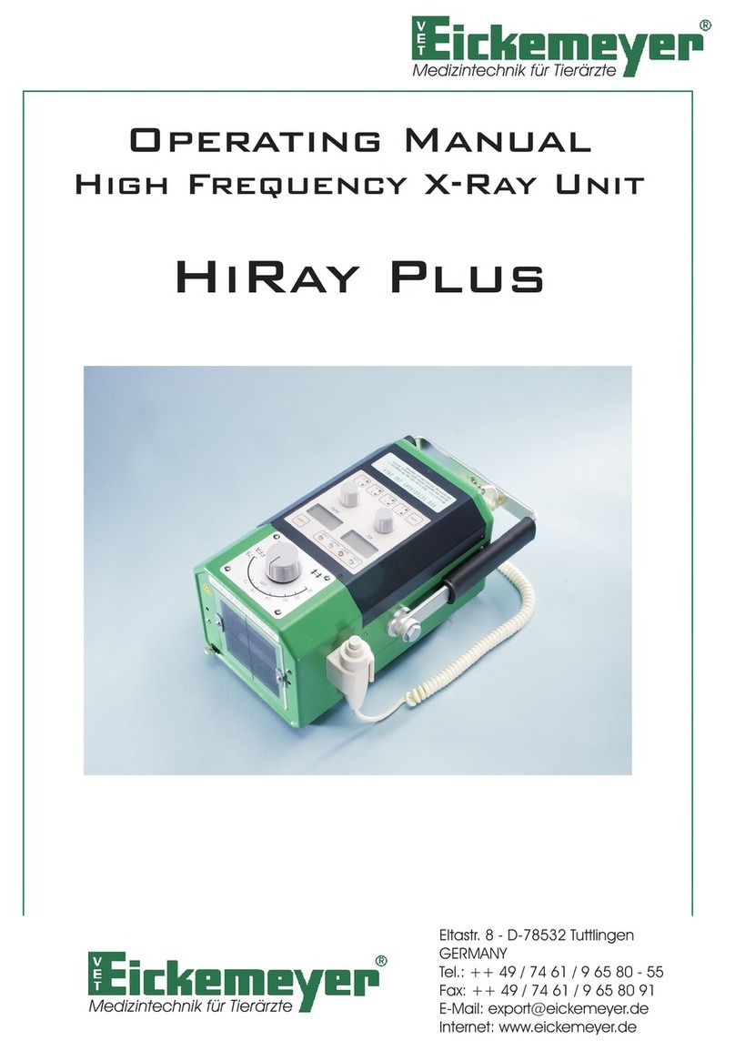
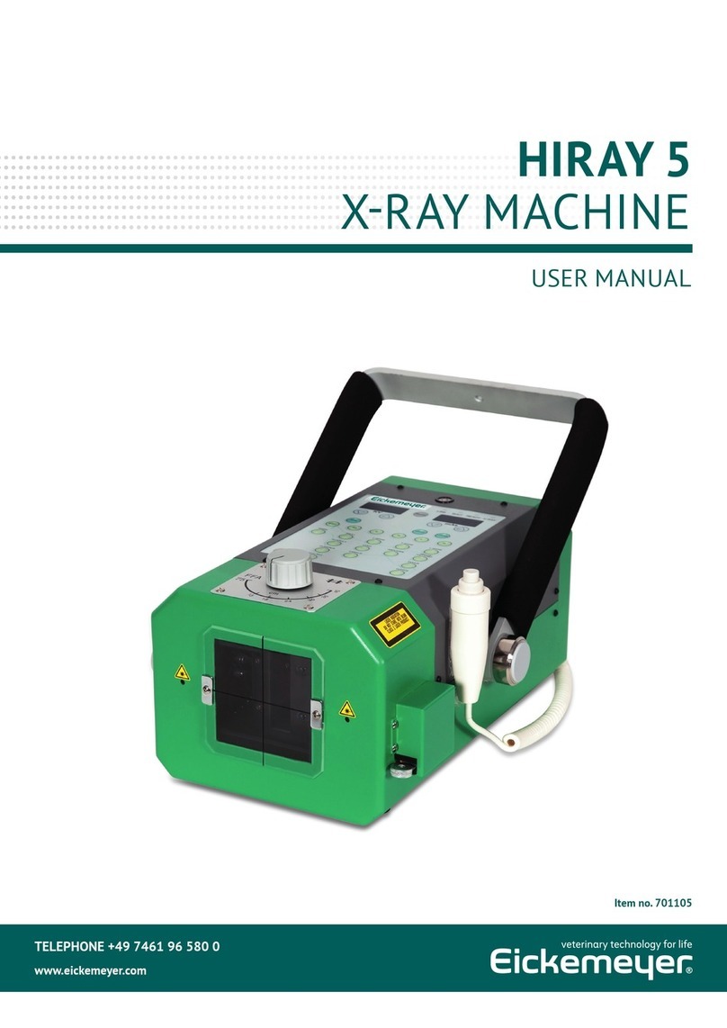
Eickemeyer HiRay Plus User manual

USER MANUAL
HIRAY PLUS
X-RAY MACHINE
Art. No. 701115
TELEPHONE +49 7461 96 580 0
www.eickemeyer.com

2
USER MANUAL HIRAY PLUS X-RAY MACHINE
T +49 7461 96 580 0 | F +49 7461 96 580 90 | E export@eickemeyer.com | www.eickemeyer.com

3
USER MANUAL HIRAY PLUS X-RAY MACHINE
T +49 7461 96 580 0 | F +49 7461 96 580 90 | E export@eickemeyer.com | www.eickemeyer.com
CONTENT
1. Description................................................................................................................................ 4
2. Panel Guide............................................................................................................................... 4
3. Operation .................................................................................................................................. 5
Assembly....................................................................................................................................................5
Before making the Exposure..............................................................................................................5
Operation ..................................................................................................................................................5
4. Specifi cations............................................................................................................................ 7
5. Exposure Schedule................................................................................................................... 9
6.1 Without Grid......................................................................................................................................9
6.2 With Grid ............................................................................................................................................9

4
USER MANUAL HIRAY PLUS X-RAY MACHINE
T +49 7461 96 580 0 | F +49 7461 96 580 90 | E export@eickemeyer.com | www.eickemeyer.com
1. DESCRIPTION
HiRay PLUS is the most advanced portable x-ray unit available both for the fi eld and clinic application.
The built-in collimator with 2 crossing lines and 2 laser pointers confi rms an easy and quick acquirement of each radiographic
position, limiting the need for remarks.
By means of the automatic mAs control technique, the real kV selected on the panel is always maintained even at the place
where the line voltage is dropped by high impedance in order to obtain the expected fi rm result.
HiRay PLUS is the mono block type, including
• Generator (includes x-ray tube in the H.T. transformer with 2 kW inverter)
• Built-in collimator and 2 laser pointers
• Line cable
• Hand switch with curled cable
• Heavy duty carrying case
2. PANEL GUIDE
1. Line switch (breaker)
2. Socket for plug of line cable
3. Socket for plug of hand switch cable
4. Handle
5. Line lamp (green
6. Wait / alarm lamp (red)
7. Ready lamp (green)
8. X-ray lamp (amber)
9. kV display window
10. mAs display window
11. kV selector
12. mAs selector
13. Collimator lamp switch
14. Collimator
15. X-ray fi eld adjustment knob
16. X-ray fi eld indicator
17. Laser pointer
18. Scale
19. Hand switch in holder
20. APR switches

5
USER MANUAL HIRAY PLUS X-RAY MACHINE
T +49 7461 96 580 0 | F +49 7461 96 580 90 | E export@eickemeyer.com | www.eickemeyer.com
3. OPERATION
Assembly
Unpack the unit and inspect it for any damage. Extend the cable out to their fullest length. Pay careful attention to the cables
and inspect them for worm or broken insulation. If they show signs of undue wear, do not plug the unit in or attempt to use
it. Connect a plug of the hand switch cable to the socket for a plug of the hand switch cable [3].
Then connect a plug of the line cable to the socket for a plug of the line cable [2] and the other plug to the power source,
confi rming the line switch [1] is at “OFF” position.
Before making the Exposure
At the fi rst installation or when the unit is used after 2 weeks or more absence from its last use, a warming up procedure of
the x-ray tube is necessary to ensure the longevity of the x-ray tube (tube aging)
A recommended warming up procedure is as below:
(1) Suffi cient radiation protection should be taken.
(2) Make the following exposures at 15 seconds intervals.
1st: 5 exposures at 50 kV / 5,0 mAs
2nd: 5 exposures at 70 kV / 5,0 mAs
3rd: 5 exposures at 100 kV / 5,0 mAs
Once this aging procedure is performed, it is not required from next time.
So the unit is ready for immediate exposure unless it is not in use for more than 2 weeks.
Operation
Turn the line switch [1] to “ON” position and confi rm the kV display [9] and mAs display [10] are lighted.
Select proper technique for the exposure, using the kV selector [11] and mAs selector [12].
In case the APR technique is desired to use:
How to store the Examination Parts and Exposure Data
Switches
APR Store
I II
APR 1
III
APR 3
IV
APR 5
V
APR 7
APR 2 APR 4 APR 6 APR 8
When either switch of II, III, IV or V is pressed, the upper lamp lights. The upper lamp of the switch II represents APR1. The
upper lamp of the switch III represents APR3, the upper lamp of the switch IV for APR5 and the upper lamp of the switch V
for APR7.
If these switches are pressed again, the lower lamp of the switch lights. The lower lamp of switch II represents APR 2, the
lower lamp of switch III APR 4, the lower lamp of switch IV APR6 and the lower lamp of switch V APR8.
After the lamp of the desired APR switch is lighted, select the kV and mAs in the display windows [9] and [10] by the rotary
selector [11] and [12] for each exam part.
Then press the switch I for “APR STORE” to store each exam. Part and its corresponding exposure data.
Immediately after releasing “APR STORE” switch, the buzzer informs the completion of the store of the parts and data.
By continuation of these steps, the data up to 8 exam. Parts are stored.

6
USER MANUAL HIRAY PLUS X-RAY MACHINE
T +49 7461 96 580 0 | F +49 7461 96 580 90 | E export@eickemeyer.com | www.eickemeyer.com
How to select the stored Data and Parts and make an Exposure
For this purpose, it is recommended to write the examination part of each APR switch (APR 1–APR8) on the panel or
somewhere to remember.
Press the desired APR switch. Then the lamp on the switch lights.
Stored exposure data of its corresponding examination part appears on the display window for confi rmation and the system
is ready for the exposure.
By pressing the collimator lamp switch [13], the halogen lamp is lighted.
Illuminated area is adjusted by turning two x-ray fi eld adjustment knobs [15]. The center of the x-ray fi eld is obtained by two
crossing lines.
Two laser pointers [17] are also lighted. Thus the crossing point (the distance between two beams at the crossing point is
within 5 mm) of these laser beam makes it possible to obtain quick and easy confi r-mation of 75 cm ±2 cm SID and almost
the center of a radiographic fi eld (which is considered to be within the range of 1.5 cm from actual center of the crossing
point of the collimator) at 75 cm of SID even under the sunlight.
Those lights are extinguished automatically after 30 seconds in order to save the life of bulbs.
To make an exposure, press the hand switch half way down. During 1.5 seconds after this fi rst press, the fi lament of the tube
is heated and the ready lamp lights.
Then depress the hand switch fully down and the x-ray lamp lights, confi rming the exposure.
The duration of the exposure is signaled by the x-ray lamp and audible buzzer sounds at the end of the exposure.
After the exposure, the wait lamp is fl ickering and next exposure is ready when this fl ickering is extinguished.
This wait lamp also works as the alarm lamp and the lamp is lighted continuously in case any trouble happens in the circuit.
Note!
1. The exposure is managed by two step depression as stated above. However full depression at one time is also
accepted. In this case, the exposure is obtained after 1.5 seconds from the full depression.
2. The exposure button of the hand switch should be depressed until the exposure is fully completed.
3. The exposure can be terminated at any time by releasing the exposure button. The mAs display blinks. To return
operation, press “LAMP” button.
4. The unit must have a certain waiting period until next exposure in order to protect the system from a damage
by an overheat which would be caused by continuous exposures.
< WAIT TIME >
When the kV setting is made at the value between 40 kV and 66 kV;
• the wait time will be between 0,3 and 5 seconds, depending on the exposure time, at the mAs
selection between 0,3 and 5 mAs.
• the wait time will be 6 seconds at the mAs selection exceeding 6,4 mAs.
When the kV setting is made at the value exceeding 68kV;
• the wait time will be between 0,45 and 4,8 seconds, depending on the exposure time, at the mAs selection
between 0,3 and 3,2 mAs.
• the wait time will be 6 seconds at the mAs selection exceeding 4 mAs.
5. The LEVEL (WATER SCALE) [21] is attached for horizontal adjustment.
6. The digital (kV-mAs) display Fascia direction can be changed.

7
USER MANUAL HIRAY PLUS X-RAY MACHINE
T +49 7461 96 580 0 | F +49 7461 96 580 90 | E export@eickemeyer.com | www.eickemeyer.com
How to change Direction of the kV-mAs Display
• A main powering switch is turned off.
• Push the APR store switch.
• A main powering switch is turned on.
• Keep pushing the APR store switch for more than three seconds at a time.
• The display direction of only digits (kV-mAs) can be changed to the contrary direction.
N.B. When you order, please specify the fascia direction, so that your ordered unit can be made at the factory according to
your fascia direction,
• The display direction00 is stored, when the power supply is turned off.
Caution!
Do not place the laser pointer for direct eye exposure since the laser beam is dangerous to the eyes.
4. SPECIFICATIONS
System Composition: X-ray control with generator (tube & H.T. transformer)
Collimator with 2 laser pointers
Power cable (6 meter long)
Hand switch with curled cable (2.5 meter long at full extension)
Performance specifi cations: Output: max. 2 kW (100 kV, 20 mA / 66 kV, 30 mA)
kV output: 40–100 kV
Stability: ±2 %
kV rising up time: within 2 msec (Until becoming up to 75% of set kV)
kV setting: 40–100 kV (2 kV step)
mAs selection: 0.3–20 mAs (32 settings):
0.3, 0.4, 0.5, 0.6, 0.7, 0.8, 0.9, 1.0, 1.1, 1.2, 1.3, 1.4, 1.5, 1.6, 1.7, 1.8, 1.9, 2.0, 2.2, 2.5, 2.8, 3.2, 4.0,
5.0, 6.4, 7.0, 8.0, 9.0, 10, 12, 16, 20 mAs
Max. deviation: kV: ±4 %
mA: ±7 %
mAs: ±15 % (0.3–16 mAs), ±20 % (20 mAs and over)
X-ray tube: Toshiba D-124
Focal spot: 1.2 x 1.2 mm
Heat unit: 20 kHU
Target angle: 16 degrees
Total fi ltration: 2.5 mm AL. eq. at 100 kV
Collimator: Single plane, double slit type
Manually operated
Complete with 30 sec timer, cross indication lines and halogen lamp.
Also 2 laser pointers are provided for easy confi rmation of the center of
a radiographic fi eld and the SID of 75 cm even under the sunlight.
Power cable: 6 m long
Hand switch: Two stage, deadman type with curled cable (2.5 meter long at full extension)

8
USER MANUAL HIRAY PLUS X-RAY MACHINE
T +49 7461 96 580 0 | F +49 7461 96 580 90 | E export@eickemeyer.com | www.eickemeyer.com
Power requirement: 200–240 V
Power capacity: 220 V, 20 A
Opt. ambient conditions: Temperature: 20 °C ±15 °C
Humidity: 69 % ±20 %
Net weight: X-ray unit: 8.8 kg
Carrying case: 4 kg
Dimensions: See drawings

9
USER MANUAL HIRAY PLUS X-RAY MACHINE
T +49 7461 96 580 0 | F +49 7461 96 580 90 | E export@eickemeyer.com | www.eickemeyer.com
5. EXPOSURE SCHEDULE
6.1 Without Grid
Thickness
in cm
Up to 5 Up to 10 Up to 15 Up to 20 Up to 25 Up to 30 Up to 35
kV mAs kV mAs kV mAs kV mAs kV mAs kV mAs kV mAs
Abdomen l-l 50–54 1,0–1,4 52–56 1,6–1,8 52–54 2,0–2,5 54–56 2,5–3,2 54–58 3,2–4,0 58–60 4,0–5,0 58–60 5,0–6,4
Spinal Column l-l
54–58 0,8–1,2 58–60 1,6–1,8 62–64 1,8–2,5 64–66 2,5–3,2 68–72 3,2–4,0 74–78 4,0–5,0 76–78 5,0–6,4
Pelvis / HD v-d 62–64 2,0–2,5 64–66 3,2–4,0 66–68 4,0–5,0 68–70 4,0–5,0 70–72 5,0–6,4 72–74 5,0–6,4
Thorax l-l 56 0,5 56–60 0,8–1,0 60–64 1,2–1,6 68–72 1,2–1,6 76–80 1,6–1,8 80–86 1,8–2,0 90–94 2,0–2,5
Extremities 50–54 0,5–0,8 54–58 1,0–1,6
6.2 With Grid
Thickness
in cm
Up to 5 Up to 10 Up to 15 Up to 20 Up to 25 Up to 30 Up to 35
kV mAs kV mAs kV mAs kV mAs kV mAs kV mAs kV mAs
Abdomen l-l 50–54 1,5–2,1 52–56 2,4–2,7 52–54 3,0–3,7 54–56 3,7–4,8 54–58 4,8–6,0 58–60 6,0–7,5 58–60 7,5–9,6
Spinal Column l-l
54–58 1,2–1,8 58–60 2,4–2,7 62–64 2,7–3,7 64–66 3,7–4,8 68–72 4,8–6,0 74–78 6,0–7,5 76–78 7,5–9,6
Pelvis / HD v-d 62–64 3,0–3,7 64–66 4,8–6,0 66–68 6,0–7,5 68–70 6,0–7,5 70–72 7,5–9,6 72–74 7,5–9,6
Thorax l-l 56 1,0 56–60 1,2–1,5 60–64 1,8–2,4 68–72 1,8–2,4 76–80 2,4–2,7 80–86 2,7–3,0 90–94 3,0–3,8
Extremities 50–54 1,0–1,2 54–58 1,5–2,4
Thickness means the amount of tissue in the center of x-ray. It should be measured correctly in order to fi nd out the right
technique. The recommendation depends on the nutritional status of the patient.
A scattered grid should be used with a thickness of more than 10 cm.
The values given here are exposure recommendations for a fi lm focus distance of 75 cm.
Film system used: green-emitting SC 400
Notes!
You might have to correct the values given in the exposure schedule depending on the fi lm intensifi er screen
system used or when used with digital systems.
For obese animals, the upper range values apply. Very slim animals should be X-rayed with the lower values. Grease is a very
good contrast medium and should usually appear light gray, while soft tissue appears dark gray. Fluid accumulations and
bones appear relatively bright on the radiograph.

10
USER MANUAL HIRAY PLUS X-RAY MACHINE
NOTES
T +49 7461 96 580 0 | F +49 7461 96 580 90 | E export@eickemeyer.com | www.eickemeyer.com

11
USER MANUAL HIRAY PLUS X-RAY MACHINE
NOTES
T +49 7461 96 580 0 | F +49 7461 96 580 90 | E export@eickemeyer.com | www.eickemeyer.com

GERMANY
EICKEMEYER KG
Eltastraße 8
78532 Tuttlingen
T +49 7461 96 580 0
F +49 7461 96 580 90
E info@eickemeyer.de
www.eickemeyer.de
SWITZERLAND
EICKEMEYER AG
Sandgrube 29
9050 Appenzell
T +41 71 788 23 13
F +41 71 788 23 14
E info@eickemeyer.ch
www.eickemeyer.ch
UNITED KINGDOM
EICKEMEYER Ltd.
3 Windmill Business Village
Brooklands Close
Sunbury-on- Thames
Surrey, TW16 7DY
T +44 20 8891 2007
F +44 20 8891 2686
E info@eickemeyer.co.uk
www.eickemeyer.co.uk
POLAND
EICKEMEYER Sp. z o.o.
Al. Jana Pawła II 27
00-867 Warszawa
T +48 22 185 55 76
F +48 22 185 59 40
E info@eickemeyer.pl
www.eickemeyer.pl
DENMARK
EICKEMEYER ApS
Lysbjergvej 6, Hammelev
6500 Vojens
T +45 7020 5019
F +45 7353 5019
E info@eickemeyer.dk
www.eickemeyer.dk
NETHERLANDS
EICKEMEYER B.V.
Bedrijventerrein Pavijen-West
Bellweg 44
4104 BJ Culemborg
T +31 345 58 9400
F +31 345 58 9401
E info@eickemeyer.nl
www.eickemeyer.nl
ITALY
EICKEMEYER S.R.L.
Via G. Verdi, 8
65015 Montesilvano (PE)
T +39 0859 35 4078
F +39 0859 35 9471
E info@eickemeyer.it
www.eickemeyer.it
CANADA
EICKEMEYER Inc.
250 Briarhill Dr.
Stratford, Ont. Canada
N5A 7S2
T +1 519 273 5558
F +1 519 271 7114
E info@eickemeyervet.ca
www.eickemeyervet.ca
Other manuals for HiRay Plus
1
This manual suits for next models
1
Table of contents
Other Eickemeyer Dental Equipment manuals
Popular Dental Equipment manuals by other brands

henke sass wolf
henke sass wolf HENKE-DENT 300 A Trio Instructions for use
orangedental
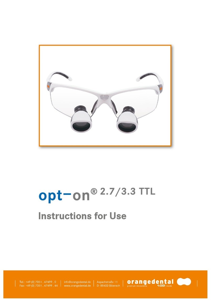
orangedental opt-on 2.7 Instructions for use
NSK
NSK Ti-Max Ti95L Repair Procedure
neopulse
neopulse NP1 MICRO user guide
Solmetex
Solmetex NXT DryVac Maintenance Kit manual
Dentsply Sirona
Dentsply Sirona Capsule Extruder 2 Instructions for use
