Pentax EG-290Kp User manual

PENTAX VIDEO GI SCOPES
EG-290Kp
OWNER’S MANUAL
EC-380LKp

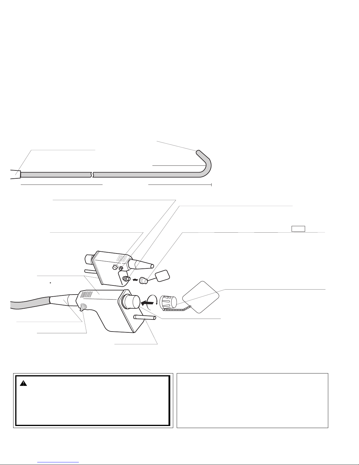
Intended Use (Gastroscope)
These instruments are intended to provide optical visualization of (via a video monitor), and therapeutic access to, the Upper Gastrointestinal Tract.
This anatomy includes, but is not restricted to, the organs; tissues; and subsystems: Esophagus, Stomach, Duodenum and Small Bowel.
The instruments are introduced via the mouth when indications consistent with the need for the procedure are observed in adult and pediatric patient
populations.
Never use the endoscope for any purpose other than that for which it has been designed.
The video endoscopes contained in this manual can only be used with PENTAX video processor, model EPK-1000 and EPK-i.
Intended Use (Colonoscopes)
These instruments are intended to provide optical visualization of (via a video monitor), and therapeutic access to, the Lower Gastrointestinal Tract.
The This anatomy includes, but is not restricted to, the organs; tissues; and subsystems: Large Bowel to the Cecum.
These instruments are introduced via the rectum when indications consistent with the need for the procedure are observed in adult and pediatric patient
populations.
Never use these endoscopes for any purpose other than that for which they have been designed.
These video endoscopes contained in this manual can only be used with PENTAX video processors, model EPK-1000 and EPK-i.
Notes
Read this manual before operating, and save this book for future reference. Failure to read and thoroughly understand the information presented in this
manual, as well as those developed for ancillary endoscopic equipment and accessories, may result in serious injury including infection by cross
contamination to the patient and/or user. Furthermore, failure to follow the instructions in this manual may result in damage to, and/or malfunction of,
the equipment.
This manual describes the recommended procedures for inspecting and preparing the equipment prior to its use and for the reprocessing and
maintenance of the equipment after its use. It does not describe how an actual procedure is to be performed, nor does it attempt to teach the beginner
the proper technique or any medical aspects regarding the use of the equipment.
It is the responsibility of each medical facility to ensure that only well educated and appropriately trained personnel, who are competent and
knowledgeable about the endoscopic equipment, antimicrobial agents/processes and hospital infection control protocol be involved in the use and the
reprocessing of these medical devices. Known risks and/or potential injuries associated with flexible endoscopic procedures include, but are not limited
to, the following: perforation, infection, hemorrhage, burns and electric shock.
Current infection control guidelines require that G.I.scopes and other semi-critical medical devices, that normally come into contact with intact mucous
membranes, such as in the gastrointestinal tract, must at least be high-level disinfected before patient use. Only the user can determine if an instrument
has undergone appropriate infection control procedures prior to each clinical use. It must be recognized that infection control practices involve many
complex and often controversial issues which are constantly evolving. PENTAX strongly recommends that user remain informed of the latest federal
and local regulations, and encourages users to follow infection control guidelines developed by various organizations for health care professionals.
The text contained in this manual is common for various types/models of PENTAX endoscopes and users must carefully follow only those sections and
instructions pertaining to the specific instrument models appearing on the front cover.
If you have any questions regarding any of the information in this manual or concerns pertaining to the safety and/or use of this equipment, please
contact your local PENTAX representative.
Sterility Statement
The instruments identified in this instructional booklet are reusable medical devices. Since they are packaged non-sterile, they must be high-level
disinfected or sterilized BEFORE initial use. Prior to each subsequent procedure, they must be subjected to an appropriate cleaning and either high-
level disinfection or sterilization process.
Conventions
Throughout this manual, the following conventions will be used to indicate a potentially hazardous situation which, if not avoided;
: could result in death or serious injury.
: may result in minor or moderate injury or property-damage.
: may result in property-damage. Also, advises owner/operator about important information on the use of this equipment.
Prescription Statement
Federal (U.S.A) law restricts this device to sale by or on the order of a physician or other appropriately licensed medical professional.
EC REP
Symbol for “MANUFACTURER”
Symbol for “DATE OF MANUFACTURE”
Symbol for “AUTHORISED
REPRESENTATIVE”

TABLE OF CONTENTS
1. NOMENCLATURE AND FUNCTION. . . . . . . . . . . . . . . . . . . . . . . . . . . . . . . . . . . . . . 1
1-1. VIDEO ENDOSCOPE . . . . . . . . . . . . . . . . . . . . . . . . . . . . . . . . . . . . . . . . . . . . . . . . . . 1
1-2. ACCESSORIES . . . . . . . . . . . . . . . . . . . . . . . . . . . . . . . . . . . . . . . . . . . . . . . . . . . . . . . 3
1-3. VIDEO PROCESSOR. . . . . . . . . . . . . . . . . . . . . . . . . . . . . . . . . . . . . . . . . . . . . . . . . . . 4
2. PREPARATION AND INSPECTION FOR USE. . . . . . . . . . . . . . . . . . . . . . . . . . . . . . 5
2-1. INSPECTION OF THE VIDEO PROCESSOR . . . . . . . . . . . . . . . . . . . . . . . . . . . . . . . 5
2-2. INSPECTION OF ENDOSCOPE. . . . . . . . . . . . . . . . . . . . . . . . . . . . . . . . . . . . . . . . . . 6
2-3. PREPARATION JUST BEFORE INSERTION OF ENDOSCOPE . . . . . . . . . . . . . . . 13
3. DIRECTIONS FOR USE
3-1. PRETREATMENT . . . . . . . . . . . . . . . . . . . . . . . . . . . . . . . . . . . . . . . . . . . . . . . . . . . . . 15
3-2. INSERTION AND WITHDRAWAL . . . . . . . . . . . . . . . . . . . . . . . . . . . . . . . . . . . . . . . 15
3-3. BIOPSY. . . . . . . . . . . . . . . . . . . . . . . . . . . . . . . . . . . . . . . . . . . . . . . . . . . . . . . . . . . . . . 17
3-4. LASER . . . . . . . . . . . . . . . . . . . . . . . . . . . . . . . . . . . . . . . . . . . . . . . . . . . . . . . . . . . . . . 18
3-5. ELECTRO-SURGERY. . . . . . . . . . . . . . . . . . . . . . . . . . . . . . . . . . . . . . . . . . . . . . . . . . 20
4. CARE AFTER USE . . . . . . . . . . . . . . . . . . . . . . . . . . . . . . . . . . . . . . . . . . . . . . . . . . . 21
4-1. ENDSCOPES . . . . . . . . . . . . . . . . . . . . . . . . . . . . . . . . . . . . . . . . . . . . . . . . . . . . . . . . . 23
4-1-1 INTERNAL CHANNELS . . . . . . . . . . . . . . . . . . . . . . . . . . . . . . . . . . . . . . . . . . . . . 23
4-1-2 PRE-CLEANING. . . . . . . . . . . . . . . . . . . . . . . . . . . . . . . . . . . . . . . . . . . . . . . . . . . . 26
4-1-3 CLEANING . . . . . . . . . . . . . . . . . . . . . . . . . . . . . . . . . . . . . . . . . . . . . . . . . . . . . . . . 27
4-1-4 HIGH-LEVEL DISINFECTION . . . . . . . . . . . . . . . . . . . . . . . . . . . . . . . . . . . . . . . . 32
4-1-5 STERILIZATION . . . . . . . . . . . . . . . . . . . . . . . . . . . . . . . . . . . . . . . . . . . . . . . . . . . 35
4-2. ENDOSCOPIC ACCESSORY INSTRUMENT (EAIs) AND ENDOSCOPE
COMPONENTS . . . . . . . . . . . . . . . . . . . . . . . . . . . . . . . . . . . . . . . . . . . . . . . . . . . . . . . 36
4-2-1 CLEANING . . . . . . . . . . . . . . . . . . . . . . . . . . . . . . . . . . . . . . . . . . . . . . . . . . . . . . . . 36
4-2-2 DISINFECTION . . . . . . . . . . . . . . . . . . . . . . . . . . . . . . . . . . . . . . . . . . . . . . . . . . . . 38
4-2-3 STERILIZATION . . . . . . . . . . . . . . . . . . . . . . . . . . . . . . . . . . . . . . . . . . . . . . . . . . . 38
4-3. WATER BOTTLE ASSEMBLY . . . . . . . . . . . . . . . . . . . . . . . . . . . . . . . . . . . . . . . . . . 40
5. POST REPROCESSING . . . . . . . . . . . . . . . . . . . . . . . . . . . . . . . . . . . . . . . . . . . . . . . 42
5-1. SERVICING . . . . . . . . . . . . . . . . . . . . . . . . . . . . . . . . . . . . . . . . . . . . . . . . . . . . . . . . . . 43
5-2. CARE AND MAINTENANCE TIPS . . . . . . . . . . . . . . . . . . . . . . . . . . . . . . . . . . . . . . . 44

- 1 -
1. NOMENCLATURE AND FUNCTION
1-1. VIDEO ENDOSCOPE
NOTE:
To avoid damaging the endoscope, do NOT twist,
rotate or bend excessively any of the rubber strain
reliefs.
ENHANCE BUTTON
Push to activate the VCR for recording live procedures.
UP/DOWN DEFLECTION LOCK
When this lever is in the “F” position, turned clockwise,
the bending section moves freely. When turned counterclockwise,
the bending section becomes progressively more stabilized.
UMBILICAL CABLE
RIGHT/LEFT DEFLECTION
CONTROL KNOB
RIGHT/LEFT DEFLECTION LOCK
Functions similar to Up/Down lock
UP/DOWN DEFLECTION CONTROL KNOB
“F” (FREEZE) BUTTON
Push to freeze an image.
“C” (COPY) BUTTON
Push to activate the hardcopy system that
was selected between “FILE” and “HARD COPY”.
MODEL DESIGNATION
SUCTION CONTROL VALVE (OF-B120)
Depress to remove fluids or air through
the instrument channel.
RUBBER INLET SEAL (OF-B190)
Allows passage of accessories while
preventing escape of fluids and air. INSTRUMENT
CHANNEL INLET
For introduction of
biopsy forceps
and other accessories
CONTROL BODY
AIR/WATER FEEDING VALVE (OF-B121)
Covering of hole in the top of the valve delivers
pressurized air. Covering of the hole and fully
depressing the valve delivers pressurized water.
U
L
R
D
F
F
BUTTON Freeze
VCR
Hardcopy
BUTTON
BUTTON
EPK-1000
Freeze
VCR
Hardcopy
EPK-i
* The function can be changed. For more details, refer
to the manual supplied with the processor
RUBBER STRAIN RELIEF

- 2 -
WARNING: Note:
Immediately after use, the metal light guide prong and the
electrical contacts/pins of the endoscope may be HOT.
To avoid burns, do not touch these areas immediately
after use. For safer handling after a procedure, grasp the
PVE connector housing.
Ensure that the soaking cap has been securely
attached (by properly rotating it) to prevent the cap from
coming off during reprocessing. Failure to securely
attach the soaking cap can result in scope damage.
INSERTION TUBE
ELECTRICAL CONTACTS
LIGHT GUIDE
Transmits light from
light source to distal
end of endoscope.
ETO GAS STERILIZATION VENTING CAP OF-C5
Provides venting of endoscope interior to equalize
internal and external pressures. This cap must be
removed before immersion.
NOTE: See important separate section
regarding the use of this cap!
SUCTION NIPPLE
For attachment to
external suction
source
PVE CONNECTOR
Can be rotated within
a 180 range
AIR/WATER PORT
To connect feeding tube from water
bottle assembly.
FEED BACK TERMINAL
Certain manufacturers’ electrosurgical units, such
as older Olympus models, require the connection
of a scope feedback cord (S-cord) to this
feedback terminal.
VENTING CONNECTOR
Accepts “RED” ETO GAS Sterilization Venting cap.
Also accepts Leakage Tester.
PVE SOAKING CAP
OE-C9
BENDING SECTION
RED
This cap must be securely attached before
immersion.
DISTAL END
(Refer to the inside rear cover of this manual)
RUBBER STRAIN RELIEF

- 3 -
1-2. ACCESSORIES
CAUTION:
• Because of the effect that accessories used through the instrument channel of the endoscope can
have on the performance of the endoscope itself, it is strongly recommended that PENTAX accesso-
ries be used with PENTAX endocopes. If a unique or highly specialized accessory is available from
another source, please contact PENTAX to arrange a test of its compatibility before using it through
the PENTAX endoscope.
• Maximum outer diameter of an endoscopic accessory instrument must be at least 0.2 mm less than
the specified instrument channel diameter in PENTAX endoscopes. Working length of an endoscopic
accessory instrument should be approximately 30 cm longer than the endoscope working length.
NOTE:
• Depending upon country and/or local PENTAX distributor, each PENTAX endoscopic accessory may
be an optional accessory.
• For patient contact endoscopic accessories, follow the specific and detailed instructions on use, care
and maintenance supplied with each product.
• To confirm the exact condition of any new accessory device, check the labeling/packaging accompany-
ing the product. Each label/package should clearly identify the contents as either sterile or non-sterile.
HANDLE
(CS6021SN)
FLEXIBLE SHAFT
WHITE BRISTLE
1) Cleaning Brush for Instrument Channel
2) Cleaning Brush for A/W Suction Valve Cylinder
( CS-C5S )
3) Biopsy Forceps
ENHANCED FLEXIBLE
PORTION
CUPS
FLEXIBLE SHAFT
GRIP
HANDLE
Pink handle defines
autoclavable forceps.

- 4 -
1-3. VIDEO PROCESSOR
CAUTION:
Please refer to the instruction supplied with the processor.
NOTE:
Do not use the new PENTAX OS-H4 water bottle cap with the older OS-H2 water container/bottle.
Although the cap may appear to fit onto the bottle, air may escape resulting in insufficient pressure and
flow of air and water during the endoscopic procedure. Both the PENTAX water bottle cap and bottle
(container) are identified by their appropriate model designation. Ensure that an OS-H4 cap is used only
with the OS-H4 water container/bottle.
Do not overtighten the bottle cap. Overtighting can cause the bottle cap to break.
WARNING:
The lifetime of the lamp in EPK-i processor is 500 hours. Prior to use, check the lamp life meter on the rear
panel to ensure the lamp life is less than 500 hours. After 500 hours of use, the image quality will
deteriorate. Excessive use of the lamp beyond 500 hours could break the lamp inside the equipment
resulting in damage to the equipment.
WARNING:
The lifetime of the lamp in EPK-i processor is 500 hours. Prior to use, check the lamp life meter on the front
panel to ensure the lamp life is less than 500 hours. After 500 hours of use, the image quality will
deteriorate. Excessive use of the lamp beyond 500 hours could break the lamp inside the equipment
resulting in damage to the equipment.
NOTE:
The lamp life could be affected by frequency of use. In which case, the lamp life might become shorter than
its respective rated hours (EPK-i: 500 hours, EPK-1000 and EPK-100p: 400 hours)
WATER BOTTLE
LAMP SWITCH
AIR PUMP SWITCH
LCD
POWER SWITCH INTERFACE SOCKET
EPK-1000
POWER SWITCH
I : ON
O: OFF
LAMP SWITCH
INTERFACE
SOCKET
BRIGHTNESS
CONTROLS
AIR PUMP SWITCH
WATER BOTTLE
EPK-i
EPK-1000

- 5 -
2. PREPARATION AND INSPECTION FOR USE
Prior to use, the endoscope, video processor and endoscopic accessory instruments must be carefully inspected for
cleanliness and proper function to determine that they are appropriate for patient use:
2-1. INSPECTION OF THE VIDEO PROCESSOR
Please refer to the Owner’s Manual of the specific model of PENTAX video processor for complete instructions.
1) Attach water bottle assembly, 2/3 filled with sterile water to the appropriate location on the left side of the
video processor.
2) Set the drain lever on the water bottle assembly to the upright position labeled A/W (air/water).
3) Plug the processor into a properly grounded receptacle with the power switch in the OFF position.
4) Make sure that the PENTAX PVE connector is aligned
with the interface socket on the front panel of the proc-
essor.
5) Connect the endoscope to the interface socket on the
processor as illustrated. Rotate the lever of the inter-
face socket clockwise after insertion.
6) Connect the air/water feeding tube from the water bot-
tle assembly to the air/water port on the side of the
PVE connector.
7) Turn the processor and air pump to the “ON” position and check for proper functioning.
8) Press the lamp switch of the processor to ignite the lamp.
NOTE:
PENTAX video endoscopes contained in this manual are only compatible with PENTAX video processor,
model EPK-1000 and EPK-i.
CAUTION:
To avoid discontinuation of endoscopic procedure, have an extra (spare) instrument available as a
standby device. Should any unforeseen event or circumstance render the original instrument inoperable
and/or unsafe for patient.
WARNING:
The addition of defoaming agents to the water supply is NOT recommended. Due to their nature, these
silicone based agents cling tenaciously to surfaces. Unless they are rinsed very thoroughly, a “barrier”
could be created which could reduce the effectiveness of the disinfection/sterilization process. Addition-
ally, repeated use of such defoamers could eventually lead to residual silicone build up resulting in
equipment malfunction such as clogged air and/or water channels.
EPK-1000
WATER BOTTLE
air/water feeding tube
CAUTION
Failure to do so may create a “miscommunication”
between the scope and processor and may result
in the loss of the system’s automatic brightness
control and in excessive light emission from the
scope. Excessive light intensity can in turn
increase the amount of delivered thermal energy
causing heating of the scope tip, specifically the
illumination (LCB) cover glass and/or objects in
contact with the distal cover glass.

- 6 -
9) Prior to each procedure, check the endoscope image quality displayed on the monitor. Confirm that the image
quality, color, automatic brightness (iris) functions are acceptable as per the instructions provided with the
PENTAX video processor.
2-2. INSPECTION OF ENDOSCOPE
Before proceeding with inspection of individual functions, PENTAX endoscopes should be tested for the integrity
of their water-tight design (example: tear in the instrument channel). This test is described in another section of
this manual entitled: “Leakage Testing.”
1) Inspection of the Insertion Tube
a) Check the entire surface of the insertion tube for abnormal conditions such as dents, crush marks, wrin-
kles, bumps, buckles, excessive bending, protrusions, bite marks, peeling of outer sheath, cuts/holes or
other irregularities. Any crush or indentation of the flexible shaft of the endoscopes can cause damage to
the internal mechanisms of the endoscopes.
b) Similarly, check the condition of the umbilical cable for outward signs of damage such as buckling, crush
marks, etc.
c) Make sure that the entire endoscope is clean and has been subjected to either a high-level disinfection or
sterilization process before each patient use.
NOTE:
As indicated elsewhere in PENTAX product labeling, endoscopes particularly the quality of the endo-
scopic image should be checked prior to patient use.
CAUTION:
If the endoscope is intended to be clinically used after testing of individual scope functions (suction, air/
water delivery, water jet, etc.) without further reprocessing, the following precaution should be exercised.
Use “fresh” distilled or sterile water during individual scope function tests to avoid recontamination of the
previously reprocessed instrument by waterborne microorganisms. Tap water, especially that which may
be left idle and uncovered for a prolonged period of time, should not be used during any inspection/test-
ing of the endoscope.
CAUTION:
Various types of endoscope leakage testers exist including manual, electro-mechanical and “automated”
versions, some of which are stand alone units and others which may be integrated into Automated Endo-
scope Reprocessors (AERs)/Washer-Disinfectors (WDs). It must be recognized that PENTAX does not
evaluate non-PENTAX leak tester systems to satisfy their specific products claims, for their effectiveness
to accurately detect leaks and/or for their compatibility with PENTAX endoscopes. Insufficient pressures
may adversely affect the endoscope, especially if pressurization occurs during automated reprocessing
at elevated temperatures. PENTAX accepts no responsibility for use of non-PENTAX leakage testers.
Users should check with the leakage tester manufacturer and confirm their specific product claims,
including compatibility with PENTAX endoscopes at various temperatures and their ability to detect leaks
with/without fluid immersion and with/without flexing of the scope’s distal bending section.
CAUTION:
To avoid further damage to the endoscope or the possibility of malfunction during a procedure, do not
use an endoscope with any abnormalities or outward signs of damage.
WARNING:
All instruments must be reprocessed prior to first time use, after any repairs/service and before every
patient use. When utilizing chemo-thermal processes for reprocessing the endoscope, the instrument
should be allowed to return to room temperature prior to use and/or further handling.

- 7 -
NOTE:
• The distal end of the endoscope as well as the electrical contacts/pins on the PVE connector must be
protected against damage from impact. Never apply excess force such as twisting, or severe bending
to the flexible portion of the endoscope.
• During pre-use inspection, ensure that the distal objective lens and the illumination (LCB) cover glass
are clean and no residues are present on these distal surfaces. If not, crisp images can NOT be dis-
played. Wipe with a gauze or the like moistened with enzymatic detergent solution.
• As indicated elsewhere in PENTAX product labelling, endoscopes particularly the quality of the endo-
scopic image should be checked prior to patient use.
• Ideally all patients should be prepped well to maximize visualization of the intended areas of interest.
Patient material and sections should be removed from the area of observation to eliminate the potential
to blur the endoscopic image and/or obscure the illumination system.
• Prior to a procedure, remove debris or secreta from observation area as much as a possible.
• Continuing use of the light guide with sticky debris might cause steam because debris is deprived of
moisture by heat. If steam is found on the light guide during a procedure, stop it immediately and with-
draw the scope carefully from a patient.
NOTE:
Flexible endoscopes and other sophisticated medical instruments are constructed of special materials,
unique parts and intricate components with strict dimensional tolerances. Specialized assembly techniques
and application of specific sealants and/or adhesives are required to ensure the watertight integrity and
maintain the functionality of these devices. It is therefore imperative that endoscopes be routinely checked
to ensure that parts used in their construction are not loose, missing or compromised that could otherwise
negatively affect the functionality of these devices. Compromised or loose components could result in device
failure, scope damage (via fluid invasion) and/or in incomplete decontamination of used instruments.
PENTAX recommends that prior to use endoscopes should be carefully inspected for their integrity and
checked for any “looseness” in the mating or joining of components including the following parts/areas:
• the channel inlet assembly (biopsy inlet port) (①)
• the suction nipple/connector (②)
• the air/water inlet port (③)
• any valve cylinder (④)
• basically, any inlet or outlet port associated with an internal channel, an indirect patient contact portion of
the endoscope
• rubber strain relief along insertion tube and umbilical cable (rotate clockwise only to tighten)
One method to check for looseness is to lightly grip
the exposed part, and while grasping the component
carefully attempt to move it in various directions. Use
of a lintfree gauze while grasping metal parts is
recommended as a protection for one’s fingers.
If any part/component remains loose (after
attempting to tighten) and/or if there is any
indication or suspicion of an abnormality or
outward signs of damage, do NOT use the
endoscope.
Contact your local PENTAX service facility.

- 8 -
2) Inspection of Deflection Controls and Locks
a) Slowly manipulate the Up/Down and the Right/Left
control knobs to see that they function smoothly. Be
certain that a full and appropriate range of deflec-
tion is possible.
b) Engage the deflection locks to be certain that the
position of the deflected tip can be stabilized.
CAUTION:
To avoid damaging the endoscopes, do NOT twist, rotate or bend excessively any of the rubber strain
relief (
①
,
②
) during inspection, clinical use, reprocessing or any handling activity. Be particularly cautious for
the insertion tube strain relief (
①
). When wiping the insertion tube and the umbilical cable, use a slow back
and forth motion to wipe them along the tube/cable. Never apply excessive force or torque on these strain
reliefs or slim tubes/cables. During ANY handling of the instrument avoid excess force, twisting, rotation and/
or bending of the actual insertion tubes and umbilical cables to prevent inadvertent damage (crush,
compression, deformity, etc.) to these parts as well as to internal components contained within the
endoscope.
U
L
R
D
F
F
U
L
R
D
F
F
U
L
R
D
F
F
Twisting or Rotating Bending
RIGHT-LEFT
UP-DOWN
NOTE:
The deflection locks are of the friction type and the degree of friction is adjustable. The degree of locking
friction depends on the rotational position of the lever.
CAUTION:
ANY lack of smooth operation of the deflection controls may be an early indication of internal damage to
and/or part(s) failure within the endoscope’s angulation system. To avoid the possibility of further endo-
scope damage or the potential for malfunction of the angulation system, do NOT use the endoscope if the
angulation mechanism does not operate properly.
Prior to use ensure that the deflection controls can rotate smoothly, that there is no grinding or excess
friction within the angulation system and that the distal bending section bends freely and smoothly.
NEVER APPLY EXCESSIVE FORCE TO THE DEFLECTION CONTROLS!
When an endoscope exhibits excessive “knob play” or if angulation is lost in any direction, do NOT use
the instrument. Excessive “knob play” can be defined as rotating of the angulation control knob(s) in any
one direction for more than 30 degrees without any corresponding distal tip deflection. The examples
above are indications that service is required to avoid more serious problems with the angulation control
system, including angle or pulley cable/wire breakage and/or the possibility of a “frozen” distal bending
section.
A “frozen” bending section can make instrument extraction from a patient more difficult.

- 9 -
3) Inspection of Air/Water Feeding
a) Prior to use, the air/water feeding valve (OF-B121)
should be inspected. Remove the valve from the
control body and make sure that the black rubber
check-valve (OE-C14) in good condition is prop-
erly attached to the bottom of the metal valve stem.
b) If the rubber check-valve is missing or not attached
properly, correctly reposition the check-valve by
turning it several times on the air/water valve stem.
For proper positioning, there should be no clear-
ance (gap) between the rubber check valve and the
metal air/water valve stem
c) To confirm that the check-valve is attached properly, visually inspect the check-valve and install the air/
water valve into the air/water cylinder on the control body. Proceed to test the air delivery function.
d) Connect the scope to the video processor. Turn air pump “ON” to desired pressure setting.
Place the scope distal tip into sterile water and confirm that no air bubbles exit the distal air nozzle.
e) To inspect air delivery, cover the hole at the top of
the air/water valve and confirm that air flows freely
from the air/water nozzle at the scope disital tip.
f) By depressing the air/water feeding valve, the water
delivery system is activated. Water should flow in a
steady stream from the air/water nozzle at the distal
tip of the endoscope. (This may take several sec-
onds on the initial attempt.) USE STERILE
WATER ONLY.
WARNING:
A worn or damaged check-valve should be replaced with a new one which has already been subjected
to a high-level disinfection or sterilization procedure (Check valve set, model OE-C15 is optionally avail-
able). For repeated use, always ensure that the check-valve has already been reprocessed. A dam-
aged, worn or missing check-valve could create continous air flow or excessive air insufflation and result
in potential patient injury such as pneumatic perforation. If the check-valve is not attached properly, it
could fall off during the endoscopic procedure and result in potential injury to the patient.
CAUTION:
If there is any doubt as to the suitability for use of the rubber check-valve, replace with a fully reprocessed
one prior to each procedure. A set of 10 pieces of the OE-C14 check-valve are available in a package as
model OE-C15.
WARNING:
If air bubbles are observed during the test, the rubber check-valve MUST BE REPLACED. Repeat the
test procedure with a new check-valve (OE-C14).
OF-B121
OE-C14
Don’t let the skirt
fold under
Action
Result Air
Feeding
Water
Feeding
–

- 10 -
g) Release the air/water valve to determine if the valve
freely returns to its OFF (neutral) position and
delivery of water (and air) ceases.
h) If the air/water valve OF-B121 does not function
properly, does not move smoothly or feels “sticky”,
remove the valve and apply a very small amount of
silicone oil lubricant onto all the O-rings. Do NOT
use excess oil, avoid “blobs”, large drops and/or
squirts of oil directly onto the metal valve stems -
instead, simply place a small droplet of oil on one’s
sterile gloved forefinger and gently swirl between
thumb and forefinger. Next place the valve with
O-ring in-between thumb and finger and gently rotate the valve so that the oil is evenly applied to the outer
edges of each rubber O-ring. Make sure the oil is applied to all O-rings and wipe off all excess.
Do NOT apply excess oil. Doing so can allow for inadvertent migration of the oil inside channels or other
areas not intended to be lubricated.
i) If air and/or water do not flow properly, NEVER attempt to clear the air or water nozzles with a needle or
any other sharp object. Instead, the following steps should be followed.
1Disconnect the endoscope from the video processor.
2Remove the air/water feeding valve.
3Using a cotton tipped applicator and alcohol, clean the valve recess (receptacle) in the control body thor-
oughly to remove any debris. Do NOT attempt to insert the applicator into the small openings within the
valve receptacle as the cotton or applicator could become lodged within these openings and cause channel
blockage.
4• Following the section of this manual for cleaning the air and water channel with detergent, flush deter-
gent through both the air and water channel.
Then flush the air several times to force any residual solution out of the channel.
• Remove the adapters and install the air/water feeding valve.
(Alternate) By leaving the air/water valve in the cylinder instead of the adapter, one may direct pressured
fluid (or air) independently to either channel to expel debris from and/or more forcefully flush solution
into either the air or water channel. This should not be attempted on a completely clogged/blocked air or
water channel/nozzle.
5Test for normal delivery of air and water. It may be necessary to repeat the above procedure if normal air
and water delivery is still not available.
OF-B121
O Ring : OF-B143
NOTE:
Excessive silicone oil (lubricant) should be avoided to prevent occlusion of the internal air or water chan-
nels/nozzles and potential impairment of the normally clear endoscopic image.
NOTE:
If blockage of the line is encountered, avoid use of excessive pressure to prevent scope damage.
NOTE:
Do NOT apply excessive force in an attempt to unblock a clogged channel as the channel and/or brush
could become damaged. Whenever channel damage is suspected, the endoscope should be leak tested.
If repeated attempts to flush the air/water system are unsuccessful, do not attempt to use the endoscope
on a patient. Contact the PENTAX service department.
NOTE:
Prior to clinical use, it is important that the entire air channel system be dry. Failure to thoroughly dry the
air system could result in an unclear or blurry image caused by very fine droplets of moisture being swept
over and/or onto the objective lens at the distal end of the scope.

- 11 -
4) Inspection of Suction Mechanism
a) Prior to use suction control valve (OF-B120)
should be inspected. Remove the value from the
control body and make sure that the rubber portion
is not damaged or worn.
b) Position the valve OF-B120 so that the small metal
tab near the base on the valve stem aligns with the
notched suction valve cylinder, also color coded in
orange. Install the valve into the suction cylinder by
gently pushing the valve into the cylinder. Never
apply excessive force to push the valve into the suc-
tion cylinder.
c) Connect suction tubing from an external suction
source to the suction nipple located on the PVE
Connector at the end of the umbilical cable. Place
the distal tip of the endoscope in a basin of sterile
water and depress the suction control valve. Water
should be rapidly aspirated into the suction system
collection container.
CAUTION:
The instrument, Air/Water channel systems are made of stainless steel, poly phenylene oxide and fluo-
rine-contained polymers. When any fluids are used with this scope, please read carefully and follow all
instructions in the manual supplied with the fluids for use and pay special attention to any reactions with
the materials identified in the intended fluid path. Only the user can determine if the fluids are appropriate
for patient use.
Silicone oil
O-Ring
OF-B127
OF-B120
Incorrect
Correct
metal
OF-B120
OF-B120
WARNING:
Improperly installed valves may not function as
originally intended. Such valves may not return
to their neutral (released) positions and/or they
may provide continuous suction. Continuous
aspiration can cause loss of air/fluid, difficulty in
maintaining proper insufflation and/or inadvertent
suctioning of tissue into the distal instrument
channel opening.
WARNING:
A worn or damaged value and/or O-ring should be replaced with a new one. The entire value mecha-
nism should be subjected to a high-level disinfection or sterilization procedure prior to use (O-ring set,
model OF-B127, is optionally available). Failure to do so could result in continuous aspiration which in
certain clinical situation can suction tissue into the distal channel opening at the scope tip and/or create
a loss of insufflated air via the suction system.
A compromised valve could also result in the potential for reflux or spit-back of patient fluids that may
present infection control risks.
SUCTION
TUBE
DEPRESS
SUCTION
NIPPLE

- 12 -
d) Release the suction control valve to determine if the valve freely returns to its OFF position and the aspira-
tion of water ceases.
e) If the suction valve does not move smoothly or feels “sticky”, remove the valve from the suction cylinder
on the control body of the endoscope. Apply a small amount of silicone oil lubricant, OF-Z11, onto rubber
part and rubber the O-ring. Place a small droplet of oil on one’s sterile gloved forefinger and gently swirl
between thumb and forefinger. Next place the valve with O-ring in-between thumb and finger and gently
rotate the valve so that the oil is evenly applied to the outer edges of the rubber O-ring.
Remove/wipe off excess lubricant with a soft gauze. Do not use excessive silicone oil.
5) Inspection of Biopsy Forceps and Instrument Channel
a) Make sure there are no kinks in the flexible shaft of
the biopsy forceps.
b) The jaws of the forceps must be free of any residual
debris. Any debris must be cleaned from the for-
ceps before they are used. Use only sterile forceps.
c) The handle mechanism on the forceps should be
operated to open and close the jaws. This mecha-
nism should operate freely.
d) Close and inspect the jaws of the forceps to make
sure the cups are in proper alignment. If the forceps
has a spike, the spike must be completely straight
and fully within the cups.
e) Any accessory should be slowly inserted through the instrument channel inlet with the endoscope in a
straight position. There should be no resistance encountered. If resistance is encountered, do not attempt
to introduce the accessory further. The instrument channel may be damaged and the endoscope should not
be used. Contact the PENTAX service department.
CAUTION:
If the instrument is to be used immediately after the inspection, use only “fresh” distilled or sterile water.
To avoid recontamination of a previously reprocessed endoscope, avoid use of idle/uncovered tap water.
NOTE:
A rubber inlet seal in good condition must be on the instrument channel inlet to prevent the loss of suction
and a risk of cross contamination to the end user due to the potential for reflux (spit-back) of patient flu-
ids. Worn seal will result in leakage and should be replaced. To ensure maximum performance of these
sealing mechanisms, consider replacing the rubber inlet seal with a new fully reprocessed one for each
procedure.
WARNING:
The use of any forceps or accessory that shows any sign of damage or difficulty of operation must be
avoided. Any malfunction of a forceps or accessory during a patient procedure could result in serious
injury to the patient. Also, the use of damaged forceps or accessories may result in serious and costly
damage to the endoscope.
WARNING:
All patient contact accessories must be thoroughly cleaned and subjected to an appropriate high-level
disinfection or sterilization process before being used for the first time and subsequently after each clin-
ical use.
CAUTION:
The instrument, Air/Water channel systems are made of stainless steel, poly phenylene oxide and fluo-
rine-contained polymers. When any fluids are used with this scope, please read carefully and follow all
instructions in the manual supplied with the fluids for use and pay special attention to any reactions with
the materials identified in the intended fluid path. Only the user can determine if the fluids are appropriate
for patient use.
CLOSE
OPEN

- 13 -
2-3. PREPARATION JUST BEFORE INSERTION OF ENDOSCOPE
1) If the endoscope has just recently been reprocessed, has been prepared or stored properly and passed all pre-
procedure inspections, the instrument should be ready to use. If necessary, the scope’s insertion tube may be
wiped down with a gauze dampehed with 70-90% ethyl or isopropyl alcohol.
2) Gently wipe the insertion tube with a gauze moistened with 70-90% ethyl or isopropyl alcohol.
3) Gently clean the objective lens with a cotton-tip applicator moistened with 70-90% ethyl or isopropyl alco-
hol. A lens cleaner (anti-fogging agent) may also be applied via gauze or other applicator.
4) Check the endoscopic image and confirm that it is of acceptable quality for clinical use. Refer also to the
owner’s manual supplied with the PENTAX video processor for inspection of the image quality.
5) (SCOPES TO BE INTRODUCED TRANSORALLY)
Prior to trans-oral insertion of the endoscope, place a bite-block (mouthpiece) into the patient’s mouth to pro-
tect the endoscope from damage during the procedure. Failure to do so can result in scratches, tears and/or
crushing of the insertion portion of the endoscope.
6) Apply a medical grade water soluble lubricant to the insertion tube. Do not use petroleum based lubricants.
NOTE:
Accessories should always be inspected and checked with the particular endoscope prior to each
procedure.
WARNING:
Do NOT clinically use the endoscope if any irregularity or abnormality is suspected. If there is any doubt
as to the suitability of use for any endoscope component, replace it with a new fully reprocessed one. An
instrument irregularity may cause scope damage and/or compromise patient or user safety.
WARNING:
Every endoscope should be properly disinfected or sterilized before being used for the first time. The
endoscope should have been properly cleaned, disinfected or sterilized after any previous use and after
being returned for any repairs/service.
Refer to the companion manual describing in detail PENTAX reprocessing instructions.
WARNING:
Current infection control guidelines require that endoscopes and their patient contact accessories either
be sterilized or at the least be subjected to high-level disinfection. Accessories which ENTER STERILE
TISSUE or THE VASCULAR SYSTEM must be sterilized before patient use. It is recommended that any
accessory intended for use in the biliary tract be subjected to an appropriate sterilization process. Only
the user can determine if an instrument has undergone appropriate infection control procedures prior to
each clinical use.
NOTE:
Contact the manufacturer and follow local regulations regarding safe use, appropriate handling and dis-
posal of alcohol products. Material Safety Data Sheets (Health and Safety Data Sheets or similar docu-
ments depending upon country) available from the alcohol manufacturer should provide guidance to end
users about composition, hazards, chemical and physical properties, first aid, handling and storage, sta-
bility, precautions, disposal, etc. associated with alcohol solution.

- 14 -
NOTE:
• The objective lens and the light guide must be kept free of the lubricant and excess lens cleaner.
• Prior to a procedure, remove debris or secreta from observation area as much as possible.
WARNING:
Never drop this equipment or subject it to severe impact as it could compromise the functionality and/or
safety of the unit. Should this equipment to be mishandled or dropped, do not use it. Return it to an
authorized PENTAX service facility for inspection or repair.

- 15 -
3. DIRECTIONS FOR USE
3-1. PRETREATMENT
1) The patient should be prepared in your normal endoscopy regimen.
3-2. INSERTION AND WITHDRAWAL
1) Slowly insert the scope under direct vision.
2)
(SCOPES TO BE INTRODUCED TRANSORALLY)
When the distal end of the scope is passed through the
pharynx, the patient should be gently biting down on the
bite block to maintain the bite block’s position during the
procedure.
3) Adjust the intensity of the video processor to obtain a
brightness level suitable for observation.
4) The deflection controls should be used as needed to position the scope. The deflection of the tip should be
done under direct vision in a gentle and deliberate manner.
5) Insufflation can be controlled by the combined use of the air valve or CO2valve to increase the amount of
insufflation and the suction control to decrease the level of insufflation. The user may elect to use CO2deliv-
ery in lieu of air delivery by using the CO2gas adapter (OF-G11) in combination with a special CO2/water
valve. (OF-B130)
WARNING:
This instrument should only be used by physicians who have thoroughly studied all the characteristics of
this instrument and who are familiar with the proper techniques of endoscopy. During the procedure,
always wear protective garments such as gloves, gowns, face masks, etc. to minimize the risk of cross
contamination.
BITE BLOCK
WARNING:
The light emission from the endoscope could cause thermal injury. To minimize the risk, use only the
minimum amount of brightness and avoid close stationary viewing and unnecessary prolonged use.
CAUTION:
ANY lack of smooth operation of the deflection controls may be an early indication of internal damage to
and/or part(s) failure within the endoscope’s angulation system. To avoid the potential for malfunction of
the angulation system, do NOT use the endoscope if the angulation mechanism does not operate prop-
erly.
Ensure that the deflection controls can rotate smoothly, that there is no grinding or excess friction within
the angulation system and that the distal bending section bends freely and smoothly.
NEVER APPLY EXCESSIVE FORCE TO THE DEFLECTION CONTROLS!
If during a procedure angulation is lost in any direction such as when “cables snap” (broken pulley wire,
broken angle wire, etc.), do NOT continue to use the instrument and do NOT rotate the deflection con-
trols. Should the angulation system fail for any reason, stop the procedure, release the lock lever and
carefully withdraw the endoscope under direct visualization.
The examples above are indications that service is required to avoid more serious problems with the
angulation control system, including the possibility of a “frozen” distal bending section.
A “frozen” bending section can make instrument extraction from a patient more difficult.

- 16 -
6) Procedures involving poorly prepped patients should be avoided as excessive patient material can negatively
affect certain scope channel functions as well as the ability to maintain a clear endoscopic view.
7) Mucous, fluids and/or other patient material should be aspirated via the instrument/suction channel and suc-
tion control valve to improve visualization. Maintain a clear view during aspiration, avoid prolonged suction
time and use the minimum level of negative pressure required to perform the clinical procedure.
8) The objective lens may be cleaned during the procedure by alternately using the air/water and suction control
valves.
9) Image capture, hard copy documentation, video recording, etc. may be carried out as necessary.
10) Before withdrawing the scope, trapped air should be suctioned to reduce patient discomfort.
11) When attempting to withdraw the scope, return the deflection locking levers to their free position. Always
withdraw the scope under direct visualization.
NOTE:
Be careful not to deliver too much air.
WARNING:
It must be recognized that variations in air flow (pressure and volume) for patient insufflation may exist
from one manufacturer’s equipment (processor and/or scope type) to another. It is, therefore, important
to closely monitor the patient at all times and to aspirate excessive air to prevent overinsufflation and
potential pneumatic perforation.
WARNING:
Do not apply excessive negative pressures (high suction settings) and/or prolonged contact of the distal
instrument channel opening (scope tip) against mucosal surfaces to avoid “suction polyps”, bleeding
and/or other trauma to the patient. During aspiration keep as clear as possible an endoscopic view of
patient anatomy and maintain some distance from scope tip to tissue to avoid suctioning of mucosa
onto/into the distal channel opening.
NOTE:
Continuing use of the light guide with sticky debris might cause steam because debris is deprived of
moisture by heat. If steam is found on the light guide during a procedure, stop it immediately, and with-
draw the scope carefully from a patient.
NOTE:
Should debris on the objective lens be difficult to clean, one can temporarily use the HIGH air pressure
setting on the processor and simultaneously press the air/water and suction control valves. Return air
pressure setting to original selection before proceeding.
UP/DOWN DEFLECTION LOCK
RIGHT/LEFT
DEFLECTION LOCK
FREE POSITION
(LOCK RELEASED)
FREE POSITION
(LOCK RELEASED)
LOCK POSITION
LOCK POSITION
WARNING:
If for any reason, the image is lost due to power
shortage, lamp or processor failure, etc. the
deflection locking levers should be released, the
scope tip should be straightened to its neutral
position, and the insertion tube should be care-
fully and slowly withdrawn from the patient.

- 17 -
3-3. BIOPSY
1) Insert the forceps through the slit in the rubber inlet seal. Be certain to hold the forceps handle in such a way
to ensure that the jaws of the forceps are in a fully closed position during insertion.
2) When a portion of the cups of the forceps becomes
visible in the viewing field, carefully advance the for-
ceps onto the target area.
3) Open the forceps cups and advance the forceps against
the target area. Carefully squeeze the forceps handle to
close the cups and obtain a specimen within the cups.
Always maintain a view of accessory during advance-
ment.
4) Withdraw the forceps slowly with the cups closed.
CAUTION:
For ALL types of endoscopic accessory instruments, always maintain a view of the accessory during
advancement, use and withdrawal of the device.
WARNING:
For safety reasons, always insert and advance the accessory in the standard, non-magnified mode.
Magnified vision reduces the depth of the viewing field making it difficult to maintain a clear view of the
accessory.
5 cm
NOTE:
When the cups are first passed through the inlet
seal, a temporary resistance will be encountered.
Hold the shaft tightly at about 5cm from the cups
and push it through.
NOTE:
During insertion, if the forceps are found hard to advance further due to resistance, decrease the deflec-
tion of the bending section to a level suitable for smooth insertion and insert the forceps again.
CAUTION:
Never apply excessive pressure when introducing
any accessory since the instrument channel may
be damaged. Malfunction of the scope as well as
costly repairs may result.
NOTE:
Because of the effect accessories used in the instrument channel of the endoscope can have on the per-
formance of the endoscope itself, it is strongly recommended that only PENTAX accessories be used
with PENTAX endoscopes. If a unique or highly specialized accessory is available from another source,
please contact PENTAX to arrange a test of its compatibility before using it through the PENTAX endo-
scope.
WARNING:
Accessories which ENTER STERILE TISSUE or THE VASCULAR SYSTEM must be sterile.
Accessories intended for use in the biliary tract should be sterilized before patient use.
This manual suits for next models
1
Table of contents
Other Pentax Medical Equipment manuals