Pentax ED-3490TK User manual

PENTAX VIDEO DUODENO SCOPE
ED-3490TK
OPERATION MANUAL
This manual describes the recommended procedures for inspecting and preparing
the equipment prior to its use. For cleaning, high-level disinfection, and sterilization,
refer to the separate Instructions for Use (Reprocessing) with the model name of the
instrument.
Only for the US

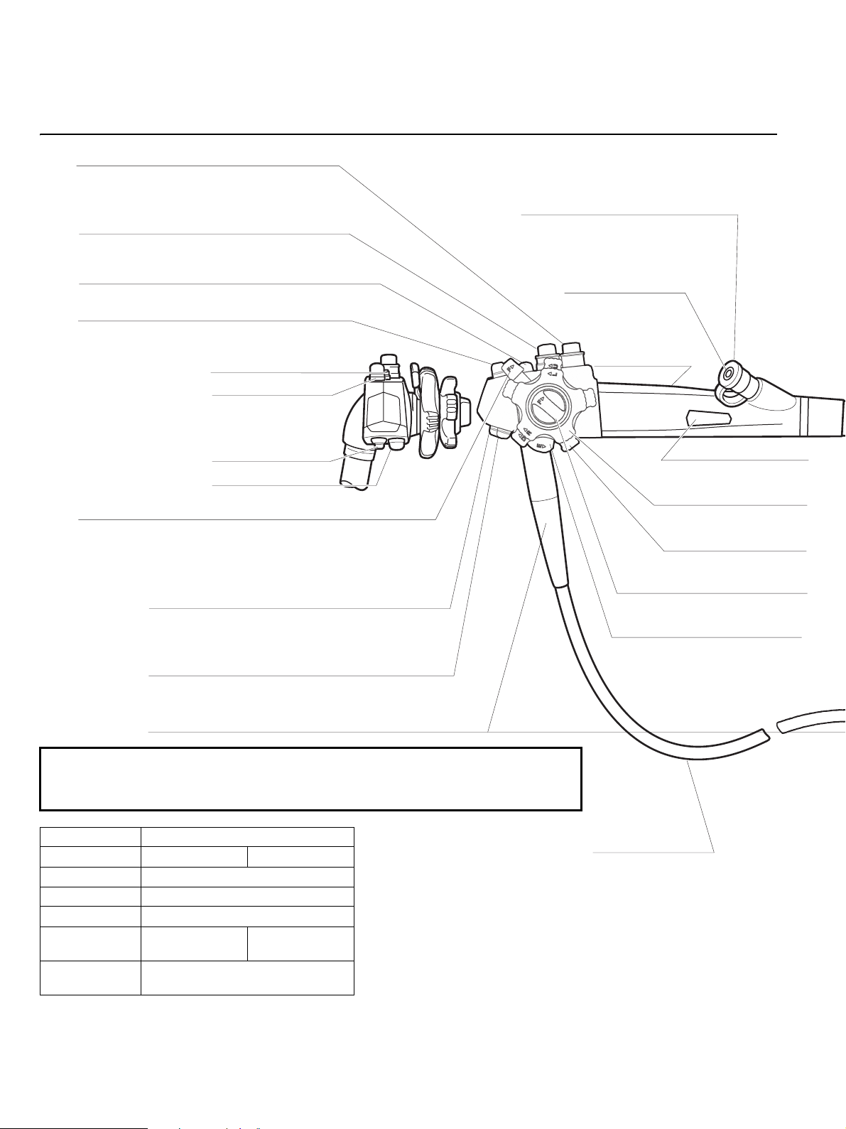
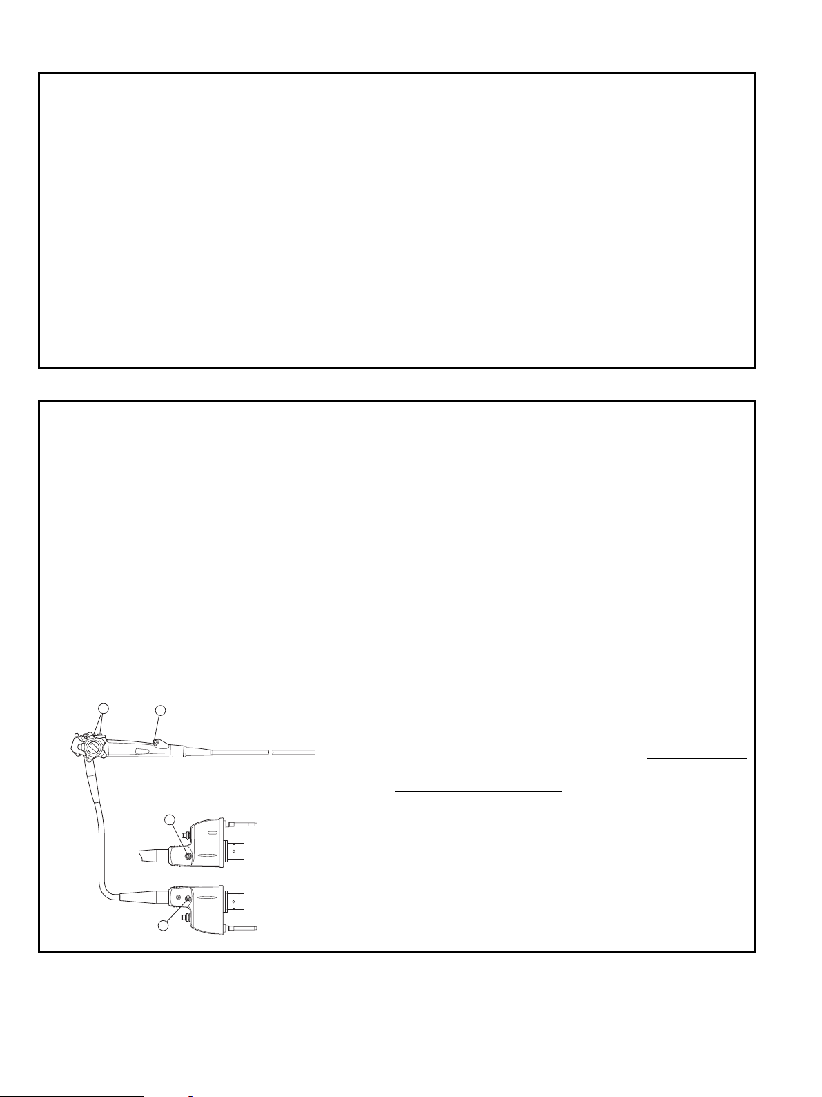
Intended Use / Indicationsfor use (Duodenoscopes)
The Video Duodenoscopes are intended to provide optical visualization of (via a video monitor), and therapeutic access to, Biliary Tract via
the Upper Gastrointestinal Tract. This anatomy includes, but is not restricted to, the organs, tissues, and subsystems: Esophagus, Stomach,
Duodenum, Common Bile, Hepatic and Cystic Ducts.
These instruments are introduced via the mouth when indications consistent with the need for the procedure are observed in adult and
pediatric patient populations.
Never use these endoscopes for any purpose other than that for which they have been designed.
The ED-3490TK can only be used with PENTAX Video Processors, Model EPK-i or EPK-1000.
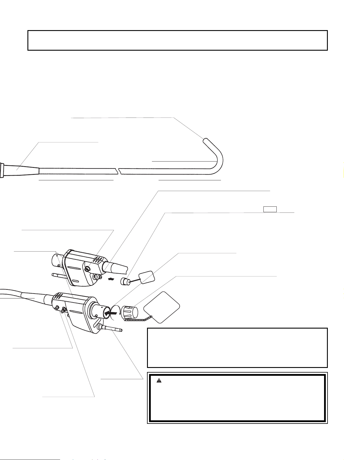
Notes
Read this manual before operating, and save this book for future reference. Failure to read and thoroughly understand the information
presented in this manual, as well as those developed for ancillary endoscopic equipment and accessories, may result in serious injury
including infection by cross contamination to the patient and/or user. Furthermore, failure to follow the instructions in this manual or the
companion Instructions for Use (Reprocessing) may result in damage to, and/or malfunction of, the equipment.
It is the responsibility of each medical facility to ensure that only well educated and appropriately trained personnel, who are competent and
knowledgeable about the endoscopic equipment, antimicrobial agents/processes and hospital infection control protocol be involved in the
use and the reprocessing of these medical devices. Known risks and/or potential injuries associated with flexible endoscopic procedures
include, but are not limited to, the following: perforation, infection, hemorrhage, burns and electric shock.
This manual describes the recommended procedures for inspecting and preparing the equipment prior to its use.
It does not describe how an actual procedure is to be performed, nor does it attempt to teach the beginner the proper technique or any
medical aspects regarding the use of the equipment. For the cleaning and maintenance after its use, please refer to the separate
“Instructions for Use (Reprocessing)”.
The text contained in this manual is common for various types/models of PENTAX endoscopes and users must carefully follow only those
sections and instructions pertaining to the specific instrument models appearing on the front cover.
If you have any questions regarding any of the information in this manual or concerns pertaining to the safety and/or use of this equipment,
please contact your local PENTAX representative.
Sterility Statement
The instruments identified in this manual are reusable semi-critical medical devices. Since they are packaged non-sterile, they must be high-
level disinfected or sterilized BEFORE initial use. Prior to each subsequent procedure, they must be subjected to an appropriate cleaning
and either high-level disinfection or sterilization process.
Refer to the companion PENTAX Instructions for Use (Reprocessing) describing in detail the recommended instructions on the care,
cleaning, disinfection and sterilization of these endoscopes.
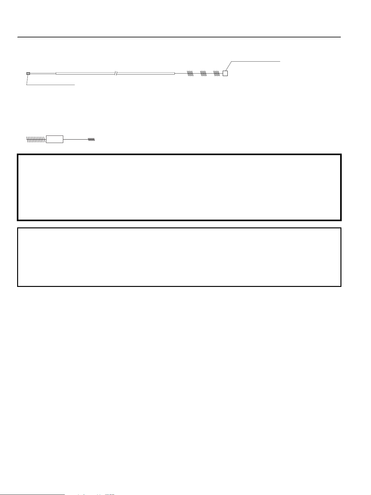
Conventions
Throughout this manual, the following conventions will be used to indicate a potentially hazardous situation which, if not avoided;
: could result in death or serious injury.
: may result in minor or moderate injury or property-damage.
: may result in property-damage. Also, advises owner/operator about important information on the use of this equipment.
Prescription Statement
Federal (U.S.A) law restricts this device to sale by or on the order of a physician or other appropriately licensed medical professional
The CE marking assures that this product complies with the requirements of the EC directive for safety.
Das CE Zeichen garantiert, daß dieses Produkt die in der EU erforderlichen Sicherheitsbestimmungen erfüllt.
Le logo CE certifie que ce produit est conforme aux normes de sécurité prévues par la Communauté Européenne.
II marchio CE assicura che questo prodotto è conforme alle direttive CE relative alla sicurezza.
La marca CE asegura que este producto cumple todas las directivas de seguridad de la CE.
WARNING
CAUTION
NOTE

TABLE OF CONTENTS
1. NOMENCLATURE AND FUNCTION ....................................................................................... 1
1-1. VIDEO ENDOSCOPE ..................................................................................................................... 2
1-2. ACCESSORIES................................................................................................................................ 3
1-3. VIDEO PROCESSOR ...................................................................................................................... 4
2. PREPARATION AND INSPECTION FOR USE ....................................................................... 5
2-1. INSPECTION OF THE VIDEO PROCESSOR............................................................................... 5
2-2. INSPECTION OF ENDOSCOPE .................................................................................................... 6
2-3. PREPARATION JUST BEFORE INSERTION OF ENDOSCOPE................................................ 15
3. DIRECTIONS FOR USE........................................................................................................... 16
3-1. PRETREATMENT........................................................................................................................... 16
3-2. INSERTION AND WITHDRAWAL............................................................................................... 16
3-3. BIOPSY ............................................................................................................................................ 18
3-4. CHOLANGIOPANCREATOGRAPHY (ERCP)............................................................................ 19
3-5. BILIARY DRAINAGE (ERBD)..................................................................................................... 19
3-6. ELECTRO-SURGERY .................................................................................................................... 20
4. CARE AFTER USE................................................................................................................... 22
SPECIFICATIONS

– 1 –
BUTTON 1
Push to freeze an image.
BUTTON 2
Push to activate the hardcopy system that
was selected between “FILE”and “HARD COPY”.
MODEL DESIGNATION
SUCTION CONTROL VALVE (OF-B120)
Depress to remove fluidsor air through
the instrument channel.
AIR/WATER FEEDING VALVE (OF-B188)
Covering of hole in the top of the valve delivers
pressurized air. Covering of the hole and fully
depressing the valve deliverspressurized water.
UP/DOWN DEFLECTION LOCK
When thislever isin the “F”position, turned clockwise,
the bending section movesfreely. When turned fully counterclockwise,
the bending section becomesprogressively more stabilized.
BUTTON 3
Push to activate the VCR for recording live procedures.
RUBBER STRAIN RELIEF
RUBBER INLET SEAL (OF-B190)
Allowspassage of accessorieswhile
preventing escape of fluidsand air.
CONTROL BODY
INSTRUMENT
CHANNEL INLET
For introduction of
biopsy forceps
and other accessories.
RIGHT/LEFT DEFLECTION
CONTROL KNOB
UP/DOWN DEFLECTION
CONTROL KNOB
RIGHT/LEFT DEFLECTION LOCK
Functionssimilar to Up/Down lock
CANNULA/FORCEPSELEVATOR
CONTROL KNOB
To g uide and direct cannulaor
forceps.
UMBILICAL CABLE
BUTTON 1
BUTTON 2
BUTTON 4
BUTTON 3
BUTTON 4
Push to select light measurement method, AVE/PEAK.
1. NOMENCLATURE AND FUNCTION
1.1 VIDEO ENDOSCOPE
*Setting at factory
NOTE:
The function of each button can be changed. For more details, refer to the
manual supplied with the EPK-i or the EPK-1000.
Scope ED-3490TK
Processor EPK-i EPK-1000
*Button 1 Freeze
*Button 2 Hard Copy
*Button 3 VCR (NTSC)/Enhance (PAL)
*Button 4 Enhance Enhance (NTSC)
AVE/PEAK (PAL)
Magnification
Control Lever
–

– 2 –
LIGHT GUIDE
Transmitslight from
light source to distal
end of endoscope.
SUCTION NIPPLE
For attachment to
external suction
source.
PVE CONNECTOR
Can be rotated
within a180
range.
AIR/WATER PORT
To connect feeding tube from water
bottle assembly.
DISTAL END
(Refer to the inside rear cover of thismanual)
BENDING SECTION
ETO GASSTERILIZATION VENTING CAP OF-C5
Providesventing of endoscope interior to equalize
internal and external pressures. Thiscap must be
removed before immersion.
NOTE: See important separate section regarding the use
of this cap!
VENTING CONNECTOR
Accepts“RED”ETO GASSterilization Venting cap.
Also acceptsLeakage Tester.
SOAKING CAP
OE-C9
ELECTRICAL CONTACTS
INSERTION TUBE
RED
Thiscap must be securely attached before
immersion. Align the black arrow on the
soaking cap with the green dot at the base
of the silver collar surrounding the
electrical contactson the PENTAX PVE
connector. Press the cap down onto the
metal collar and turn clockwise to secure.
FEED BACK TERMINAL
RUBBER STRAIN RELIEF
To connect the OL-Z3cable
From the PENTAX video
processor, model EPK-i/1000.
See detail information on
page 21.
NOTE:
To avoid damaging the endoscope, do NOT twist, rotate or bend excessively any of the rubber strain reliefs.
NOTE:
Ensure that the soaking cap has been securely attached (by
properly rotating it) to prevent the cap from coming off during
reprocessing. Failure to securely attach the soaking cap can
result in scope damage.
WARNING:
Immediately after use, the metal light guide prong and the
electrical contacts/pinsof the endoscope may be HOT. To
avoid burns, do not touch these areas immediately after use.
For safer handling after aprocedure, grasp the PVE
connector housing.

– 3 –
1-2. ACCESSORIES
1) Cleaning Brush for Suction System (Instrument Channel, Suction Tube)
2) Cleaning brush for recessed areas, scope tip, valve/selector cylinders and channel ports
(including A/W, Suction, Forward Water Jet, surrounding the elevator mechanism, etc.)
CAUTION:
•Because of the effect that accessoriesused through the instrument channel of the endoscope can have on the
performance of the endoscope itself, it isstrongly recommended that PENTAX accessoriesbe used with
PENTAX endocopes. If aunique or highly specialized accessory isavailable from another source, the
accessory manufacturer should be consulted to confirm compatibility with PENTAX endoscopesbefore use.
•Maximum outer diameter of an endoscopic accessory instrument must be at least 0.2 mm less than the
specified instrument channel diameter in PENTAX endoscopes. Working length of an endoscopic accessory
instrument may be approximately 30 cm longer than the endoscope working length.
NOTE:
• Depending upon country and/or local PENTAX distributor, each PENTAX endoscopic accessory may be an
optional accessory.
•Forpatient contact endoscopic accessories, follow the specific and detailed instructionson use, care and
maintenance supplied with each product.
• To confirm the exact condition of any new accessory device, check the labeling/packaging accompanying the
product. Each label/package should clearly identify the contentsas either sterile or non-sterile.
BACK END (BLUE)
Proximal End (White)
This brush is provided non-sterile for one time use. Never reuse this disposable brush on more than one instrument.
(CS6021T)
( CS-C9S )

– 4 –
1-3. VIDEO PROCESSOR
EPK-i
EPK-1000
WARNING:
The lifetime of the lamp in EPK-i processor
is500 hours. Prior to use, check the lamp life
meter on the rear panel to ensure the lamp
life isless than 500 hours. After 500 hoursof
use, the image quality will deteriorate.
Excessive use of the lamp beyond 500 hours
could break the lamp inside the equipment
resulting in damage to the equipment.
WARNING:
The lifetime of the lamp in EPK-1000
processor is400 hours. Prior to use, check
the lamp life meter on the front panel to
ensure the lamp life isless than 400 hours.
After 400 hoursof use, the image quality will
deteriorate. Excessive use of the lamp
beyond 400 hourscould break the lamp
inside the equipment resulting in damage to
the equipment.
NOTE:
The lamp life could be affected by frequency of use. In which case, the lamp life might become shorter than its
respective rated hours(EPK-i: 500 hours, EPK-1000: 400 hours)
CAUTION:
Please refer to the instruction supplied with the processor.
NOTE:
Use an air/water delivery system that iscompatible with PENTAX endoscopesand video processors, and ensure
that the cap assembly issecurely fitted to the corresponding water bottle with matched model. Failure to properly
secure the cap assembly to the water bottle may result in insufficient flowsof air and water during the endoscopic
procedure.
NOTE:
Software update may be required depending on the software version of the EPK-i processor. If the software isnot
updated, the image will not be displayed. If the imagesare not displayed correctly, please contact your local
PENTAX service facility.
WATER BOTTLE
LAMP SWITCH
AIR PUMP SWITCH
LCD
POWER SWITCH INTERFACE SOCKET
EPK-1000
POWER SWITCH
I : ON
O: OFF
LAMP SWITCH
INTERFACE
SOCKET
BRIGHTNESS
CONTROLSAIR PUMP SWITCH
WATER BOTTLE

– 5 –
2. PREPARATION AND INSPECTION FOR USE
Prior to use, the endoscope, video processor and endoscopic accessory instruments must be carefully inspected for cleanliness and
proper function to determine that they are appropriate for patient use:
2-1. INSPECTION OF THE VIDEO PROCESSOR
Please refer to the Owner’s Manual of the specific model of PENTAX video processor for complete instructions.
1) Mount the water bottle assembly within reach of the air pump fitting on the video processor and the air/water port on the PVE
connector of the endoscope.
6) Connect the air/water feeding tube from the water bottle assembly to the air/water port on the side of the PVE connector.
7) Turn the processor and air pump to the “ON” position and check for proper functioning.
8) Press the lamp switch of the processor to turn ON the lamp.
9) Prior to each procedure, check the endoscope image quality displayed on the monitor. Confirm that the image quality, color,
automatic brightness (iris) functions are acceptable as per the instructions provided with the PENTAX video processor.
NOTE:
PENTAX 90K seriesvideo endoscopescontained in thismanual are only compatible with PENTAX video
processor model EPK-i or EPK-1000.
CAUTION:
To avoid discontinuation of the endoscopic procedure, have an extra(spare) instrument available as astandby
device. Should any unforseen event or circumstance render the original instrument inoperable and/or unsafe for
patient.
WARNING:
The addition of defoaming agentsto the water supply isNOT recommended. Due to their nature, these silicone
based agentscling tenaciously to surfaces. Unless they are rinsed very thoroughly, a“barrier” could be created
which could reduce the effectiveness of the disinfection/sterilization process. Additionally, repeated use of such
defoamerscould eventually lead to residual silicone build up resulting in equipment malfunction such as clogged
air and/or water channels.
2) Plug the processor into a properly grounded receptacle with
the power switch in the OFF position.
3) Make sure that the PENTAX PVE connector is aligned with
the interface socket on the front panel of the processor.
4) Connect the endoscope to the interface socket on the
processor as illustrated.
5) Rotate the lever of the interface socket clockwise after
insertion.
CAUTION:
After connecting the endoscope to the EPK-i video
processor, alwaysmake sure that the endoscope is
firmly secured to the scope receptacle by turning the
locking lever to the “lock” position.
air/water feeding tube

– 6 –
2-2. INSPECTION OF ENDOSCOPE
Before reprocessing and/or immersion in any fluids, PENTAX endoscopes should be tested for the loss of integrity in their
watertight construction by using PENTAX brand leak testers. For specific details on PENTAX recommended leak detection
procedures, please refer to the instructions supplied with PENTAX leak testers.
1) Inspection of the Insertion Tube
a) Check the entire surface of the insertion tube for abnormal conditions such as dents, crush marks, wrinkles, bumps, buckles,
excessive bending, protrusions, bite marks, peeling of outer sheath, cuts/holes or other irregularities. Any crush or indentation
of the flexible shaft of the endoscopes can cause damage to the internal mechanisms of the endoscopes.
b) Similarly, check the condition of the umbilical cable for outward signs of damage such as buckling, crush marks, etc.
c) Make sure that the entire endoscope is clean and has been subjected to either a high-level disinfection or sterilization process
before each patient use.
CAUTION:
If the endoscope isintended to be clinically used after testing of individual scope functions(suction, air/water
delivery, water jet, etc.) without further reprocessing, the following precaution should be exercised.
Use sterile water during individual scope function teststo avoid recontamination of the previously reprocessed
instrument by waterborne microorganisms. Tap water, especially that which may be left idle and uncovered for a
prolonged period of time, should not be used during any inspection/testing of the endoscope.
CAUTION:
Various typesof endoscope leakage testersexist including manual, electro-mechanical and “automated”
versions, some of which are stand alone unitsand otherswhich may be integrated into Automated Endoscope
Reprocessors(AERs)/Washer-Disinfectors(WDs). It must be recognized that PENTAX doesnot
evaluate non-PENTAX leak tester systemsto satisfy their specific productsclaims, for their effectiveness
to accurately detect leaksand/or for their compatibility with PENTAX endoscopes. Insufficient pressures
may adversely affect the endoscope, especially if pressurization occursduring automated reprocessing
at elevated temperatures. PENTAX acceptsno responsibility for use of non-PENTAX leakage testers.
Usersshould check with the leakage testermanufacturer and confirm their specific product claims, including
compatibility with PENTAX endoscopesat various temperaturesand their ability to detect leakswith/
without fluid immersion and with/without flexing of the scope’sdistal bending section.
CAUTION:
To avoid further damage to the endoscope or the possibility of malfunction during aprocedure, do not use any
endoscope with any abnormalitiesor outward signsof damage.
WARNING:
All instrumentsmust be reprocessed prior to first time use, after any repairs/service and before every patient
use. When utilizing chemo-thermal processesfor reprocessing PENTAX endoscopes, the instrumentsshould be
allowed to return to room temperature prior to use and/or further handling.

– 7 –
NOTE:
• The distal end of the endoscope as well as the electrical contacts/pinson the PVE connector must be protected
against damage from impact. Never apply excess force such as twisting, or severe bending to the flexible
portion of the endoscope.
•Asindicated elsewhere in PENTAX product labeling, endoscopesparticularly the quality of the endoscopic
image should be checked prior to patient use.
•During pre-use inspection, ensure that the distal objective lensand the illumination (LCB) cover glass are clean
and no residuesare present on these distal surfaces. If not, crisp imagescan NOT be displayed. Wipe them
with agauze or the like moistened with amild detergent solution (e.g. enzymatic detergent or cleaning solution
specially formulated to clean endoscopes).
•Ideally all patientsshould be prepped well to maximize visualization of the intended areas of interest. Patient
material and secretionsshould be removed from the areaof observation to eliminate the potential to blur the
endoscopic image and/or obscure the illumination system.
• Prior to aprocedure, remove debrisor secretafrom observation areaas much as possible.
•Continuing use of the light guide with sticky debrismight cause steam because debrisisdeprived of moisture
by heat. If steam isfound on the light guide during aprocedure, stop it immediately and withdraw the scope
carefully from apatient.
NOTE:
Flexible endoscopesand other sophisticated medical instrumentsare constructed of special materials, unique
partsand intricate componentswith strict dimensional tolerances. Specialized assembly techniquesand
application of specific sealantsand/or adhesivesare required to ensure the watertight integrity and maintain the
functionality of these devices. It istherefore imperative that endoscopesbe routinely checked to ensure that parts
used in their construction are not loose, missing or compromised that could otherwise negatively affect the
functionality of these devices. Compromised or loose componentscould result in device failure, scope damage
(viafluid invasion) and/or in incomplete decontamination of used instruments.
PENTAX recommendsthat prior to use endoscopesshould be carefully inspected for their integrity and checked
for any “looseness” in the mating or joining of componentsincluding the following parts/areas:
•thechannel inlet assembly (biopsy inlet port) (①)
•thesuction nipple/connector (②)
•theair/water inlet port (③)
•any valve cylinder (④)
•basically, any inlet or outlet port associated with an internal channel, an indirect patient contact portion of the
endoscope
•rubber strain relief along insertion tube and umbilical cable (rotate clockwise only to tighten)
One method to check for looseness isto lightly grip the
exposed part, and while grasping the component carefully
attempt to move it in various directions. Use of alintfree
gauze while grasping metal partsisrecommended as a
protection for one’sfingers.
If any part/component remains loose (after attempting
to tighten) and/or if there is any indication or
suspicion of an abnormality or outward signs of
damage, do NOT use the endoscope. Contact your
local PENTAX service facility.
U
L
R
D
F
F
41
3
2

– 8 –
CAUTION:
To avoid damaging the endoscopes, do NOT twist, rotate or bend excessively any of the rubber strain relief
(
①
,
②
)during inspection, clinical use, reprocessing or any handling activity. Be particularly cautious for the insertion
tube strain relief (
①
). When wiping the insertion tube and the umbilical cable, use aslow back and forth motion to
wipe them along the tube/cable. Never apply excessive force or torque on these strain reliefsor slim tubes/cables.
During ANY handling of the instrument avoid excess force, twisting, rotation and/or bending of the actual insertion
tubesand umbilical cablesto prevent inadvertent damage (crush, compression, deformity, etc.) to these partsas
well as to internal componentscontained within the endoscope.
2) Inspection of Deflection Controls and Locks
a) Slowly manipulate the Up/Down and the Right/Left
control knobs to see that they function smoothly. Be
certain that a full and appropriate range of deflection is
possible.
b) Engage fully the deflection locks to be certain that the
position of the deflected tip can be stabilized.
CAUTION:
ANY lack of smooth operation of the deflection controlsmay be an early indication of internal damage to and/or
part(s) failure within the endoscope’sangulation system. To avoid the possibility of further endoscope damage or
the potential for malfunction of the angulation system, do NOT use the endoscope if the angulation mechanism
doesnot operate properly.
Prior to use ensure that the deflection controlscan rotate smoothly, that there isno grinding or excess friction
within the angulation system and that the distal bending section bendsfreely and smoothly. NEVER APPLY
EXCESSIVE FORCE TO THE DEFLECTION CONTROLS!
When an endoscope exhibitsexcessive “knob play” or if angulation islost in any direction, do NOT use the
instrument. Excessive “knob play” can be defined as rotating of the angulation control knob(s) in any one direction
for more than 30 degreeswithout any corresponding distal tip deflection. The examplesabove are indicationsthat
service isrequired to avoid more serious problemswith the angulation control system, including angle or pulley
cable/wire breakage and/or the possibility of a“frozen” distal bending section.
A “frozen” bending section can make instrument extraction from apatient more difficult.
Tw i sting or Rotating Bending
U
L
R
D
F
F
U
L
R
D
F
F
U
L
R
D
F
F
RIGHT-LEFT
UP-DOWN
F
U
L
R
D
F

– 9 –
a) Install the valve into the A/W cylinder by gently pushing the valve into the cylinder. Never apply excessive force to push
the valve into the A/W cylinder.
b) Connect the scope to the video processor. Turn air pump “ON” to desired pressure setting. Place the scope distal tip into
sterile water and confirm that no air bubbles exit the distal air nozzle.
3) Prior to use, the air/water feeding valve (OF-B188) should be
inspected. Remove the air/water valve from the control body and
ensure that rubber O-rings (OF-B192) in good condition are
properly attached.
CAUTION:
Since the rubber check-valve can NOT be replaced by
the end user, replace the entire A/W valve with anew
one if the check valve isdamaged/ missing.
WARNING:
Worn or damaged valves(O-ringsin particular) should be replaced with new oneswhich have already been
subjected to ahigh-level disinfection or sterilization procedure (O-ring set, model OF-B192, isoptionally
available). Failure to do so could create arisk of cross contamination to the end usersdue to the potential for
reflux or spit-back of patient fluidsout of the air/water valve. It could also create continuous air flow or excessive
air insufflation and result in the potential patient injury such as pneumatic perforation.
WARNING:
If air bubblesare observed during the test, the valve MUST BE REPLACED. Repeat the test procedure with a
new valve (OF-B188).
O-Ring
Check Valve
Cap
OF-B188
O-Ring SET
OF-B192

– 10 –
f) If air and/or water do not flow properly, NEVER attempt to clear the air or water nozzles with a needle or any other sharp
object. Instead, the following steps should be followed.
①Disconnect the endoscope from the video processor.
②Remove the suction control valve and the air/water feeding valve.
③Using a cotton tipped applicator and alcohol, clean the valve recess (receptacle) in the control body thoroughly to remove
any debris. Do NOT attempt to insert the applicator into the small openings within the valve receptacle as the cotton or
applicator could become lodged within these openings and cause channel blockage.
④• Following the companion Instructions for Use (Reprocessing) for chemical cleaning of the air and water channel with
detergent, flush detergent through both the air and water channels.
Rinse the air and water channel(s) with sterile water.
Then flush the air several times to force any residual solution out of the channel.
• Remove the adapters and install the air/water feeding valve.
(Alternate) By leaving the air/water valve in the cylinder instead of the OF-B153 adapter, one may direct pressured fluid
(or air) independently to either channel to expel debris from and/or more forcefully flush solution into either the air or
water channel. This should not be attempted on a completely clogged/blocked air or water channel/nozzle.
⑤Test for normal delivery of air and water. It may be necessary to repeat the above procedure if normal air and water delivery
is still not available.
g) If the air/water valve does not function properly, does not move smoothly or feels “sticky”, remove the valve and apply a
very small amount of silicone oil lubricant onto all the O-rings. Do NOT use excess oil, avoid “blobs”, large drops and/or
squirts of oil directly onto the metal valve stems - instead, simply place a small droplet of oil on one’s sterile gloved
forefinger and gently swirl between thumb and forefinger. Next place the valve with O-ring in-between thumb and finger
and gently rotate the valve so that the oil is evenly applied to the outer edges of each rubber O-ring. Make sure the oil is
applied to all O-rings and wipe off all excess.
Do NOT apply excess oil. Doing so can allow for inadvertent migration of the oil inside channels or other areas not intended
to be lubricated.
c) To inspect air delivery, cover the hole at the top of the air/
water valve and confirm that air flows freely from the air/
water nozzle at the scope distal tip.
d) By depressing the air/water feeding valve, the water
delivery system is activated. Water should flow in a steady
stream from the air/water nozzle at the distal tip of the
endoscope. (This may take several seconds on the initial
attempt.) USE STERILE WATER ONLY.
e) Release the air/water valve to determine if the valve freely
returns to its OFF (neutral) position and delivery of water
(and air) ceases.
NOTE: If blockage of the line isencountered, avoid use of excessive pressure to prevent scope damage.
NOTE: Do NOT apply excessive force in an attempt to unblock aclogged channel as the channel and/or brush could become
damaged. Whenever channel damage issuspected, the endoscope should be leak tested.
If repeated attemptsto flush the air/water system are unsuccessful, do not attempt to use the endoscope on apatient. Contact
the PENTAX service facility.
NOTE: Prior to clinical use, it isimportant that the entire air channel system be dry. Failure to thoroughly dry the air system could
result in an unclear or blurry image caused by very fine dropletsof moisture being swept over and/or onto the objective lens
at the distal end of the scope.
Action
Result Air
Feeding
Water
Feeding
–

– 11 –
4) Inspection of Elevator
a) Make sure that the entire surfaces of the elevator
(mechanism and surrounding area) are clean and have been
properly reprocessed.
b) This is the control that will guide and direct either the
biopsy forceps or other accessory during a procedure. To
inspect, push elevator control knob forward with thumb of
the left hand. The elevator in the distal tip should elevate
in proportion to the distance the control knob is moved.
The motion of the elevator and the knob should be smooth
and easy without any “play” involved.
5) Inspection of Suction Mechanism
a) Prior to use, the suction control valve (OF-B120) should
be inspected. For easier identification, a orange colored
indicator is placed on top of the OF-B120 valve
mechanism. Remove the valve from the control body and
make sure that rubber portions of the valve are not
damaged or worn.
WARNING:
A worn or damaged valve and/or O-ring should be replaced with anew one. The entire valve mechanism should be
subjected to ahigh-level disinfection or sterilization procedure prior to use (O-ring set, model OF-B127, isoptionally
available). Failure to do so could result in continuous aspiration which in certain clinical situationscan suction tissue
into the distal channel opening at the scope tip and/or create aloss of insufflated air viathe suction system.
A compromised valve could also result in the potential for reflux or spit-back of patient fluidsthat may present
infection risks.
U
L
R
D
F
F
OF-B120

– 12 –
b) Position the valve OF-B120 so that the small metal tab
near the base on the valve stem aligns with the notched
suction valve cylinder, also color coded in orange.
Install the valve into the suction cylinder by gently
pushing the valve into the cylinder. Never apply excessive
force to push the valve into the suction cylinder.
WARNING:
Improperly installed valvesmay not function as
originally intended. Such valvesmay not return to
their neutral (released) positionsand/or they may
provide continuous suction. Continuous aspiration
can cause loss of air/fluid, difficulty in maintaining
proper insufflation and/or inadvertent suctioning of
tissue into the distal instrument channel opening.
c) Connect suction tubing from an external suction source to
the suction nipple located on the PVE Connector at the end
of the umbilical cable. Place the distal tip of the endoscope
in a basin of sterile water and depress the suction control
valve. Water should be rapidly aspirated into the suction
system collection container.
d) Release the suction control valve to determine if the valve
freely returns to its OFF position and the aspiration of
water ceases.
e) If the suction valve does not move smoothly or feels “sticky”,
remove the valve from the suction cylinder on the control
body of the endoscope. Apply a small amount of silicone oil
lubricant, OF-Z11, onto rubber part and the rubber O-ring.
Place a small droplet of oil on one’s sterile gloved
forefinger and gently swirl between thumb and forefinger.
Next place the valve with O-ring in-between thumb and
finger and gently rotate the valve so that the oil is evenly
applied to the outer edges of the rubber O-ring.
Remove/wipe off excess lubricant with a soft gauze. Do
not use excessive silicone oil.
CAUTION:
If the instrument isto be used immediately after the inspection, use only sterile water. To avoid recontamination of a
previously reprocessed endoscope, avoid use of idle/uncovered tap water during any inspections.
NOTE:
A rubber inlet seal in good condition must be on the instrument channel inlet to prevent the loss of suction and a
risk of cross contamination to the end user due to the potential for reflux (spit-back) of patient fluids. Worn seals
will result in leakage and should be replaced. To ensure maximum performance of these sealing mechanisms,
consider replacing the rubber inlet seal with anew fully reprocessed one for each procedure.
Suction Valve
OF-B120 (orange)
1
2
SUCTION
TUBE
DEPRESS
SUCTION
NIPPLE
F
U
L
R
D
F
F
OF-B120
Incorrect
Correct
metal
OF-B120
O-Ring
OF-B127
Silicone oil
OF-B120

– 13 –
d) Close and inspect the jaws of the forceps to make sure the cups are in proper alignment. If the forceps has a spike, the spike
must be completely straight and fully within the cups.
e) Any accessory should be slowly inserted through the instrument channel inlet with the endoscope in a straight position.
There should be no resistance encountered. If resistance is encountered, do not attempt to introduce the accessory further.
The instrument channel may be damaged and the endoscope should not be used. Contact the PENTAX Service Department.
6) Inspection of Biopsy Forceps and Instrument Channel
a) Make sure there are no kinks in the flexible shaft of the
biopsy forceps.
b) The jaws of the forceps must be free of any residual debris.
Any debris must be cleaned from the forceps before they
are used. USE ONLY STERILE FORCEPS.
c) The handle mechanism on the forceps should be operated
to open and close the jaws. This mechanism should
operate freely.
WARNING:
The use of any forcepsor accessory that showsany sign of damage or difficulty of operation must be avoided.
Any malfunction of aforcepsor accessory during apatient procedure could result in serious injury to the patient.
Also, the use of damaged forcepsor accessoriesmay result in serious and costly damage to the endoscope.
CAUTION:
Endoscopic accessory instruments(EAIs) may be used with PENTAX flexible endoscopes. It should be
understood that special care and caution must be exercised when using accessories, particularly non-
PENTAX productsthrough the instrument/suction channel of an endoscope. Thisisespecially true when
attempting to pass accessoriesthrough narrow channelswhen curved in atight bending radius.
Please note that damage to the endoscope and/or accessory instrument ispossible if excessive force is
applied during insertion (or withdrawal) of the EAI. To prevent equipment damage or device failure, please
adhere to the following precautions:
• Never apply too much pressure or excessive force during insertion through the instrument channel.
• Never attempt to force endoscopic accessories, such as biopsy forcepsthrough afully angulated distal bending
section.
• Prior to using accessoriesfrom another source (non-PENTAX products), contact the manufacturersof the
accessoriesto confirm if the device has been checked for compatibility.
Failure to follow these recommendationscan result in scope and/or accessory damage/failure, including but
not limited to:
• Channel puncture/leakage
• Fluid invasion
• Fiber breakage
• Other internal component failure
Should resistance be encountered when inserting an accessory, STOP!If resistance isat the scope tip, slightly
withdraw the accessory, reduce the angulation (within the distal bending section), then slowly and carefully
advance the accessory under direct vision.
Several factorscan affect the ease/difficulty of accessory passage through the endoscope channel:
• Outside diameter of accessory compared to inside channel diameter
• Non-flexible (rigid) portionsof an accessory
• The curve or bend (bending radius) within achannel through which the accessory will pass
• Damaged accessory
Due to the variablesabove, prior to each procedure, it isimportant to check the particular accessory intended
to be used to satisfy the clinical procedure to be performed. Such pre-use inspectionswill allow for
uninterrupted and more expeditious examinations.
To confirm the absence of severe channel damage affecting the watertight integrity of the endoscope,
perform appropriate leak testing of the scope per PENTAX instructions.
CLOSE
OPEN

– 14 –
WARNING:
All patient contact accessoriesmust be thoroughly cleaned and subjected to an appropriate high-level
disinfection or sterilization process before being used for the first time and subsequently after each clinical use.
CAUTION:
The instrument, A/W and the water jet channel systemsare made of stainless steel, poly phenylene oxide and fluorine-
contained polymers. When any fluidsare used with thisscope, please read carefully and follow all instructionsin the
manual supplied with the fluidsfor use and pay special attention to any reactionswith the materialsidentified in the
intended fluid path.
NOTE:
Accessoriesshould alwaysbe inspected and checked with the particular endoscope prior to each procedure.
WARNING:
Do NOT clinically use the endoscope if any irregularity or abnormality issuspected. If there isany doubt as to
the suitability of use for any endoscope component, replace it with anew fully reprocessed one. An instrument
irregularity may cause scope damage and/or compromise patient or user safety.

– 15 –
2-3. PREPARATION JUST BEFORE INSERTION OF ENDOSCOPE
1) If the endoscope has just recently been reprocessed, has been prepared or stored properly and passed all pre-procedure
inspections, the instrument should be ready to use. If necessary, the scope’s insertion tube may be wiped down with a gauze
dampened with 70-90% ethyl or isopropyl alcohol.
2) Gently clean the objective lens with a cotton-tip applicator moistened with 70-90% ethyl or isopropyl alcohol. A lens cleaner
(anti-fogging agent) may also be applied via gauze or other applicator.
3) Check the endoscopic image and confirm that it is of acceptable quality for clinical use. Refer also to the owner’s manual
supplied with the PENTAX video processor for inspection of the image quality.
4) Prior to trans-oral insertion of the endoscope, place a bite-block (mouthpiece) into the patient’s mouth to protect the endoscope
from damage during the procedure. Failure to do so can result in scratches, tears and/or crushing of the insertion portion of the
endoscope.
5) Apply a medical grade water soluble lubricant to the insertion tube. Do not use petroleum based lubricants.
WARNING:
Every endoscope should be properly disinfected or sterilized before being used for the first time. The endoscope
should have been properly cleaned and disinfected or sterilized after any previous use and after being returned
for any repairs/service.
Refer to the companion Instructionsfor Use (Reprocessing) describing in detail PENTAX reprocessing
instructions.
WARNING:
Current infection control guidelinesrequire that endoscopesand their patient contact accessorieseither be
sterilized or at the least be subjected to high-level disinfection. Accessorieswhich ENTER STERILE TISSUE or
THE VASCULAR SYSTEM must be sterilized before patient use. Only the user can determine if any instruments
and accessorieshave undergone appropriate infection control proceduresprior to each clinical use.
NOTE:
Contact the manufacturer and follow local regulationsregarding safe use, appropriate handling and disposal of
alcohol products. Material Safety DataSheets(Health and Safety DataSheetsor similar documentsdepending
upon country) available from the alcohol manufacturer should provide guidance to end usersabout composition,
hazards, chemical and physical properties, first aid, handling and storage, stability, precautions, disposal, etc.
associated with alcohol solution.
NOTE:
The objective lensmust be kept free of the lubricant and excess lenscleaner.
WARNING:
Never drop thisequipment or subject it to severe impact as it could compromise the functionality and/or safety
of the unit. Should thisequipment to be mishandled or dropped, do not use it. Return it to an authorized PENTAX
service facility for inspection or repair.

– 16 –
3. DIRECTIONS FOR USE
3-1. PRETREATMENT
1) The patient should be prepared in your normal endoscopy regimen.
3-2. INSERTION AND WITHDRAWAL
4) The deflection controls should be used as needed to position the scope. Deflection of the tip should be performed under direct
vision in a gentle and deliberate manner.
WARNING:
Thisinstrument should only be used by physicianswho have thoroughly studied all the characteristicsof this
instrument and who are familiar with the proper techniquesof endoscopy. During the procedure, alwayswear
protective garmentssuch as gloves, gowns, face masks, etc. to minimize the risk of cross contamination.
NOTE:
Do not use awater supply device that can exert 30kPaor greater of water pressure to the suction channel (suction
valve) during endoscopic examination.
1) Slowly insert the scope under direct vision.
2) When the distal end of the scope is passed through the
pharynx, the patient should be gently biting down on the bite
block to maintain the bite block’s position during the
procedure.
3) Adjust the intensity of the video processor to obtain a
brightness level suitable for observation.
WARNING:
The light emission from the endoscope could cause thermal injury. To minimize the risk, use only the minimum
amount of brightness and avoid close stationary viewing and unnecessary prolonged use.
CAUTION:
ANY lack of smooth operation of the deflection controlsmay be an early indication of internal damage to and/or
part(s) failure within the endoscope’sangulation system. To avoid the potential for malfunction of the angulation
system, do NOT use the endoscope if the angulation mechanism doesnot operate properly.
Ensure that the deflection controlscan rotate smoothly, that there isno grinding or excess friction within the
angulation system and that the distal bending section bendsfreely and smoothly.
NEVER APPLY EXCESSIVE FORCE TO THE DEFLECTION CONTROLS!
If during aprocedure angulation islost in any direction such as when “cablessnap” (broken pulley wire, broken
angle wire, etc.), do NOT continue to use the instrument and do NOT rotate the deflection controls. Should the
angulation system fail for any reason, stop the procedure, release the lock lever and carefully withdraw the
endoscope under direct visualization.
The examplesabove are indicationsthat service isrequired to avoid more serious problemswith the angulation
control system, including the possibility of a“frozen” distal bending section.
A “frozen” bending section can make instrument extraction from apatient more difficult.
BITE BLOCK
U
L
R
D
F
F

– 17 –
5) Insufflation shoud be controlled by the combined use of the air/water valve to increase the amount of insufflation and the suction
control to decrease the level of insufflation.
6) Procedures involving poorly prepped patients should be avoided as excessive patient material can negatively affect certain scope
channel functions as well as the ability to maintain a clear endoscopic view.
7) Mucous, fluids and/or other patient material should be aspirated via the instrument/suction channel and suction control valve to
improve visualization. Maintain a clear view during aspiration, avoid prolonged suction time and use the minimum level of
negative pressure required to perform the clinical procedure.
8) The objective lens may be cleaned during the procedure by alternately using the air/water and suction control valves.
9) Image capture, hard copy documentation, video recording, etc. may be carried out as necessary.
10)Before withdrawing the scope, trapped air should be suctioned to reduce patient discomfort.
11) When attempting to withdraw the scope, return the deflection locking levers to their free position. Always withdraw the scope
under direct visualization.
CAUTION:
Be careful not to deliver too much air.
WARNING:
It must be recognized that variationsin air flow (pressure and volume) for patient insufflation may exist from one
manufacturer’sequipment (light source, video processor and/or scope type) to another. It is, therefore, important
to closely monitor the patient at all timesand to aspirate excessive air to prevent overinsufflation and potential
pneumatic perforation.
WARNING:
Do not apply excessively negative pressures(high suction settings) and/or prolonged contact of the distal
instrument channel opening (scope tip) against mucosal surfacesto avoid “suction polyps”, bleeding and/or
other traumato the patient. During aspiration keep as clear as possible an endoscopic view of patient anatomy
and maintain some distance from scope tip to tissue to avoid suctioning of mucosa onto/into the distal channel
opening.
NOTE:
Should debrison the objective lensbe difficult to clean, one can temporarily use the HIGH air pressure setting
on the processor and simultaneously press the air/water and suction control valves. Return air pressure setting to
original selection before proceeding.
WARNING:
If for any reason, the image islost due to power
shortage, lamp or processor failure, etc. the
deflection locking leversshould be released, the
scope tip should be straightened to itsneutral
position, and the insertion tube should be carefully
and slowly withdrawn from the patient.
F
UP/DOWN DEFLECTION LOCK
FREE POSITION
(LOCK RELEASED)
To p spoke of angulation knobs in thisposition
correspondsto neutral distal tip orientation
LOCK POSITION
RIGHT/LEFT
DEFLECTION LOCK
FREE POSITION
(LOCK RELEASED)
LOCK POSITION
Table of contents
Other Pentax Medical Equipment manuals