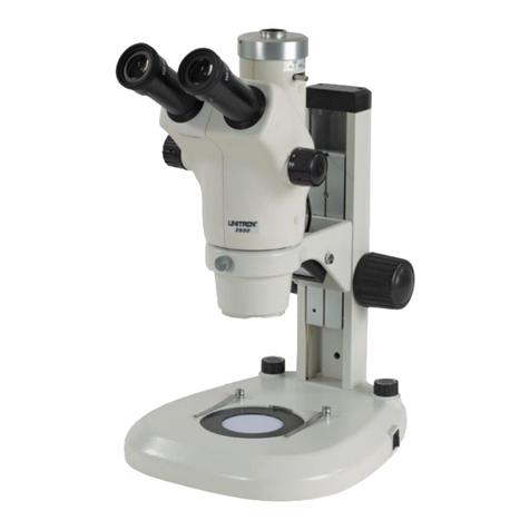
Unitron CFM Series User manual

73 Mall Drive, Commack, NY 11725 • 631-543-2000 (P) • 631-589-6975 (F)
www.unitronusa.com • info@unitronusa.com
CFM SERIES
COMPARISON FORENSIC
MICROSCOPE
MANUAL

COMPARISON FORENSIC MICROSCOPE
UNITRON® 73 Mall Drive, Commack, NY 11725 • 631-543-2000 • www.unitronusa.com 2
CONTENTS
SAFETY NOTES .................................................................................................................. 3
CARE AND MAINTENANCE ................................................................................................ 3
INTRODUCTION .................................................................................................................. 4
TECHNICAL PARAMETERS................................................................................................ 4
INSTRUMENT STRUCTURE .......................................................................................... 5-10
SET UP............................................................................................................................... 11
FUNCTION AND OPERATION ..................................................................................... 12-19
USING THE POLARIZER ........................................................................................ 12
LIGHT CONTROL PANEL ....................................................................................... 13
USING THE SEPARATION LINE ............................................................................ 14
ADJUSTING INTERPUPILLARY DISTANCE .......................................................... 15
ADJUSTING THE STAGE ....................................................................................... 15
ADJUSTING THE FOCUS ....................................................................................... 15
ADJUSING THE MAGNIFICATION ......................................................................... 16
CAMERA CENTRATION ......................................................................................... 16
USING THE BULLET HOLDER .......................................................................... 17-19
SELECTING PROPER MAGNIFICATION OF DIGITAL CAMERA .......................... 20
TROUBLESHOOTING........................................................................................................ 21
MAINTENANCE.................................................................................................................. 22
SERVICE............................................................................................................................ 22
WARRANTY ....................................................................................................................... 22

COMPARISON FORENSIC MICROSCOPE
UNITRON® 73 Mall Drive, Commack, NY 11725 • 631-543-2000 • www.unitronusa.com 3
SAFETY NOTES
1. Open the shipping carton carefully -- your microscope arrived packed in a molded shipping carton.
Do not discard the carton: the shipping carton should be retained for reshipment of your
microscope if needed.
2. Carefully remove the microscope from the shipping carton and place the microscope on a flat,
vibration-free surface.
3. Avoid placing the microscope in dusty surroundings, in high temperature or humid areas as mold and
mildew can form. Carefully remove the microscope from the shipping carton and place the
microscope on a flat, vibration-free surface.
4. Please check the complete microscope, spare parts and consumable parts according to the packing
list.
5. All electrical connectors (power cord) should be inserted into an electrical surge protector to prevent
damage due to voltage fluctuations.
NOTE: Always plug the microscope power cord into a suitable grounded electrical outlet. A
grounded 3-wire cord is provided.
CARE AND MAINTENANCE
1. Do not attempt to disassemble any component including eyepieces, objectives or the focusing
assembly.
2. Keep the instrument clean; remove dirt and debris regularly. Accumulated dirt on metal surfaces
should be cleaned with a damp cloth. More persistent dirt should be removed using a mild soap
solution. Do not use organic solvents for cleansing.
3. The outer surface of the optics should be inspected and cleaned periodically using an air bulb. If dirt
remains on the optical surface, use a soft, lint free cloth or cotton swab dampened with a lens
cleaning solution (available at camera stores). All optical lenses should be swabbed using a circular
motion. A small amount of absorbent cotton wound on the end of a tapered stick makes a useful tool
for cleaning recessed optical surfaces. Avoid using an excessive amount of solvent as this may
cause problems with optical coatings or cemented optics or the flowing solvent may pick up grease
making cleaning more difficult.
4. Store the instrument in a cool, dry environment. Cover the microscope with a dust cover when not in
use.
5. UNITRON®microscopes are precision instruments which require periodic servicing to maintain
proper performance and to compensate for normal wear. A regular schedule of preventative
maintenance by qualified service personnel is highly recommended. Your authorized UNITRON
distributor can arrange for this service.
COMPARISON FORENSIC MICROSCOPE
UNITRON® 73 Mall Drive, Commack, NY 11725 • 631-543-2000 • www.unitronusa.com 4
INTRODUCTION
Congratulations on the purchase of your new UNITRON microscope. UNITRON microscopes are
engineered and manufactured to the highest quality standards. Your microscope will last a lifetime if
used and maintained properly. UNITRON microscopes are carefully assembled, inspected and tested by
our staff of trained technicians in our New York facility. Careful quality control procedures ensure each
microscope is of the highest quality prior to shipment.
TECHNICAL PARAMETERS
Objective Magnification: 16205 & 16206:0.4x, 1.0x, 1.5x, 2.0x, 3.0x, 4.0x
Optional auxiliary lens: 2X
Eyepieces with Standard Binocular Head:CWF 10x/22mm (standard)
CWF 20x/13mm (optional)
CWF 16x/16mm (optional)
Working Distance: 152 mm
Adjustment of
Interpupillary Distance: 55-75mm
Stage:
•Two Universal Holders
•Two flat stages
•Tiltable
•25° gradient adjustment in different directions
•Combined operation for two stages: horizontal movement range -- 55mm;
elevation and subsidence 80mm
•Independent operation for two stages: horizontal movement range
of X and Y: 55mm x 55mm; elevation and subsidence -- 55mm
C- Mount Camera Adapter: Optional: 0.4x, 1.0x
Illumination: •Input voltage: 100V – 240V; Output voltage: 12V 5A
•2.5W circular LED light (42 LED ring)
•Gooseneck LED spotlight (White LED light)
Optional: UV, Green, Red LED Light
•Gooseneck fluorescent light
•Remote phosphor gooseneck light
•3W transmitted light (48 LED ring)
•Coaxial illuminator: high power, 1W LED

COMPARISON FORENSIC MICROSCOPE
UNITRON® 73 Mall Drive, Commack, NY 11725 • 631-543-2000 • www.unitronusa.com 7
CAT# 16206 Item Identification
1. Base
2. Light Control Panel
3. Height Adjustment Focus Knob
4. Lateral Adjustment Focus Knob
5. Socket for Gooseneck Fluorescent Light
6. Focus Knob
7. Transformer
8a. Gooseneck Fluorescent Light or Remote Phosphor Gooseneck Light
9. LED Ring Light
10. Set Screw
11. Set Screw
12. Bridge Body
13. Separation Line Adjusting Knob
14. C-mount Camera Adapter
15. Digital Camera
16. n/a
17. n/a
18. Eyepiece
19. Binocular Head
20. Tightening Set Screw
21. Separation Adjustment Screw
22. Magnification Adjustment Knob
23. Magnification Changer Knob
24. Locking Screw
25. Tightening Knob
26. Universal Holder Stage
27. Front and Back Adjustment Knob
28. Left and Right Adjustment Knob
29. Socket for LED Ring Light
30. Set Screw
31. Long Case Holder
32. Observe Knob Changer

COMPARISON FORENSIC MICROSCOPE
UNITRON® 73 Mall Drive, Commack, NY 11725 • 631-543-2000 • www.unitronusa.com 10
CAT# 16205 Item Identification
1. Base
2. Light Control Panel
3. Height Adjustment Focus Knob
4. Lateral Adjustment Focus Knob
5. Socket for Gooseneck LED Light
6. Focus Knob
7. Transformer
8. Gooseneck LED Light
9. LED Ring Light
10. Set Screw
11. Set Screw
12. Bridge Body
13. Separation Line Adjusting Knob
14. C-mount Camera Adapter
15. Digital Camera
16. n/a
17. n/a
18. Eyepiece
19. Binocular Head
20. Tightening Set Screw
21. Separation Adjustment Screw
22. Magnification Adjustment Knob
23. Magnification Changer Knob
24. Locking Screw
25. Tightening Knob
26. Universal Holder Stage
27. Front and Back Adjustment Knob
28. Left and Right Adjustment Knob
29. Socket for LED Ring Light
30. Set Screw
31. Long Case Holder
32. Observe Knob Changer

COMPARISON FORENSIC MICROSCOPE
UNITRON® 73 Mall Drive, Commack, NY 11725 • 631-543-2000 • www.unitronusa.com 11
SET UP
Please review INSTRUMENT STRUCTURE on pages (5-6) before attempting to set up the microscope.
MAIN BODY
1. Place the microscope on a suitable stable or
motorized worktable.
2. Remove the dust cap in the middle of bridge
and install the Binocular Head (19). Lock it
in with the Set Screw (20).
3. Install the Bridge Body (12) into arm of stand
and lock it with the Lock Screw (24).
4. Remove the dust caps on the Binocular
Head (19) and insert the Eyepieces (18) into
the tubes.
5. Insert the power cable of the transformer into
the Base (1), and plug the other end into a
grounded AC110V outlet.
ILLUMINATION
Ring Light
Set up the Ring Light (9) by plugging it into the Socket (29) and securing it by tightening the Set Screw
(10).
Gooseneck LED Light or Gooseneck Fluorescent Light or Remote Phosphor Gooseneck
Light
See Figure 1 and Figure 2, the Gooseneck Fluorescent Light (Figure 1, 8a) or the Gooseneck LED Light
(Figure 2, 8) is secured to Socket (5).

COMPARISON FORENSIC MICROSCOPE
UNITRON® 73 Mall Drive, Commack, NY 11725 • 631-543-2000 • www.unitronusa.com 12
FUNCTION AND OPERATION
ILLUMINATION
Using a Polarizer (Optional)
Using a Polarizer will eliminate scattered stray light and glare for better image quality.
Connect the Polarizer with a Spot Lamp or a Transmitted Lamp, then screw the Analyzer on.
Adjust the brightness and change the polarizing angle by rotating the Analyzer to get a polarizing effect.
Figure 3
Analyzer
Polarizer

COMPARISON FORENSIC MICROSCOPE
UNITRON® 73 Mall Drive, Commack, NY 11725 • 631-543-2000 • www.unitronusa.com 13
FUNCTION AND OPERATION (Continued)
ILLUMINATION (continued)
Light Control Panel -- (Figure 6)
The REFL and RING can control the light from all three-pin plugs such as Gooseneck LED Spot Light,
LED Ring Light, and Fluorescent Light.
Figure 6

COMPARISON FORENSIC MICROSCOPE
UNITRON® 73 Mall Drive, Commack, NY 11725 • 631-543-2000 • www.unitronusa.com 14
USING THE SEPARATION LINE
The separation line should be a thin, black and straight, as is shown in Figure 7-(c). Turning the
Separation Line Adjusting Knob (13) can move the comparison line continuously to have a single, cutting
or overlapping view field.
If the separation line appears as is shown in Figure 7-(a) or Figure 7-(b), this means the line has
changed out of shape and needs to be adjusted by the following steps:
1. Insert the screwdriver that comes with the microscope into the screw slit in the calibration hole
(21).
2. Observe the separation line through the eyepiece and slightly rotate the screwdriver until
separation line is in the shape as shown in Figure 7-(c).
a. If the separation line is like in Figure 7-(a), adjust the screw in the right hole.
b. If it is like the line in Figure 7-(b), adjust the screw in the left hole.
Figure 7

COMPARISON FORENSIC MICROSCOPE
UNITRON® 73 Mall Drive, Commack, NY 11725 • 631-543-2000 • www.unitronusa.com 15
FUNCTION AND OPERATION (Continued)
ADJUSTING INTERPUPILLARY DISTANCE
To adjust the interpupillary distance, hold the left
and right eyetubes while observing a specimen.
Rotate the eyetubes around the central axis until
the fields of view of both eyetubes coincide
completely. A complete circle should be seen in
the viewing field when viewing the specimen
slide. An improper adjustment will cause
operator fatigue and will disrupt the objective
parfocality.
Where “·” ①on the eyepiece tube lines up,
then that is the number for the interpupillary
distance. Range:55~75mm. (Fig. 8).
Remember your interpupillary for future
operation.
ADJUSTING THE STAGE
Use Knobs (27) and (28) to adjust the stage movement from front to back and left to right. The Stage
(26) can be rotated 360°. Move the Stage (26) to adjust it in different directions. The Lateral Adjustment
Focus Knob (4) can link the two stages to make the same movements.
ADJUSTING THE FOCUS
To ensure that you obtain sharp images with both eyes (since eyes vary especially for those wearing
glasses) any eyesight variation can be corrected in the following manner:
1. Set both diopter collars on the eyepieces to “0.”
2. Set the magnification on the microscope to 4.0x
3. Set the indicator line to be viewed on the right
side only.
4. Place the enclosed stage micrometer on the right
side stage.
5. Adjust the focus of the microscope to bring the
micrometer to its sharpest focus using your left
eye only to observe.
6. Rotate the diopter collar to obtain the sharpest
focus.
7. Now using your right eye only obtain the same
sharp focus by rotating the right diopter collar
until the sharpest image appears.
8. Repeat the above procedures by changing the
Indicator Line to view the specimen from the left
side only.
9. Repeat these procedures several times going
from the maximum to minimum magnification to
ensure you get a sharp image at all
magnifications.
Figure 8

COMPARISON FORENSIC MICROSCOPE
UNITRON® 73 Mall Drive, Commack, NY 11725 • 631-543-2000 • www.unitronusa.com 16
FUNCTION AND OPERATION (Continued)
ADJUSTING THE MAGNIFICATION
To get the highest quality image, set both the left and right objectives at the same magnification; rotate
the Magnification Adjustment Knob (25) to change the objective magnification; under the nominal
magnification ratio, the right-side magnification still needs to make fine adjustment. The steps of fine
adjustment on the magnification are below:
Separately place the stage micrometer on the left and right stage surface, observe the scales image by
the eyepiece, move the stage micrometer to keep the reticle scales matched; if the two objective
magnifications are not identical, all scales in the view field will not match. Rotate the Magnification
Adjustment Knob [Figure 1-(23)] in a clockwise or counter-clockwise direction. Using the fine focus
adjustment knob, refocus it until the image is clear and move the stage micrometer to overlap the scales.
Repeat the above procedures until the magnifications of both left and right objectives are identical.
Shown as Figure 6 the same adjustment should be done when the objective magnification changes.
CAMERA CENTRATION
Centering a digital camera that is connected to the microscope and observed with a monitor. The C-
Mount Adapter was pre-centered during final inspection so the monitor image coincides with the
eyepiece image.The following procedure is provided for reference.
1. Set the B/T selector (located on the back of the CFM Main Bridge) to the “T” position.
2. Set the Beam Selector Knob (located on the front of the Main Bridge) to the Left Fieldview
position (knob rotated fully CCW).
3. Place one of the Calibration Slides on the left stage. Note: Placing a piece of white paper under
the slide will increase contrast of the scale.
4. Select the 1.0x objective of the Magnification Changer.
5. Using the left Stage X/Y movement controls and left side Focus Knob, focus on the scale of
Calibration Slide.
6. Position the center of the Calibration scale (the numeral 5) in the center of the eyepiece field of
view (FOV). The 5 on the calibration Slide will from now on (in this procedure) be called the
Target.
7. Increase the magnification from 1.0x up to 4.0x. If the Target shift—remember the direction it
moved (See Fig. b).
8. Set the magnification back to 1.0x and move the Target in the opposite direction of the movement
observed in the previous step.
9. Repeat steps 6. & 7 until the Target does not move.
10. Next loosen the three set screws of C-Mount adapter and move the camera until the image of
target is in the center of the monitor FOV.
11. Sequentially tighten each of the 3 centering set screws so as to maintain the Target image in the
center of the monitor FOV.

COMPARISON FORENSIC MICROSCOPE
UNITRON® 73 Mall Drive, Commack, NY 11725 • 631-543-2000 • www.unitronusa.com 17
12. The center of the eyepieces FOV and the center of the monitor FOV now coincide with each other
through the CFM’s magnification range.
The schematic diagram is below:
USING THE BULLET HOLDER
1. Insert the spring-loaded bullet holder into
casing shell and turn to expand, using the
appropriate size. (Figure 9 and 12)
2. Install the Universal Holder base (6) onto the
Mechanical Stage and secure it using the two
screws (3) as shown in Figure 10.
Monitor
Use low magnification to move
target to center of monitor.
Target moves away
from center.
Adjust the C-Mount adapter until
the target is at the center and
image remains centered when
changing magnifications/zooming.
Move target in opposite direction at low
magnification. Repeat movements until
the target remains at the same point
when changing magnifications/zooming.
Magnification from
low to high
Magnification from
high to low
Figure 9
Universal Holder
Mechanical Stage
Figure 10

COMPARISON FORENSIC MICROSCOPE
UNITRON® 73 Mall Drive, Commack, NY 11725 • 631-543-2000 • www.unitronusa.com 18
FUNCTION AND OPERATION (Continued)
USING THE BULLET HOLDER (Continued)
3. Viewing Samples (Figures 11 & 12)
To view trace on the bottom of the bullet
shell, thread the wire brush holder with
bullet shell into the base in an upright
position (Figure 11).
To view trace on the side of the bullet
shell, put (25) on the end of the bullet shell
and secure its position by locking it in place
with (29) (Figure 11).
To inspect a sample with a large
diameter, unscrew (11) and remove (30)
(Figure 12).
Figure 12
Figure 11
Direction 1
Direction 2
Hole for the bullet holder

COMPARISON FORENSIC MICROSCOPE
UNITRON® 73 Mall Drive, Commack, NY 11725 • 631-543-2000 • www.unitronusa.com 19
FUNCTION AND OPERATION (Continued)
USING THE BULLET HOLDER (Continued)
4. Positioning Samples (Figures 13, 14, 15)
Rotate the base of the Universal Holder by
loosening the locking screw (4a) (Figure 14).
To move the bullet into a horizontal or
inclined setting, loosen the locking screw
(4a) and slide the bullet holder along the
groove. Secure into place by tightening the
locking screw.
To adjust the big diameter sample, unscrew
(9) and move (8) (Figure 12) forwards or
backwards until you obtain a suitable
position.
11
29
4b
14
20
25
11
29
4b
14
20
25
4a
9
9
8
Figure 13
Figure 14
Figure 15

COMPARISON FORENSIC MICROSCOPE
UNITRON® 73 Mall Drive, Commack, NY 11725 • 631-543-2000 • www.unitronusa.com 20
FUNCTION AND OPERATION (Continued)
SELECTING PROPER MAGNIFICATION
OF DIGITAL CAMERA
Formulas for Magnification Calculation
Total Magnification = magnification of body x Magnification of digital camera x Digital magnification
(x magnification of optional auxiliary lens)
Diameter of Object view field = length of digital camera sensor target surface diagonal line/magnification
of objective/magnification of digital camera/ (x magnification of optional auxiliary lens)
Sensor Size of Digital Camera (Unit: mm)
Digital magnification = length of monitor diagonal line/ camera sensor target surface diagonal line
For example:
Digital magnification of 8” monitor with 1/3” camera sensor =
6
4.258 ×
= 33.87
Digital magnification of 17” monitor with 1/3” camera sensor =
6
4.2517 ×
= 71.97
8.8
6.6
2/3"
6.4
4.8
1/2"
4.8
3.6
1/3"
11 86
Table of contents
Other Unitron Microscope manuals

Unitron
Unitron Z650HR Series User manual

Unitron
Unitron Z730 series User manual

Unitron
Unitron 12100 Series User manual

Unitron
Unitron Versamet 4 User manual

Unitron
Unitron EXAMET-4 User manual

Unitron
Unitron MEC4 User manual

Unitron
Unitron MEC2 User manual

Unitron
Unitron LX Microscopes MIDAS User manual

Unitron
Unitron Z10 User manual

Unitron
Unitron Z850 Series User manual