Trust
The challenge: to develop a new optical design
that once again substantially improves depth of
field, light and working distance.
Bringing it all within reach
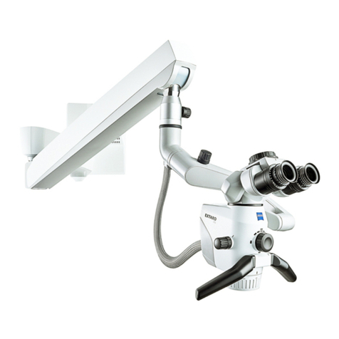
State-of-the-art apochromatic optics deliver the
legendary crystal-clear images, sharp details and
natural colors associated with Carl Zeiss. The
innovative optical design, with a new Varioskop™,
provides a larger working range and virtually
ensures comfortable working conditions – even
when using long instruments. The depth of field
has been increased by 17% and can be doubled
by activating the DeepView function.
Bringing Light to the Darkness
Surgeons now have 20% more light to work
with, the assistant even more. Spot illumination
precisely adjusts the light cone – all without
reflections. The patented, two-channel illumination
system delivers light wherever it’s needed – a
distinct advantage, which provides higher-contrast
images in narrow and deep canals.
Focus at the Push of a Button
The high-speed autofocus automatically delivers
razor-sharp images at all times, regardless of
magnification. And when focusing manually?
An intuitive laser-focusing aid assists in selecting
the exact focal point – particularly helpful when
using the mouth switch.
Stress Free
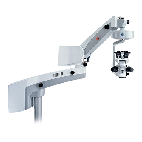
Whether the surgeon remains standing or seated,
the 30% more compact design of OPMI Pentero
allows for freedom of hand and instrument
movement while providing a short distance to the
surgical field.
Thanks to its overhead design, the suspension
system can be placed in any position – even
behind the surgeon.
“I have been using Carl Zeiss microscopes for the last
two and half decades. They have provided the best in
optics and have been constantly upgrading the technol-
ogy. My team and I trust Carl Zeiss optics.”
Dr. A. S. Hegde, Chairman Neurosciences, Sri Sathya
Sai Institute of Higher Medical Sciences, Bangalore,
India
“With the tremendous improvements to the co-obser-
vation tube, the OPMI Pentero really helps the assis-
tant. The tube provides a better view of the operating
field and does not move when the microscope is reposi-
tioned.”
J.J. van Overbeeke M.D., Ph.D., St. Elisabeth Hospital,
Tilburg, The Netherlands
9
