manuals.online logo
Brands
  1. Home
  2. •
  3. Brands
  4. •
  5. Olympus
  6. •
  7. Microscope
  8. •
  9. Olympus VS110 Fluorescence User manual

Olympus VS110 Fluorescence User manual

Multi-Channel Fluorescence Solution for Virtual Microscopy
Microscopy & Imaging
VS110TM Fluorescence
Virtual slide systems
biomolecular processes. Fluorescent markers have become
one of the main methods of generating clear contrast in
light microscopy. Furthermore, they have allowed very
specific identification of molecules, enabling ultra-precise
localization and transport studies. These capabilities
are essential for numerous applications throughout
many different research areas. Many experiments use
multiple sections of a sample to show the distribution of
a certain molecule. Analyzing these can take up many
hours of the researcher’s time and block the microscope
for other users. By scanning the sections on the VS110
fluorescence system, the researcher can view their virtual
slide image data away from the microscope and even
perform analysis using dedicated imaging software.
All VS110 fluorescence system components are designed
to interact seamlessly, producing a fully automated, high-
speed multi-channel fluorescence scanning system
with excellent flexibility and simple operation. These
components ensure that users gain the best results for
all types of specimens with minimum effort. All users are
able to review the same sample, yet control the position
and magnification individually, as if it were a real slide. This
ability to acquire complete slides at high magnification
and resolution virtually, delivers significant benefits to
researchers, as well as students in professional education.
VS110 Fluorescence Acquisition Wizard
ENHANCING THE CAPABILITIES OF VIRTUAL MICROSCOPY
VS110™ FLUORESCENCE SYSTEM
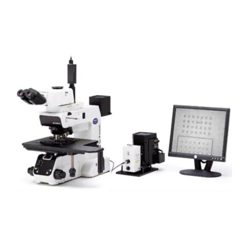
The Olympus VS110 Virtual Microscopy System com-
bines microscopy seamlessly with digital imaging,
allowing the creation of a virtual slide that is a digital
copy of the original specimen. The virtual slide can be
shared in real time with multiple persons across the
globe. The VS110 generates a high resolution image
of the whole specimen, which can be viewed and ana-
lyzed from the overview image at low magnification
up to maximum magnification by simply panning and
zooming. All samples can be evaluated instantly and
simultaneously as they are stored electronically on a
central server. As a result, the VS110 is ideal for use
in a number of areas including research, bio/pharma
and education.
The Olympus VS110 can be extended to scan fluore-
scence images. Such a ‘virtual fluorescence system’
can be used with fluorescent samples, giving a com-
pletely unique perspective – a slide recorded in full
fluorescence can be thoroughly examined without any
risk of bleaching or damage to the cells.
Microscopy has advanced rapidly over the last few years
in terms of the microscope hardware and the techniques
used to produce contrast or markers developed to identify
molecules, organelles and cell types. Today, we have a
much more accurate way of investigating cellular and
A multi-labled sagittal section of a rat embryo. Fluorescence image
courtesy of Dr. Dragan Maric, NINDS, National Institutes of Health.
VS110™ FLUORESCENCE SYSTEM
ENHANCING THE CAPABILITIES OF VIRTUAL MICROSCOPY
The VS110 fluorescence solution
The real power of the VS110 software lies in its functionality. It
is designed to provide all the features needed to make research
use as intuitive as possible. These functions are presented
to the user via an intuitive and easy-to-use interface.
The user is guided step by step through the virtual slide
acquisition process by the Scan Wizard. Full-slide scanning
can be set up with just three mouse clicks. With the VS110, it is
also possible to review the overview slide images and assign
individual magnifications, scan areas and other parameters to
the slides, even in a batch scan. Although several modes are
predefined, the advanced software allows experienced users to
be in full control of the details of the scanning process. Therefore,
different scan settings can be assigned to individual slides,
saving significant scanning time, as well as offering the possibility
of only scanning areas of interest, reducing the data file size.
For scanning fluorescence images the scan mode is enlarged
by a special Fluorescence tab, which has been adapted to fulfill
the special requirements of multi-fluorescence microscopy.
The acquisition process starts with generating a brightfield
or fluorescence overview image of the entire slide. It includes
the automatic detection of the specimen to avoid time-
consuming scanning of areas where there is no sample on
the slide. As a second step, the high-magnification scan is
automatically performed over the whole slide or over smaller,
predefined areas of interest using the preselected objective.
Multi-Channel Fluorescence
Configured with high-speed, 8 position excitation and emission
filter wheels, the VS110FL provides increased flexibility in the
choice of fluorochromes as well as extremely fast acquisition
of multi-channel images. Individual configuration of each of the
color channels helps to ensure the optimum focus for each
channel. Image acquisition is performed using a high per-
formance cooled 14bit monochrome CCD camera and stored
as full 16bit grayscale data.
Virtual-ZTM for Fluorescence Imaging
The VS110 fluorescence system is also capable of scanning
multiple large specimens in up to 15 z-planes. By selecting
the scan mode, the reviewer can just simply focus through
the sample, as well as examine regions of interest in different
dimensions. At each position of the whole sample,
the VS110 system acquires a series of fluorescence images
at various focus positions, and these fluorescence z-stacks
are stored within the images and can be focused virtually
on the monitor. The image is displayed without delay on the
screen while being scanned and can be immediately viewed
without affecting the acquisition. This is due to the high-
performance and unique multi-threading Olympus software.
Advanced Features
The optional VS-Desktop Review Station software contains
advanced imaging tools to further enhance the acquired
virtual slide images. Included in these tools are smoothing
and sharpening filters, min mean and maximum projections,
as well as no neighbor and nearest neighbor deconvolution.
Additional tools in VS-Desktop include separation of
each of the color channels, as well as exporting this data
to other 3rd party software for further image analysis.
Specifications are subject to change without any obligation on the part of the manufacturer.
Soft Imaging System is a trademark of Olympus Soft Imaging Solutions GmbH.
www.olympus-sis.com
www.soft-imaging.net
Olympus America Inc.
3500 Corporate Parkway
P.O. Box 610
Center Valley, PA 18034-0610
www.olympusamerica.com
General VS110™ Specifications
Fully automated virtual slide scanner
Motorized BX®microscope frame
Objectives included: 2x PLAPON, 10x, 20x, 40x UPLSAPO, other objectives may be added optionally
Illumination: brightfield with 100 W halogen lamphouse
VS110-S1 with slide holder for one 1” x 3” slide
VS110-S5 with slide holder for five 1” x 3” slides or for two 2” x 3” slides
VS110-L100 with slide loader for 100 3” x 1’’ slides, with integrated bar code scanner
Digital CCD camera: 2/3” CCD camera, 6.45 µm x 6.45 µm pixel size, high sensitivity, high resolution
Peltier-cooled, 14-bit dynamic range
Workstation with Dual XEON™ technology and 500 GB data storage capacity,
network interface 100/1,000 Mbit/s Ethernet, 4 GB RAM and high-resolution TFT monitor
Data storage solution (optional)
CE compliant
Resolution
High-resolution scanning (standard 1x camera adapter): 0.32 µm/pixel (20x/NA 0.75)
and 0.16 µm/pixel (40x/NA 0.9)
Fast scanning camera using 0.5x camera adapter: 0.34 µm/pixel (20x/NA 0.75)
and 0.17 µm/pixel (40x/NA 0.9)
Scanning speed (brightfield)
15 x 15 mm sample area with 20x objective: standard scanning mode < 3 min
Image acquisition
Acquisition scanning wizard
Automatic sample and tissue finding
Autofocus and manual focus routines
Multilayer scanning: multiple scans in one image
Seamless stitching
Virtual-Z™ acquisition
Single button for automatic scanning of up to 100 slides (with VS110-L100)
Reviewed batch scan
Scan directly to .vsi, .tif or .btf formats
Software functions and modules
View images locally, zoom, crop and rotate images
Compress images and save images in different file formats
Insert text annotations and drawings; create views of regions of interest, record audio annotations
(sound card, microphone and speakers required)
Net Image Server software for remote access to virtual slides with VS110 software, OlyVIA viewer
or Web browser and management of virtual slides and user access to images
Conferencing module for simultaneous and synchronized viewing of virtual slides, conference access
control and user management, minute-taking tool, chat function
TMA module to acquire individual images of TMA cores, create virtual TMA’s
NetImage Server and OlyVIA™ can support Hamamatsu .ndpi file format
Analysis
Integrated measurement functions: distances, areas, circumferences, point count, export results
to Microsoft Excel
Analysis with software packages, e.g. phase analysis to measure shares of tissue types, particle
analysis to identify content of tumor cells
638
519
720
625
174.5*
588
598
359 483
341
174.5*
334
514
487
209
VS110-S1/VS110-S5 VS110-L100
Weight: 52 kg, power consumption: 952 W Weight: 100 kg, power consumption: 1,024 W
* This measurement may vary according to interpupillary distance. The measurement given
pertains to a distance of 62 mm.
Dimensions (mm)
VS 110 Fluorescence Features
Dual cameras support brightfield and fluorescence
14-bit monochrome camera (XM10)
Support of 16bit grayscale data
Multi-channel imaging (> 3 channels)
Dedicated fluorescence acquisition wizard
incl. monochrome or color overview acquisition and 10x overview image option for greater detail
incl. auto exposure for optimal exposure time
Virtual-Z™ fluorescence acquisition
Optional: advance filtering eg deblur, max intensity projection (available in optional VS-Desktop Software)
Optional: oil scanning up to 100x
Olympus VS110 systems are for research and educational use only.
©2010 Olympus America Inc. All rights reserved. Printed in the United States of America. Olympus, VS, Virtual-Z, OlyVIA, and BX are trademarks or registered
trademarks of Olympus Corporation, Olympus America Inc. and/or their affiliates. All other trademarks are the property of their respective owners. OLYVS110FL.11.01
SPECIFICATIONS

Other Olympus Microscope manuals

Olympus CX33 User manual

Olympus

Olympus CX33 User manual

Olympus BH2 Series User manual

Olympus

Olympus BH2 Series User manual

Olympus BX-URA2 User manual

Olympus

Olympus BX-URA2 User manual

Olympus VMF User manual

Olympus

Olympus VMF User manual

Olympus CHS User manual

Olympus

Olympus CHS User manual

Olympus DP73 User manual

Olympus

Olympus DP73 User manual

Olympus BX53 User manual

Olympus

Olympus BX53 User manual

Olympus IX73 User manual

Olympus

Olympus IX73 User manual

Olympus BX51-P User manual

Olympus

Olympus BX51-P User manual

Olympus BX3M-CB User manual

Olympus

Olympus BX3M-CB User manual

Olympus BXC-CBB User manual

Olympus

Olympus BXC-CBB User manual

Olympus BX51WI User manual

Olympus

Olympus BX51WI User manual

Olympus DP22 User manual

Olympus

Olympus DP22 User manual

Olympus bx40 User manual

Olympus

Olympus bx40 User manual

Olympus DP80 User manual

Olympus

Olympus DP80 User manual

Olympus VANOX Operating instructions

Olympus

Olympus VANOX Operating instructions

Olympus MX50A/T User manual

Olympus

Olympus MX50A/T User manual

Olympus BHT User manual

Olympus

Olympus BHT User manual

Olympus CK2 User manual

Olympus

Olympus CK2 User manual

Olympus BX43 User manual

Olympus

Olympus BX43 User manual

Olympus SZX9 User manual

Olympus

Olympus SZX9 User manual

Olympus FV3000 User guide

Olympus

Olympus FV3000 User guide

Olympus BX53-P User manual

Olympus

Olympus BX53-P User manual

Olympus Fluoview FV1000 Instruction Manual

Olympus

Olympus Fluoview FV1000 Instruction Manual

Popular Microscope manuals by other brands

Levenhuk DTX 90 user manual

Levenhuk

Levenhuk DTX 90 user manual

Saxon NK103-B instruction manual

Saxon

Saxon NK103-B instruction manual

Digital Blue QX5 user guide

Digital Blue

Digital Blue QX5 user guide

Swift Stereo SM80 Use and care

Swift

Swift Stereo SM80 Use and care

Leica MC170 HD user manual

Leica

Leica MC170 HD user manual

Firefly RW180 user manual

Firefly

Firefly RW180 user manual

Bresser 5803100 instruction manual

Bresser

Bresser 5803100 instruction manual

Accu-Scope 3052-LED manual

Accu-Scope

Accu-Scope 3052-LED manual

Andonstar AD205 user manual

Andonstar

Andonstar AD205 user manual

Zeiss Axio Scope.A1 operating manual

Zeiss

Zeiss Axio Scope.A1 operating manual

LEUCHTTURM DM1 User instructions

LEUCHTTURM

LEUCHTTURM DM1 User instructions

Zeiss Axiolab 5 Quick reference guide

Zeiss

Zeiss Axiolab 5 Quick reference guide

Thermo Scientific Nicolet RaptIR user guide

Thermo Scientific

Thermo Scientific Nicolet RaptIR user guide

ViTiny UM30 user guide

ViTiny

ViTiny UM30 user guide

Agilent Technologies 7500 AFM datasheet

Agilent Technologies

Agilent Technologies 7500 AFM datasheet

OPTO-EDU A13.2603 instruction manual

OPTO-EDU

OPTO-EDU A13.2603 instruction manual

National 158-LED instructions

National

National 158-LED instructions

3B SCIENTIFIC PHYSICS BS-200 instruction manual

3B SCIENTIFIC PHYSICS

3B SCIENTIFIC PHYSICS BS-200 instruction manual

manuals.online logo
manuals.online logoBrands
  • About & Mission
  • Contact us
  • Privacy Policy
  • Terms and Conditions

Copyright 2025 Manuals.Online. All Rights Reserved.