Hadeco Bidop ES-100V3 User manual


TABLE OF CONTENTS
Cautions
1. Introduction......................................................................................................................1
1-1. Features..................................................................................................................2
1-2. Clinical applications.................................................................................................3
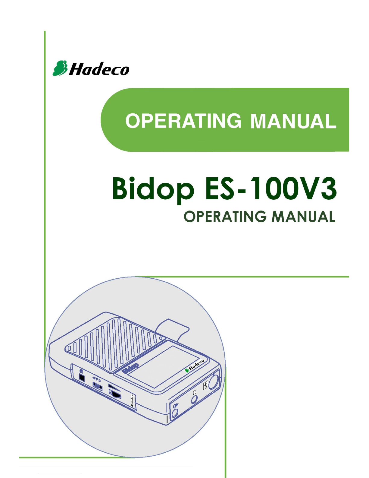
2. Appearance......................................................................................................................4
2-1. Front view................................................................................................................4
2-2. Back side view and Probe.......................................................................................5
3. Quick start........................................................................................................................6
3-1. Turning the unit ON / OFF.......................................................................................6
3-2. Checking battery level and replacing battery...........................................................7
3-3. Measuring blood velocity.........................................................................................8
3-3-1.Normal mode.......................................................................................................8
3-3-2.Site guidance mode...........................................................................................10
3-4. Measuring heart rate (2MHz only).........................................................................12
3-5. 2 MHz BEEP mode ...............................................................................................14
4. Menu and Mode settings................................................................................................15
4-1. Menu.....................................................................................................................15
4-1-1. Menu operation ................................................................................................15
4-1-2. MENU for Blood Velocity Measurement mode .................................................16
4-1-3. MENU for Blood Velocity Freeze mode............................................................16
4-1-4. MENU for Heart Rate mode (Measurement and Freeze).................................17
4-1-5. MENU for 2MHz BEEP Measurement mode....................................................18
4-1-6. MENU for 2MHz BEEP Freeze mode...............................................................18
4-2. Mode setting details ..............................................................................................19
a. MEMORY - STORE................................................................................................19
b. MEMORY - READ...................................................................................................19
c. MEMORY - CLEAR.................................................................................................20
d. MODE (Baseline mode)..........................................................................................20
e. DIR (Flow direction)................................................................................................20
f. TIME (Time scale)....................................................................................................21
g. DISP (DISP, OTHERS –DISP, DISP/BEEP) ..........................................................21
h. SOUND (Beep sound for HR).................................................................................21
i. UPPER (Upper limit for HR).....................................................................................21
j. LOWER (Lower limit for HR)....................................................................................21
k. LIMIT-1 (Limit for 2MHz BEEP mode).....................................................................22
l. LIMIT-2 (Maximum / Average for 2MHz BEEP mode)..............................................22
m. OTHERS - LANGUAGE.........................................................................................22

n. OTHERS - UNIT (cm/s / kHz).................................................................................22
o. OTHERS - FILTER (Arterial / Venous filter)............................................................22
p. OTHERS - SMOOTH (Smoothing filter)..................................................................22
q. OTHERS - CAL (Calibration)..................................................................................23
r. OTHERS - BACKLIGHT ..........................................................................................23
s. OTHERS - AUTO-OFF (Automatic shut-off)............................................................23
t. OTHERS - FREEZE ................................................................................................23
u. OTHERS - PRB20MHz...........................................................................................23
v. FLOW (Blood volume flow) / w. DIAMETER (Estimated vessel diameter)
.....................24
5. Maintenance..................................................................................................................25
5-1. Performance check by user...................................................................................25
5-2. Cleaning................................................................................................................25
5-3. Probe sterilization..................................................................................................25
5-3-1. Sterilizable probes (Amplifier required) ............................................................25
5-3-2. Sterilization.......................................................................................................26
5-4. Warranty................................................................................................................29
6. Supplemental information..............................................................................................30
6-1. Basic mode .............................................................................................................30
6-2. LCD display...........................................................................................................30
6-2-1. Blood Velocity mode.........................................................................................30
6-2-2. Heart Rate mode (Only 2 MHz probe)..............................................................32
6-2-3. 2 MHz BEEP mode ..........................................................................................34
6-3. Numerical data......................................................................................................35
6-4. External outputs ....................................................................................................36
6-5. Symbol list.............................................................................................................36
6-6. Contents in package..............................................................................................36
7. Options..........................................................................................................................37
7-1. Probe selection......................................................................................................37
7-2. Others ...................................................................................................................38
7-3. Photoplethysmograph ...........................................................................................39
7-4. Foot Temperature (Except European Union Countries).........................................44
8. Technical information.....................................................................................................46
8-1. Principles...............................................................................................................46
8-2. Block diagram........................................................................................................46
8-3. Specifications........................................................................................................46
8-4. Safety standards ...................................................................................................48

Cautions
Please read the following important points carefully before you operate the
unit.
1. Only skilled persons should operate the unit.
2. Use the unit for measuring blood flow.
3. Do not apply any modification to the unit.
4. Device placement
(1) Follow the requirements for storage and operating environments.
(2) Do not place it near water.
(3) Dot not place it where atmospheric pressure, temperature, humidity,
ventilation, sunlight, dust, salt, sulfur and so forth will not affect the
unit adversely.
(4) Pay attention to the stability conditions such as inclination, vibration,
and shock during transportation and installation work.
(5) Do not place it where chemicals are stored, or where gas may be
generated.
(6) Do not place it where the unit tends to fall.
(7) Do not place it on or adjacent other electronic device.
5. Before use:
(1) Make sure that the unit operates safely and correctly by following the
maintenance procedures mentioned in "§ 5-1. Performance check by
user".
(2) Make sure that all cables are connected correctly and safely.
(3) Using it with other equipment together may cause a misdiagnosis or
danger to patient due to a malfunction.
(4) Double check that all the cables do not obstruct any external
connection to the patient.
(5) Do not sterilize the main unit, non-sterilizable probes and amplifiers to
prevent any damage.
(6) Sterilizable probes (optional) should be sterilized before use. (See
§5-3. Probe sterilization)
6. Operation
(1) Do not use the unit simultaneously with an electric cautery,
cardioverter, other ultrasonic device or mobile phone.

(2) Be careful not to exceed time and volume of diagnosis treatment
required.
(3) Always make sure the unit and patient are not under abnormal
conditions.
(4) When any abnormality is found on the unit or the patient, take proper
action such as stopping operating the unit in a manner safe to the
patient.
(5) Do not let the patient touch the unit.
(6) Use the designated components only such as the probe.
(7) Do not use the components for other devices.
(8) Use the unit under the operating environments specified on the
specifications.
(9) Use the Bidop as specified in the Operating manual.
(10) Do not use the unit in a strong electromagnetic field or it may cause
incorrect measurement.
7. After use
(1) Turn the unit off the way specified.
(2) Do not pull the cable(s) too much while disconnecting or it may cause
damage.
(3) Clean the unit, cables and probes and place them in right place for
the next use.
8. Storage
(1) Follow the caution (2) to (6) of section # 4 Device placement in the
previous page.
(2) Clean the unit, probes and place them in right place for the next use.
(3) When using the unit next time, perform the maintenance to make
sure it works properly and safety.
9. Maintenance
(1) Do the periodical maintenance by following the procedures
mentioned in "§ 5-1. Performance Check by user".
(2) The maintenance must be done at least once a year.
10. Probes
(1) Clean the probe using dump cloth before use. Using alcohol or
thinner may damage the probe.
(2) The probe transducer tip is very thin and delicate. Please handle with
great care and use the probe cap when not in use.

(3) Optional sterilizable probe (reusable & disposable) can be sterilized
in the manner described in “§5-3. Probe sterilization”. However, only
one time sterilization is possible for disposable probe, and do not
reuse it. Except optional ACP probe, do not sterilize probes by steam
autoclave.
11. Ultrasonic gel
(1) Do not apply ultrasonic gel to the probe body other than the tip of
probe.
(2) Using other materials may damage the probe.
(3) The ultrasonic gel enclosed is non-sterile and do not use it for
surgeries.
(4) Incidence of allergy: Discontinue use of gel if an allergic reaction
occurs.
12. Battery
(1) When battery is extremity low, the LCD display will not operate. Also
there will be no speaker sounds. Replace the battery.
(2) Use a 9 volt alkaline square type battery. A non-alkaline may cause a
shortage of power.
(3) When not using the unit for a long time, remove the battery.
13. Repair services
(1) When the unit gets out of order, contact the dealer for repair from
whom you purchased the unit.
(2) Only authorized persons should perform the repair services.
14. Do not disassemble the Bidop.
15. Destruction
(1) In case of destruction of the unit, follow the instructions for disposition
of the destruction appointed by each country or local government.
(2) Do not place battery in fire or it may cause an explosion and injury.
16. Any connected computer is not allowed to be in the patient area
according to IEC60601-1.

1
1. Introduction
Thank you very much for choosing the Bidop ES-100V3.
The Hadeco Bidop ES-100V3 is a uniquely designed bi-directional pocket
Doppler with LCD display. It detects arterial and venous blood flows in
extremities.
The Bidop displays velocity waveform and numerical data.
Please read this manual carefully to acquaint yourself with the Bidop
operation.
This medical device can be used by doctor for the purposes mentioned in
"§1-2. Clinical Applications" for patient in hospital and clinic.
For the use with computer, please refer to the operating manual for Windows
linking software optional.

2
1-1. Features
•BI-DIRECTIONAL HAND HELD DOPPLER WITH LCD DISPLAY
Displays real-time waveforms, numerical data on backlit LCD.
•Hadeco DESIGNED SMART MICROPROCESSOR
Various mode settings are available for optimal measurement with the
menu displayed on the LCD and unique side Shuttle Button.
30 waveform memories.
•CONVENIENT PROBE ACTIVATION BUTTON
Freezes waveform and numerical data for notation.
Turns Bidop ON and OFF.
•MULTIPROBE SELECTION of 2, 4, 5, 8, 10 and 20 MHz
•USB COMPUTER INTERFACE
Stores waveforms and numerical data in your computer for data analyzing
and filing. Communication cable and Smart-V-Link Windows software are
optional.
•PHOTOPLETHYSMOGRAPH (PPG) PROBE OPTIONAL
Expands arterial & venous testing.

3
1-2. Clinical applications
•Detections of arterial and venous blood flow velocity for vascular
disease
Probes to be used: BT2M20S8C (2 MHz)
BT4M05S8C (4 MHz)
BT5M05S8C (5 MHz)
BT8M05S8C (8 MHz)
BT10M5S8C (10 MHz)
* PEAK & MEAN blood velocity determinations
* Detection of arterial and venous blood flow velocity for vascular
disease

4
2. Appearance
2-1. Front view
1. Probe connector: To connect probe.
2. Headset: To connect headset. It cuts off the speaker.
3. Serial port: To connect computer. (USB)
4. Volume control: To adjust sound volume.
5. Shuttle Button: To turn the unit ON/OFF, press it inside.
To select menu, turn it up or down.
To set the mode or execute command, press it inside.
To display next memory data, turn it up and down.
Press inside
Turn up
Turn down

5
6. BACK button: To quit menu mode.
To go back to previous menu.
To change Display mode from WAVE to DATA and
vice versa.
To get out of Site guidance mode.
7. Speaker: Outputs Doppler sounds.
8. LCD display: Displays waveform, numerical data, Heart Rate and
menu for mode settings. See “§ 6-2. LCD Display” for
details.
2-2. Back side view and Probe
9. Probe holder: For probe placement when not in use.
10. Strap holes: To connect neck strap (Option).
11. Battery cover: For battery placement.
12. Probe button: To turn the unit ON.
To freeze the waveform & numerical data when
power is ON.
To turn the unit OFF, press it longer than 2 sec.

6
3. Quick start
3-1. Turning the unit ON / OFF
(1) Set the alkaline battery in the unit.
(See “§ 3-2. Checking battery level and
replacing battery”.)
(2) Connect the probe to the Bidop so that
the polarity mark will be placed under
mark.
(3) Press the probe button or shuttle button
to turn the unit ON. Press button the
second time longer than 2 sec. to turn it
OFF.
Low battery:
When battery is low, low battery indicator
appears. Replace the battery for
further use.
Automatic shut-off:
When the AUTO-OFF is ON, if the unit is left on, the power is
automatically shut off after following time passes:
(a) 15 minutes when in measurement.(35 min. for FHR waveform mode)
(b) 2 minutes when no signal.
(c) 5 minutes when on freeze mode.
Note: If Automatic shut-off functions while on Freeze mode, Bidop will revert to
Freeze mode and display the frozen waveform on LCD by turning the unit on.
Polarity mark
Low battery indicator

7
Error message:
In case any inappropriate probe is
connected to the unit, error message as
shown right will be displayed.
3-2. Checking battery level and replacing battery.
(1) Turn the unit on and press Shuttle button
to display Menu and battery level
indicator will be shown on top right of
MENU.
It shows 4 levels as shown below.
When battery is low, replace the battery
with new one as follows.
(2) When you replace battery, turn the unit
off and open the battery cover as shown
right.
(3) To prevent any damage to the battery
terminal, please insert the battery to
battery box as shown right.
(4) Use a 9 volt ALKALINE square type
battery. A non-alkaline may cause a
shortage of power.
Set the battery in the unit ensuring that
the positive and negative terminals
correspond to the + and - marks on the
label in battery box, respectively.
Note: Use the battery composed in the package as a standard component for
performing operation check only.
Low
Full

8
3-3. Measuring blood velocity
3-3-1. Normal mode
This section explains the fundamental use of measuring blood velocity.
Refer to “§ 4. MENU and Mode settings” for various uses.
(1) Put ultrasonic gel on the probe top or
patient skin.
(2)Put the probe on the measurement area
and move it slowly to locate the point
where the maximum Doppler sounds are
heard. An ideal probe angle to the vessel
is approximately 45° to 60°.
(3)When the waveform becomes rhythmical
and stable, wait more than 5 sec. without
moving probe and press the probe
button to freeze the waveform.
Note: Pressing probe button longer than 2 sec.
will turn the unit OFF.
(4)To get numerical data, press BACK
button or go to menu and change the
DISP mode to DATA.
Note: See “§ 4-2-g. DISP" for operation. Also,
see “§6-3. Numerical Data” for the
parameters.
Probe button

9
(5)Headset can be used to listen to Doppler sounds. It will cut off the
speaker.
To store the data:
If you wish to store the waveform and numerical data on the memory, do
the following procedures;
1.Press the Shuttle Button to display
MENU and press it again on MEMORY
to go to MEMORY menu.
2.Press the Shuttle button on STORE
and next memory number available for
storage will be displayed as shown in
the right.
Turn Shuttle Button up and down to
change the memory number, if necessary.
3.Press the Shuttle button to store the data.
To display stored data;
1.Press the Shuttle button to go to
MEMORY menu.
2.Press the Shuttle button on READ and
select the memory number with "*" by
turning the Shuttle Button up and
down.
3.Press the Shuttle button to show the
waveform. To show next waveform,
turn the Shuttle button up and down.

10
LCD Backlighting time;
LCD backlighting time can be adjusted on BACKLIGHT mode setting.
See “§4-2-r. OTHERS –BACKLIGHT”for details.
Press BACK button longer than 2 sec. to turn it ON / OFF instantly
during testing.
3-3-2. Site guidance mode
This mode allows you to easily proceed
multiple Smart-V-Link testing by just pressing
probe button without connecting Smart-V-
Link.
Register Abbreviated site & test names on
the unit through Smart-V-Link to activate this
mode. Once the names are registered, the
unit will show each of names at the beginning
of each testing to let you know where to test
next.
Preparation for site guidance mode:
(1) Connect the unit to the computer with the USB cable and start
Smart-V-Link.
(2) Go to Site Screen and input abbreviated site & test name for each
waveform memory and then store the names on the unit.
(3) Go to MEMORY menu and clear ALL memory data on the unit
before newly starting the site guidance mode.
See the section “§.4-1-4. Site” on Smart-V-Link, V4.1 or over,
operating manual for more details.
<Site Screen of Smart-V-Link>

11
Site guidance mode procedures:
(1) Turn the unit off and on and the 1st
guidance with memory number and
abbreviated site & test name will
appear as shown in the right.
Note: The first memory number available
will be selected automatically.
(2) Press the probe button to start
monitoring waveform.
Press Back button to get out of “Site
guidance mode” for normal mode
operation.
(3) Press the probe button the 2nd time to
freeze the waveform when it becomes
stable and the 2nd guidance “STORE?”
as shown in the right will appear.
(4) Press probe button the 3rd time to store
the frozen waveform data on the
designated memory number.
(5) The 1st guidance for the next testing will
appear as shown in the right. Repeat
steps #(5) to #(7) until all testing is
completed.
Press probe button when the message
shown right is displayed upon
completion of all testing and the unit
will get out of site guidance mode.
Probe button
Probe button
Memory number
Abbreviated site & test name

12
3-4. Measuring heart rate (2MHz only)
This section explains the fundamental use of measuring heart rate. Refer to
“§ 4. Menu and Mode settings” for various uses.
(1) Connect the 2 MHz probe to the Bidop and turn the unit ON.
(2) Press the Shuttle button to display MENU
and set DISP mode for WAVE or DATA.
If necessary, change the upper and lower
limits by turning the Shuttle button up and
down between 60 and 220 BPM at 5 BPM
intervals and press the Shuttle button to
set them. See “§4-2-i. UPPER” and “§4-2-
j. LOWER” for details.
(3) Put ultrasonic gel on the probe top or
patient skin surface.
(4) Put the probe on the measurement area
and move it slowly to locate the point
where the maximum sounds are heard.
(5) When the heart rate becomes stable,
press the probe button, or BACK button
while using waterproof probe, to freeze it.
In case FHR exceeds the upper and lower
limits, LCD will start flashing and also,
beep sounds will be heard if SOUND
mode is set for ON. See “§4-2-h. SOUND”
for details.
Gel
Skin surface
Probe button

13
To store the monitoring waveform data on the memory, follow the step
“To store the data”in “§3-3-1. Normal mode”.
Note1: Numerical heart rate can’t be stored on the memory.
Note2: If the asterisk (*) is shown above the frozen heart rate as a calculation error
indicator, press probe button and do the testing again.
Manual counting mode is available if heart rate cannot be calculated, see “§
6-2-2. Heart Rate mode”.
(6) Headset can be used to listen to Doppler sounds. It will cut off the
speaker.
Lower limit
Upper limit
WAVE mode
DATA mode
Table of contents
Other Hadeco Medical Equipment manuals
Popular Medical Equipment manuals by other brands

Cardinal Health
Cardinal Health SVED 6701132 Clinician Quick Reference Guide

3B Medical
3B Medical Luna G3 BPAP 25A Quick operation manual

Zimmer
Zimmer Z Field Dual Instructions for use

Moog
Moog Infinity Orange Quick reference instructions

GE
GE Healthcare Brivo XR118 Service manual

Atmos
Atmos C 341 operating instructions

Spacelabs Healthcare
Spacelabs Healthcare Lifecard CF Operation manual

ParAid
ParAid TRANSPORTER+ user manual

WY'EAST MEDICAL
WY'EAST MEDICAL DENALI600 Instructions for use

Promeba
Promeba STRETCHER user guide

Cardiac Science
Cardiac Science Powerheart G5 user guide

Acacia
Acacia Pharm Pump II Frequently asked questions