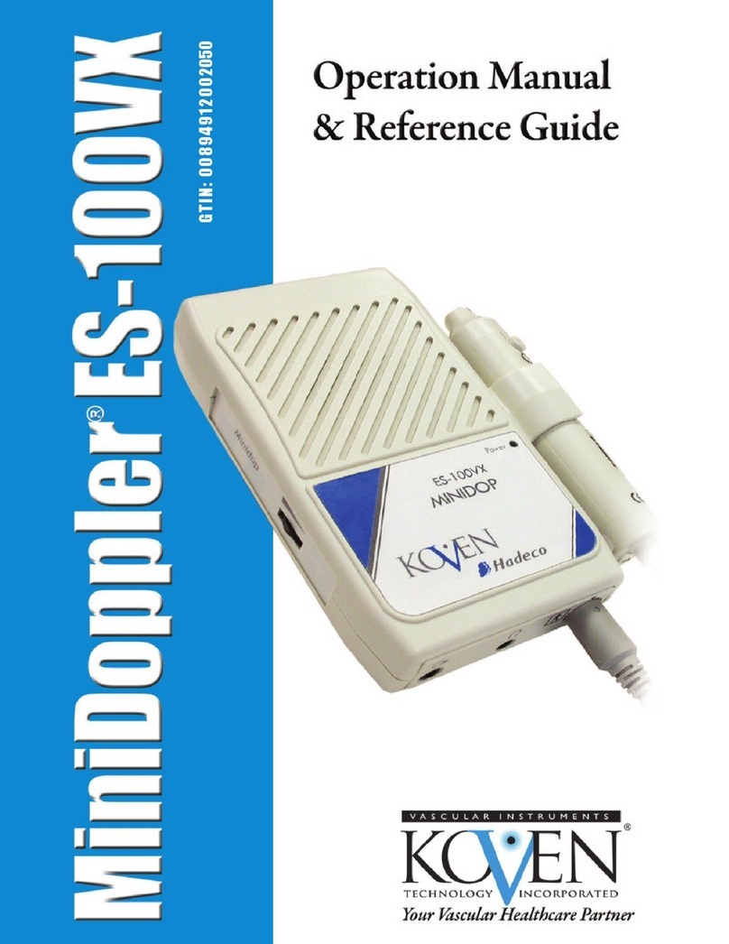
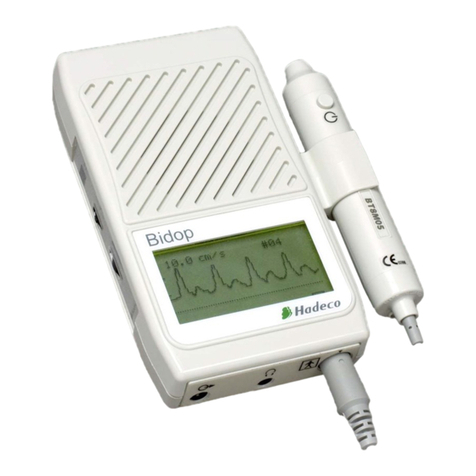
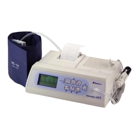
Hadeco Smartdop 45 User manual


TABLE OF CONTENTS
Cautions
1. Introduction ......................................................................................................................1
1-1. Features....................................................................................................................1
1-2. Clinical applications...................................................................................................3
1-3. Probe selection .........................................................................................................3
1-4. Contents of package .................................................................................................4
2. Quick start ........................................................................................................................5
2-1. Turning the unit ON / OFF.........................................................................................5
2-2. Charging / Discharging battery..................................................................................6
2-3. Checking battery level...............................................................................................7
2-4. Setting printer paper..................................................................................................8
2-5. Measuring blood velocity...........................................................................................9
2-6. Measuring fetal heart rate (2 MHz only) ..................................................................10
3. Appearance and mode settings......................................................................................12
3-1. Operating controls...................................................................................................12
3-2. Mode settings..........................................................................................................15
3-2-1. Basic Modes.....................................................................................................15
3-2-2. Menu ................................................................................................................16
a. Menu structure ....................................................................................................16
b. Menu operation ...................................................................................................16
c. Menu for Blood Velocity Measurement mode......................................................18
d. Menu for Blood Velocity Freeze mode ................................................................19
e. Menu for Fetal Heart Rate mode (Measurement and Freeze).............................20
3-2-3. Mode Setting Details ........................................................................................21
a. MEMORY - STORE.............................................................................................21
b. MEMORY - READ ...............................................................................................21
c. MEMORY - CLEAR .............................................................................................22
d. MODE (Baseline mode) ......................................................................................22
e. TIME (Time scale) ...............................................................................................23
f. DIR (Flow direction) .............................................................................................23
g. DISP / OTHERS - DISP (Waveform / Data) ........................................................23
h. UPPER (Upper limit for FHR)..............................................................................24
i. LOWER (Lower limit for FHR) ..............................................................................24
j. PATIENT (Patient data input)................................................................................24
k. OTHERS - LANGUAGE ......................................................................................26
l. OTHERS - UNIT (cm/s / kHz)...............................................................................26
m. OTHERS - FILTER (Arterial / Venous filter)........................................................26
n. OTHERS - SMOOTH (Smoothing filter) ..............................................................26
o. OTHERS - CAL (Calibration)...............................................................................26
p. OTHERS - AUTO-OFF (Automatic shut-off)........................................................26

q. OTHERS - KEYBOARD (External)......................................................................27
r. OTHERS - P.ID PRT (Patient data print) ..............................................................27
s. OTHERS - PRB-KEY (Probe button function) .....................................................27
t. OTHERS - DATE (Date and time setting).............................................................27
u. OTHERS - PRINT (Printer activation) .................................................................27
3-3. LCD display.............................................................................................................28
3-3-1. Blood Velocity Mode.........................................................................................28
3-3-2. Fetal Heart Rate (FHR) mode (Only 2 MHz probe) ..........................................29
3-4. Printing waveforms and data...................................................................................31
3-5. Numerical data ........................................................................................................33
3-6. External outputs ......................................................................................................34
3-7. Symbol list...............................................................................................................34
4. Maintenance ..................................................................................................................35
4-1. Performance check by user ....................................................................................35
4-2. Cleaning..................................................................................................................35
4-3. Warranty..................................................................................................................35
5. Options ..........................................................................................................................36
5-1. Probe selection .......................................................................................................36
5-2. Sterilizable doppler probes......................................................................................36
5-3. Photoplethysmograph .............................................................................................39
5-4. Pneumoplethysmograph .........................................................................................45
6. Technical information .....................................................................................................51
6-1. Principles ................................................................................................................51
6-2. Block diagram .........................................................................................................52
6-3. Specifications ..........................................................................................................53
6-4. Safety standards .....................................................................................................55

CAUTIONS
Please read the following important points carefully before you operate the unit.
1. Only skilled persons should operate the unit.
2. Use the unit for measuring blood flow.
3. Do not apply any modification to the unit.
4. Device placement
(1) Follow the requirements for storage and operating environments.
(2) Do not place it near water.
(3) Dot not place it where atmospheric pressure, temperature, humidity, ventilation,
sunlight, dust, salt, sulfur and so forth will affect the unit adversely.
(4) Pay attention to the stability conditions to avoid too much inclination, vibration,
shock and so on during transportation and installation of the unit.
(5) Do not place it where chemicals are stored or gas may be generated.
(6) Do not place it where the unit tends to fall.
5. Before use:
(1) Make sure that the unit operates safely and correctly by implementing
performance check mentioned in "§ 4-1. Performance Check by User".
(2) Make sure that all cables are connected correctly and safely.
(3) Using it with other equipment together may cause a misdiagnosis or danger to
patient due to a malfunction.
(4) Double-check that all the cables do not obstruct any external connection to the
patient.
(5) Do not sterilize the main unit by gas, autoclave or so on to prevent any damage.
(6) Sterilizable probes should be sterilized before use.
6. Operation
(1) Do not use the unit simultaneously with either electric cautery, cardioverter, other
ultrasonic device or mobile phone.
(2) Be cautious not to exceed too much time and volume required for the
measurement.
(3) Always make sure the unit and patient are not under abnormal conditions.
(4) When any abnormality is found on the unit or the patient, take proper action such
as stopping use of the unit in a manner safe for the patient.
(5) Do not let the patient touch the unit.
(6) Use the designated components only.
(7) Do not use the components for other devices.
(8) Use the unit under the operating environments specified on the specifications.
(9) Use the unit as specified in the operation manual.

(10)Do not use the unit in a strong electromagnetic field or it may cause incorrect
measurements.
7. After use:
(1) Turn the unit off the way specified.
(2) Do not pull the cable(s) too much while disconnecting or it may cause damage.
(3) Clean the unit, AC adaptor, cables and probes and place them in right place for
the next use.
8. Storage
(1) Follow the caution (2) to (6) of section #4 Device placement in the previous page.
(2) When using the unit next time, perform the maintenance to make sure it works
properly and safety.
9. Maintenance and inspection
(1) Do the periodical maintenance by following the procedures mentioned in "§ 4-1.
Performance Check by User".
(2) The maintenance must be done at least once a year.
10. Probes
(1) Clean the probe using damp cloth before use. Using alcohol or thinner may
damage the probe.
(2) The probe transducer tip is very thin and delicate. Please handle with great care
and use the probe cap when not in use.
(3) Optional sterilizable probe (reusable & disposable) can be sterilized in the manner
described in § 5-2. Sterilizable Doppler probes. However, only one time
sterilization is possible for disposable probe, and do not reuse it.
Except optional ACP probe, do not sterilize probes by steam autoclave.
11. Ultrasonic gel
(1) Do not apply ultrasonic gel to the probe body other than the tip of probe.
(2) Using other materials such as baby oil and cream may damage the probe.
(3) The ultrasonic gel enclosed is non-sterile and do not use it for surgeries.
(4) Incidence of allergy: Discontinue use of gel if any allergic reaction occurs.
12. Battery
(1) When battery is extremely low, the LCD display will not operate. Also there will be
no speaker sounds. Charge the battery.
(2) Battery life is 300 full charges. When full charging life is obviously short, contact
your dealer for replacing battery.
13. For transportation of the unit, it should be packed properly to protect against shock.

14. Repair services
(1) When the unit gets out of order, contact the dealer for repair from whom you
purchased the unit.
(2) Only authorized persons should perform the repair services.
15. Do not disassemble the unit.
16. Destruction
(1) In case of destruction of the unit, follow the instructions for disposal of the
destruction appointed by each country or local government.
(2) Do not place battery in a fire or it may cause an explosion and injury.
17. Any connected computer is not allowed to be in the patient area according to
IEC60601-1.

1
1. Introduction
Thank you very much for choosing the Smartdop 45.
The Hadeco Smartdop 45 is a uniquely designed bi-directional handheld Doppler with LCD
display and fast printer. It detects arterial and venous blood flows in extremities as well as
fetal heart sounds.
The Smartdop displays velocity waveforms, numerical data and fetal heart rate. Also it
prints them out.
Please read this manual carefully to acquaint yourself with the Smartdop operation.
This medical device can be used by doctor for the purposes mentioned in "1-2. Clinical
applications" for patient in hospital and clinic.
For the use with computer, refer to the operating manual for Windows linking software
optional.
1-1. Features
* BI-DIRECTIONAL HANDHELD DOPPLER WITH LCD DISPLAY and FAST PRINTER
Displays real-time velocity waveforms, numerical data and heart rate.
Prints frozen waveform, numerical data and monitored heart rate as well as patient data.
* HADECO DESIGNED SMART MICROPROCESSOR
Various mode settings are available for optimal measurement with the menu displayed
on the LCD and unique Scroll Button. 30 waveform memory
* CONVENIENT PROBE ACTIVATION BUTTON
Freezes and prints waveform and numerical data for notation.
Button function can be changed on probe key mode setting, PRB-KEY.
* MULTIPROBE SELECTION of 2, 4, 5, 8, 10 and 20 MHz.
(When connecting 20 MHz probe, only kHz (Doppler frequency shift) is available in Unit
mode. See “§ 3-2-3. l. Others - Unit” for the details)
* USB COMPUTER INTERFACE
Stores waveforms and numerical data in your computer for data analyzing and filing.

2
Communication cable and Windows software are optional.
* PHOTOPLETHYSMOGRAPH (PPG) AND PNEUMOPLETHYSMOGRAPH (PV)
PROBES OPTIONAL
Expands arterial & venous testing.

3
1-2. Clinical applications
1-2-1. Detections of fetal heart rate
Probe to be used BT2M20S8C (2 MHz)
* Evaluation of fetal heart rate and sounds throughout pregnancy except where fetal
heart is not developed sufficiently during the first trimester.
1-2-2. Detections of arterial and venous blood flow velocity for
vascular disease
Probes to be used BT4M05S8C (4 MHz)
BT5M05S8C (5 MHz)
BT8M05S8C (8 MHz)
BT10M5S8C (10 MHz)
*ABI studies
* PEAK & MEAN blood velocity determinations.
* Peripheral vascular procedures.
* Blood pressure segmental studies.
* Venous compressions.
* Penile & digit systolic pressures.
* Flow velocities in recovery room.
1-3. Probe selection
The frequency of diagnostic ultrasound is inversely proportional to depth of penetration.
The Smartdop has 5 interchangeable probes with different frequencies. Use those probes
depending on your applications.
Note: One of these probes comes with Smartdop 45 as a standard accessory. Other probes are
available as optional accessories.
BT2M20S8C (2 MHz): Fetal heart rate and sounds
BT4M05S8C (4 MHz): Deep peripheral blood velocity and flow
BT5M05S8C (5 MHz): Deep peripheral blood velocity and flow
BT8M05S8C (8 MHz): Superficial blood velocity and flow
BT10M5S8C (10 MHz): Superficial blood velocity and flow

4
1-4. Contents of package
Main unit 1
Probe 1
AC adaptor 1
Ultrasonic gel 1
Paper 1
Carrying case 1

5
2. Quick start
For the first time of use and in case the unit has been not used for a while, fully charge the
internal battery.
2-1. Turning the unit ON / OFF
(1) Connect the probe to the probe connector so that
the polarity mark will be placed under
mark as shown in the right (12 o’clock).
(2) Press the power button to turn the unit OFF.
(3) When Smartdop is turned ON first time, the
message as shown in the picture right will
appear, press Enter button to proceed.
(4) When battery is low, low battery indicator
appears as shown in the right. Smartdop can be
used for a few more minutes. Recharge the
battery as soon as possible for further use. Low battery
Polarity mark
Power button
Enter button
Right button
Back button

6
(5) AUTOMATIC POWER OFF
When the AUTO-OFF mode is ON, if the unit is left on, the power is automatically
shut off after following time passes:
(a) 35 minutes when in measurement. (Only Fetal Heart Rate WAVE mode)
(b) 15 minutes when in measurement. (Except Fetal Heart Rate WAVE mode)
(c) 5 minutes when no signal.
(d) 10 minutes when on freeze mode.
Note: If Automatic shut-off functions while on Freeze mode, Smartdop will revert to
Freeze mode and display the frozen waveform on LCD by turning the unit on.
2-2. Charging / Discharging battery
(1) Turn the unit off and plug the AC adaptor to the
unit to charge battery. The charging indicator will
go solid orange while charging is in progress.
Note: Use the designated adaptor, see "§
6-3.Specifications".
(2) When the battery is fully charged, the charging
indicator will go solid green. Unplug the AC
adaptor.
(3) Discharging battery:
Using and charging the battery repeatedly
without fully discharging may cause a shorter full
charge battery life. Fully discharge battery before
charging every once in a while as follows:
• Turn the unit off and plug the AC adaptor.
• Press Back Button and hold it, and press
Enter button to display battery level.
• While holding Back Button, press Right button
to discharge the battery.
Charging indicator
AC adaptor connector
Enter button
Right button
Back button

7
• The “DISCHARGE” will be displayed on the
LCD.
• After the discharging completed, charge
process will start automatically and the
“DISCHARGE” will disappear, then charging
indicator will go solid orange.
Note:It takes about 3 hours to fully charge battery.
Battery life is 300 full charges. Contact your dealer
for replacing battery.
2-3. Checking battery level
Battery level indicator shows upper right of the menu
screen.
(1) Turn the unit on and press Enter button to display
the menu.
(2) Battery level indicator shows the battery level in 4
steps as shown right.
(3) Charge the battery when in low.
Low battery indicator
When the battery is low, the low battery indicator will
be shown on bottom right of LCD. Recharge the
battery.
See "2-2. Charging / Discharging Battery"
If battery gets extremely low, the message shown
right will appear upon turning it on to indicate
memory data have been cleared for waveform data,
Battery level indicator
Low battery indicator
70%
Full
Low
30%

8
Date setting, and Patient data excluding other Menu
mode settings.
(1) Press Enter button to go to Measurement mode
and charge the battery.
2-4. Setting printer paper
(1) Press Cover Open Button to open the paper
cover and remove the roll shaft.
(2) Insert the roll shaft into the paper roll.
(3) Set them in the paper compartment as shown in
the right. Pull the paper edge out a few inches
and close the paper cover tightly.
Cover Open Button
Roll shaft

9
2-5. Measuring blood velocity
This section explains the fundamental use of measuring blood velocity. Refer to “3.
Appearance and Mode Settings” for various uses.
(1) Connect the probe to the probe connector so that
the polarity mark will be placed under
mark as shown in the right (12 o’clock).
(2) Put ultra ultrasonic gel on the probe top or patient
skin.
(3) Press the power button to turn the unit on. Turn
the volume control to maximum.
If you wish to enter the patient data, see “3-2-3-j.
Patient Data Input”.
(4) Put the probe on the measurement area and
move it slowly to locate the point where the
maximum Doppler sounds are heard. An ideal
probe angle to the vessel is approximately 45 to
60 degrees.
(5) When the waveform becomes rhythmical and
stable, wait more than 5 sec without moving the
probe, and then press the probe button to freeze
the waveform. Press Print Button to print the
waveform of last 5 sec. if necessary.
Note:Probe button function can be selected from Freeze &
Print on PRB-KEY mode setting.
Polarity mark
Power button
Volume control

10
(6) Headset (Option) can be used to listen to
Doppler sounds. It will cut off the speaker.
2-6. Measuring fetal heart rate (2 MHz only)
This section explains the fundamental use of measuring fetal heart rate. Refer to “3.
Appearance and Mode Settings” for various uses.
(1) Connect the probe to the probe connector so
that the polarity mark will be placed under
mark as shown in the right (12 o’clock).
(2) Put ultrasonic gel on the probe top or the skin
surface.
(3) Press Power button to turn the unit on. Turn the
volume control to maximum.
(4) If you wish to monitor heart rate waveform, see
“3-2-3-g. DISP (Waveform / Data)” to change the
mode.
(5) Put the probe on the middle of the abdomen at
right angle to the skin surface, and move it
slowly to locate the point where the maximum
heart beat Doppler sounds are heard.
Caution: Verify the fetal heart rate.
(Maternal heart rates match the maternal pulse
rates.)
Headset connector
Polarity mark
Power button
Volume control

11
(6) When the heart rate becomes stable, press the
probe button or Right button to freeze it.
Note:If the stable signals are not being detected, the “*”
mark will be shown above “HR”.
(7) Headset (Option) can be used to listen to
Doppler sounds. It will cut off the speaker.

12
3. Appearance and mode settings
3-1. Operating controls
3-1-1. Front left view
1. Paper cover: For printer paper.
2. Cover Open Button To open the paper cover.
3. LCD display: Displays waveform, numerical data, heart rate and
menu for mode settings.
4. Speaker: Outputs Doppler sounds.
5. Probe connector: To connect probe
6. Power button: To turn the unit on / off.
7. Volume control: To adjust sound volume.
8. Strap hole: To attach hand strap.
9. Headset: To connect headset. It cuts off the speaker.
2
3
4
5
6
7
8
8
9
10
1
12
13
19
1
<Scroll Button>
14
15
16
17
18
Back Button

13
10. AC adaptor connector: To connect the designated AC adaptor.
11. Keyboard connector: To connect PS2 keyboard, optional.
12. Charging indicator: Indicates battery status.
Orange : Charging
Green : Fully charged
13. Scroll Button: Consists of 5 internal buttons and has following
functions.
14. Up: To select upper menu item.
To increase waveform memory number in Freeze
mode.
To move cursor up on the on-screen keyboard.
15. Right: To move cursor right on the on-screen keyboard.
To go to submenu.
16. Down: To select lower menu item.
To decrease waveform memory number in Freeze
mode.
To move cursor down on the on-screen keyboard.
17. Left: To move cursor left on the on-screen keyboard.
To go back to main menu from submenu or get out
from menu.
18. Enter: To go to menu mode.
To perform the selected command on the menu.
19. Print / Back Button: To activate / deactivate the printer.
To go back to previous screen.

14
3-1-2. Front right view and Probe
1. Probe holder: For probe placement when not in use.
2. Communication port: To connect computer. (USB)
3. Probe: Multi-probe selection of 2, 4, 5, 8 and 10 MHz
4. Probe button: To freeze and unfreeze the waveform.
To activate and deactivate the printer.
Note: The function is defined in the menu PRB-KEY. See “3-2-3-s, OTHERS - PRB-KEY” for
the details.
5. Probe cap: To protect the probe transducer tip when probe is not
in use.
4
53
2
1
Table of contents
Other Hadeco Medical Equipment manuals