Abbe Condenser
A two-lens sub-stage condenser located below the stage of a
microscope and functions to collect light and direct it onto the
object being examined. Its high numerical aperture makes it
particularly suited for use with most medium- and high-magni-
fication objectives.
Aperture, Numerical (N.A.)
The numerical aperture is an important factor determining the
efficiency of the condenser and objective. It is represented by
the formula: (N.A. = ηsinα), where ηis the refractive index of a
medium (air, water, immersion oil etc.) between the objective
and the specimen or condenser, and αis half of the maximum
angle at which light enters or leaves the lens from or to a
focused object point on the optical axis.
Cover Glass Thickness
Transmitted light objectives are designed to image specimens
that are covered by a thin cover glass (cover slip). The thick-
ness of this small glass piece is now standardized at 0.17 mm
for most applications.
Diaphragm, Condenser
A diaphragm, which controls the effective size of the condenser
aperture. A synonym for the condenser illuminating aperture
diaphragm.
Magnification
The number of times by which the size of the image exceeds
the original object. Lateral magnification is usually meant. It is
the ratio of the distance between two points in the image to the
distance between the two corresponding points in the object.
Micrometer: um
A metric unit of length measurement
= 1x10⁻⁶ meters or 0.000001 meters
Nanometer (nm)
A unit of length in the metric system equal to 10-9 meters.
Phase–contrast (microscopy)
A form of microscopy, which converts differences in object
thickness and refractive index into differences in image ampli-
tude and intensity.
Real Viewfield
The diameter in millimetres of the object field.
Real Viewfield = Eyepiece Field of View /Objective Magnification
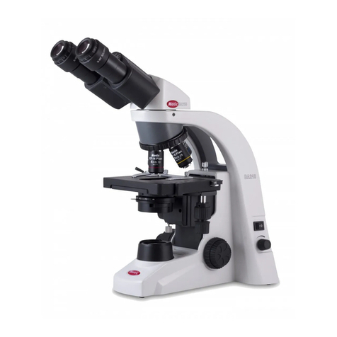
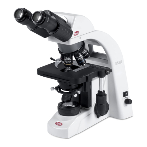
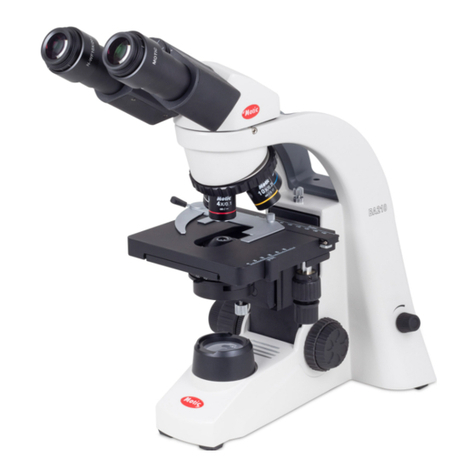
For example BA210:
Eyepiece field of view = 20mm
Objective magnification = 10X
Microscope Terminology
Diameter of the object field = 20/10 = 2.0mm
Diopter adjustment
The adjustment of the eyepiece of an instrument to provide ac-
commodation for the eyesight differences of individual observers.
Depth of Focus
The axial depth of the space on both sides of the image plane
within which the image is sharp. The larger the N.A. of objec-
tive, the shallower the depth of focus.
Field of View (F.O.V.)
That part of the image field, which is imaged on the observer’s
retina, and hence can be viewed at any one time. The field
of view number is now one of the standard markings of the
eyepiece.
Filter
Filters are optical elements that selectively transmit light. It
may absorb part of the spectrum, or reduce overhaul intensity
or transmit only specific wavelengths.
Immers ion Oil
Any liquid occupying the space between the object and micro-
scope objective. Such a liquid is usually required by objectives
of 3-mm focal length or less.
Resolving Power
A measure of an optical system’s ability to produce an image
which separates two points or parallel lines on the object.
Resolution
The result of displaying fine details in an image
Total Magnification
The total magnification of a microscope is the individual magni-
fying power of the objective multiplied by that of the eyepiece.
Working Distance
This is the distance between the objective front lens and the
top of the cover glass when the specimen is in focus. In most
instances, the working distance of an objective decreases as
magnification increases.
X–axis
The axis that is usually horizontal in a two-dimensional coor-
dinate system. In microscopy X-axis of the specimen stages is
considered that which runs left to right.
Y–axis
The axis that is usually vertical in a two-dimensional coordinate
system. In microscopy Y-axis of the specimen stages is consid-
ered that which runs front to back.
