Optopol REVO nx User manual

SOCT User Manual Version 10.0 rev. A
Manufacturer:
OPTOPOL Technology Sp. z o. o.
ul. Żabia 42
42-400 Zawiercie
POLAND
www.optopol.com
User Manual
REVO nx
REVO nx 130
SOCT Copernicus REVO
SOCT Copernicus
REVO 60
REVO 80
REVO FC
Software Version 10.0
User Manual Rev. A
0197
Make sure you read this manual before using
the instrument. Keep this manual in a safe
place so that you can use it in the future.

2 / 374
SOCT User Manual Version 10.0 rev. A
Copyright
© 2021 OPTOPOL Technology Sp. z o. o. All rights reserved. No part of this publication may be stored
in a retrieval system, transmitted or reproduced in any way, including but not limited to photocopy,
photograph, magnetic or other record, without the prior agreement and written consent of OPTOPOL
Technology Sp. z o. o.
User Manual Version 10.0 rev. A
Date of issue of manual: February 2021
Date of review: February 2021
For Customer Service or Technical Service contact your authorized dealer of OPTOPOL Technology
Sp. z o. o. Contact details are available on www.optopol.com.
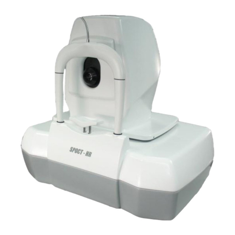
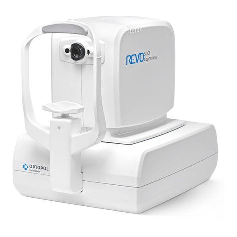
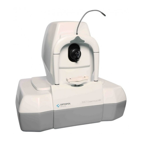
Note: The SOCT Copernicus REVO/SOCT Copernicus/REVO 60/REVO 80/REVO nx
REVO nx 130/REVO FC is hereinafter referred to as the “SOCT”.
The name of application which controls the scanning device is SOCT.
Federal (U.S.A.) law restricts this device to sale, distribution and use by or on the order of a
physician. Proper procedures and techniques are the responsibility of the medical professional.
License and use of the SOCT is intended only for trained medical personnel in accordance with the
license agreement –all other usage is prohibited –warranty restrictions and possible claim
limitations apply.

3 / 374
SOCT User Manual Version 10.0 rev. A
1DESCRIPTION OF THE DEVICE .............................................................................................13
INTENDED USE ......................................................................................................................13
INTENDED USER....................................................................................................................14
THE MINIMUM KNOWLEDGE ............................................................................................14
EDUCATION NEEDED FOR OPERATING THE TOMOGRAPH..........................................................14
OPERATING SKILLS .........................................................................................................14
OCCUPATIONAL SKILLS.....................................................................................................15
JOB REQUIREMENTS FOR THE USER ....................................................................................15
PLACES OF USE.....................................................................................................................15
PATIENT POPULATION.............................................................................................................15
PROPER INSTRUMENT USE.......................................................................................................15
CONTRAINDICATION...............................................................................................................16
INSTRUCTION MANUAL AVAILABILITY.................................................................................16
INSTRUCTION MANUAL APPLICABILITY ...............................................................................16
DISPOSAL .....................................................................................................................17
PROTECTIVE MEASURES FOR IT SYSTEMS ...................................................................................17
CYBERSECURITY FUNCTIONS ....................................................................................................17
SYSTEM OVERVIEW ........................................................................................................17
AUTHENTICATION OF USERS.............................................................................................18
AUTO-LOGOFF ..............................................................................................................18
ENSURE TRUSTED CONTENT.............................................................................................18
CYBERSECURITY EVENT ....................................................................................................18
RECOVER......................................................................................................................18
OTHER IMPLEMENTED MECHANISMS..................................................................................19
2TECHNICAL DATA................................................................................................................20
TECHNICAL DATA REVO NX 130 / REVO NX .............................................................................20
SOCT COPERNICUS REVO/ SOCT COPERNICUS / REVO 60/REVO 80 ........................................21
TECHNICAL DATA REVO FC ....................................................................................................22
DEVICE CLASSIFICATION ..........................................................................................................23
MINIMUM COMPUTER SYSTEM REQUIREMENTS...........................................................................23
CAPTURE STATION ..........................................................................................................23
REVIEW STATION............................................................................................................24
3SAFETY ..............................................................................................................................25
SAFETY INFORMATION ............................................................................................................25
PRODUCT LABEL ...................................................................................................................26
SAFETY STANDARDS ...............................................................................................................28
WARNINGS..........................................................................................................................30
CAUTIONS ...........................................................................................................................34
GENERAL NOTES ...................................................................................................................36
NOTES ON USE .....................................................................................................................38
4 / 374
SOCT User Manual Version 10.0 rev. A
BEFORE USE..................................................................................................................38
AFTER USE....................................................................................................................38
4UNPACKING AND INSTALLATION ........................................................................................39
UNPACKING .........................................................................................................................39
CONNECTING CABLES .............................................................................................................40
DEVICE CONNECTION .............................................................................................................42
FACTORY DEFAULT CALIBRATION AND CONFIGURATION ..................................................................44
5SOCT SOFTWARE ...............................................................................................................45
RUNNING SOCT APPLICATION .................................................................................................45
SOCT APPLICATION STRUCTURE ...............................................................................................45
6PATIENT WINDOW .............................................................................................................46
PATIENT LIST VIEW.................................................................................................................46
CUSTOMIZATION OF THE PATIENT LIST VIEW.........................................................................47
REGISTERING NEW PATIENTS ....................................................................................................47
EDITING PERSONAL DATA.........................................................................................................49
UNREGISTERING PATIENTS .......................................................................................................49
EXAMINATION LIST ................................................................................................................49
DELETING EXAM/S................................................................................................................50
CONNECTING SCANS CONNECTED TO THE WRONG PATIENT.............................................................51
EXPORTING EXAMINATIONS .....................................................................................................51
EXPORT WITH ANONYMIZATION ........................................................................................51
IMPORTING EXAMINATIONS .....................................................................................................51
FILTER .............................................................................................................................52
OUTPUT ..........................................................................................................................52
WORK LIST.......................................................................................................................53
7EXAMINATION ACQUSITION TAB........................................................................................54
SELECTION OF SCAN PATTERN MODE..........................................................................................59
SELECTION OF SCANNING PROGRAM..........................................................................................60
OCT BIOMETRY PROGRAMS.............................................................................................61
OCT TOPOGRAPHY PROGRAMS ........................................................................................61
OCT ANGIOGRAPHY PROGRAMS*.....................................................................................61
FUNDUS CAMERA PROGRAMS*........................................................................................62
OCT + FUNDUS PHOTOGRAPHY*......................................................................................62
SELECTION OF PROTOCOL........................................................................................................63
DEVICE HEAD MOVEMENT CONTROLS ........................................................................................64
EYE PREVIEW........................................................................................................................65
FUNDUS PREVIEW .................................................................................................................66
PSLO LIVE FUNDUS PREVIEW............................................................................................66

5 / 374
SOCT User Manual Version 10.0 rev. A
IR PREVIEW..................................................................................................................66
OPERATION ON THE FUNDUS PREVIEW.......................................................................................68
MOVING THE SCANNING AREA .........................................................................................68
ROTATION OF THE SCANNING ANGLE..................................................................................68
MOVING THE INTERNAL FIXATION TARGET...........................................................................68
CHANGING THE SCAN WIDTH ............................................................................................68
CHANGING THE SCANS DISTANCE .......................................................................................69
FIXATION TARGET CHANGE.......................................................................................................69
CUSTOMIZING SCAN PARAMETERS.............................................................................................69
LIVE OCT PREVIEW............................................................................................................72
ITRACKING ....................................................................................................................73
ACCEPTANCE WINDOW FOR A 3D SCAN..............................................................................73
8CONDUCTING EXAMINATION.............................................................................................76
PREPARATION FOR EXAMINATION..............................................................................................76
ACQUISITION MODES DESCRIPTION ...........................................................................................77
FULL AUTO MODE ..........................................................................................................77
SEMI AUTO MODE..........................................................................................................77
MANUAL MODE.............................................................................................................79
SCANNING PROGRAMS DESCRIPTION .........................................................................................80
RETINA EXAMINATION.....................................................................................................80
CENTRAL EXAMINATION...................................................................................................80
DISC AREA EXAMINATION.................................................................................................81
OCT ANGIOGRAPHY EXAMINATION ...................................................................................82
OCT ANGIOGRAPHY MOSAIC MODE ..................................................................................83
ANTERIOR MEASUREMENT...............................................................................................85
WIDE ANTERIOR PROGRAMS............................................................................................86
BIOMETRY PROGRAM......................................................................................................89
TOPOGRAPHY PROGRAM .................................................................................................89
FUNDUS IMAGE REVO FC (REVO FC ONLY)......................................................................89
FULL RANGE EXAMINATION MODE ............................................................................................94
ANTERIOR FULL RANGE EXAMINATION MODE ......................................................................94
CENTRAL FULL RANGE EXAMINATION MODE ........................................................................95
EXTERNAL FIXATION ...............................................................................................................96
CHORIORETINAL/VITREORETINAL MODE ....................................................................................96
EXAMINATION TIPS ................................................................................................................97
TIPS FOR AUTOMATIC EYE ALIGNMENT...............................................................................97
TIPS FOR MANUAL EYE ALIGNMENT ..................................................................................97
AUTO C-GATE ISSUES......................................................................................................98
TIPS TO SUCCESSFULLY SCAN DIFFICULT PATIENTS ...............................................................101
CRITERA FOR EXAM ACCEPTANCE ...........................................................................................103

6 / 374
SOCT User Manual Version 10.0 rev. A
EVALUATION OF OCT FUNDUS RECONSTRUCTION FOR POSTERIOR 3D EXAMS..........................103
EVALUATION OF OCT TOMOGRAMS FOR POSTERIOR AND ANTERIOR SCANS .............................107
OCT IMAGES ..............................................................................................................109
FUNDUS PHOTOGRAPHY ................................................................................................110
3D EXAMINATION ACCEPTANCE CRITERIA ................................................................................110
SACCADES ..................................................................................................................110
BANDING ...................................................................................................................110
WEAK LOCAL SIGNAL ........................................................................................................111
BLINKS.......................................................................................................................111
FLOATERS ...................................................................................................................111
CROPPED IMAGE..........................................................................................................111
OCT ANGIOGRAPHY ACCEPTANCE CRITERIA .........................................................................111
SEGMENTATION ERRORS ................................................................................................111
IMAGE QUALITY...........................................................................................................112
9RESULT REVIEW ...............................................................................................................114
TYPE OF VIEW MODE...........................................................................................................114
[SINGLE] TAB SCREEN....................................................................................................114
[BOTH EYES] TAB SCREEN ..............................................................................................114
[COMPARISON] TAB SCREEN ...........................................................................................114
[PROGRESSION] TAB SCREEN ..........................................................................................114
LOCK FUNCTION...........................................................................................................114
TYPES OF ANALYSIS..............................................................................................................115
SINGLE RETINA ANALYSIS ...............................................................................................115
AI DENOISE.......................................................................................................................115
3D TAB .....................................................................................................................116
10 POSTERIOR ANALYSIS..................................................................................................117
RETINA THICKNESS ANALYSIS .............................................................................................117
SINGLE TAB ................................................................................................................117
BOTH EYES TAB ............................................................................................................121
COMPARISON..............................................................................................................125
PROGRESSION .............................................................................................................126
OPTIC NERVE HEAD ANALYSIS ....................................................................................129
ADVANCED VIEW – RETINA AND OPTIC NERVE HEAD ANALYSIS.................................................138
STRUCUTRE & FUNCTION – COMBINED OCT AND VF REPORT ..........................................139
RELATIONSHIPS BETWEEN VF AND RNFL/GANGLION MAPS .................................................142
REQUIREMENTS THAT MUST BE MET FOR LOADING PERIMETER TESTS IN THE SOCT APPLICATION ..142
STRUCTURE & FUNCTION – VF RESULTS LAYER IN COMBINED VIEW .......................................143
STRUCTURE & FUNCTION – VF LOCATIONS LAYER ..............................................................145
CENTRAL EXAMINATION ....................................................................................................148
CENTRAL 3D EXAMINATION...........................................................................................148

7 / 374
SOCT User Manual Version 10.0 rev. A
2D SCAN PROGRAMS RESULTS REVIEW.................................................................................149
SINGLE B-SCAN EXAMINATION REVIEW.............................................................................150
RASTER EXAMINATION RESULTS REVIEW............................................................................151
RADIAL EXAMINATION RESULTS REVIEW............................................................................153
CROSS EXAMINATION RESULTS REVIEW.............................................................................154
3D VISUALIZATION...........................................................................................................155
MANIPULATION OF THE 3D CUBE ....................................................................................155
SELECTION OF DISPLAYED LAYERS .....................................................................................155
OPERATION PANEL........................................................................................................156
[SOLID] VIEW .............................................................................................................158
[VOLUME VIEW]..........................................................................................................159
11 ANTERIOR SEGMENT ANALYSIS....................................................................................160
ANTERIOR RADIAL ...........................................................................................................163
[SINGLE] TAB SCREEN....................................................................................................163
[BOTH EYES] TAB SCREEN..............................................................................................165
[COMPARISON] TAB SCREEN ...........................................................................................165
[PROGRESSION] TAB SCREEN ..........................................................................................166
EDIT ANTERIOR SURFACE................................................................................................168
AOD MEASUREMENT ...................................................................................................169
ANGLE MEASUREMENT TOOL..........................................................................................170
CALIPER TOOL .............................................................................................................171
TOMOGRAM REVIEW ANALYSIS .......................................................................................171
12 FULL SCREEN WINDOW ...............................................................................................172
FUNDUS RECONSTRUCTION, EYE PREVIEW OR PSLO................................................................172
IMAGING TOOLS ..............................................................................................................173
MEASUREMENT TOOLS AND ANNOTATIONS ...........................................................................173
BRIGHTNESS AND CONTRAST ADJUSTMENT............................................................................174
FULL SCREEN MODE EXIT ...................................................................................................175
TOMOGRAM WINDOW MANIPULATION ................................................................................175
EDITION OF RECOGNIZED LAYERS.........................................................................................176
MANUAL DISC CONTOUR EDITION .......................................................................................177
REDRAW THE DISC CONTOUR ..........................................................................................178
13 PRINT..........................................................................................................................180
POSTERIOR SEGMENT EXAMINATION REPORTS/OUTPUTS .........................................................180
RETINA 3D.................................................................................................................180
DISC 3D ....................................................................................................................183
OPTIC NERVE HEAD ANALYSIS REPORTS .............................................................................185
TOPOGRAPHY EXAMINATION REPORTS..............................................................................191
FUNDUS EXAMINATION REPORTS.........................................................................................195

8 / 374
SOCT User Manual Version 10.0 rev. A
ANGIOGRAPHY EXAMINATION REPORTS................................................................................197
DISC OCT-A EXAMINATION REPORTS...................................................................................201
BIOMETRY EXAMINATION REPORTS......................................................................................205
MULTI B-SCAN REPORT.....................................................................................................206
MANUALLY SELECT TOMOGRAMS ....................................................................................207
MULTI B-SCAN REPORT FOR BOTH AND COMPARISON VIEW..................................................207
SINGLE TOMOGRAM REPORT..............................................................................................208
SELECT DESIRED PRINTER...................................................................................................208
14 OUTPUT......................................................................................................................209
15 SELECTING FUNDUS PHOTO.........................................................................................210
ADDING FUNDUS PHOTO TO THE EXAMINATION......................................................................210
FUNDUS IMAGE REGISTRATION CORRECTION..........................................................................212
LINKING A FUNDUS PHOTO TO AN EXAMINATION....................................................................212
16 EXAMINATIONS CORRELATION ....................................................................................214
OCT-OCT REGISTRATION .................................................................................................214
AUTOMATIC CORRELATION .............................................................................................214
MANUAL REGISTRATION ................................................................................................215
FUNDUS CORRELETION .....................................................................................................215
AUTOMATIC CORRELATION .............................................................................................215
MANUAL CORRELATION.................................................................................................217
MOVING THE OVERLAID OCT IMAGE ...............................................................................218
CLOSING FUNDUS AND OCT CORRELATION WINDOW ..........................................................219
EXTRACTING TOMOGRAMS FROM A 3D EXAM .......................................................................219
FUNDUS CAMERA – RESULT REVIEW..........................................................................220
COLOR FUNDUS PHOTO [SINGLE] VIEW X 1........................................................................220
COLOR FUNDUS PHOTO [SINGLE] VIEW X 4........................................................................221
COLOR FUNDUS PHOTO FULL SCREEN VIEW........................................................................221
COLOR FUNDUS PHOTO [BOTH] VIEW...............................................................................222
COLOR FUNDUS PHOTO [COMPARISON] VIEW ....................................................................222
17 ANGIOGRAPHY OCT ....................................................................................................223
RETINA OCT-A.............................................................................................................223
[SINGLE] VIEW – STANDARD ..........................................................................................223
[SINGLE] VIEW - DETAILED.............................................................................................224
ANGIO OCT ANALYSIS..................................................................................................226
QUANTIFICATION TOOLS: FAZ, VFA, NFA............................................................................234
FAZ TOOL ..................................................................................................................234
VFA TOOL ..................................................................................................................237
NFA TOOL..................................................................................................................238

9 / 374
SOCT User Manual Version 10.0 rev. A
QUANTIFICATION MAPS [DENSITY AND SKELETON].............................................................240
ANGIO OCT ANALYSIS TABLE .........................................................................................244
[BOTH] VIEW..............................................................................................................244
[COMPARISON] VIEW....................................................................................................246
[PROGRESSION] VIEW ...................................................................................................247
MOTION CORRECTION......................................................................................................248
MOTION CORRECTION IN 3D EXAMINATIONS ....................................................................249
DISC OCT-A..................................................................................................................250
[SINGLE] VIEW............................................................................................................250
[BOTH] VIEW ..............................................................................................................251
ANGIO DISC COMPARISON VIEW .....................................................................................253
[PROGRESSION] VIEW ...................................................................................................253
MOSAIC ........................................................................................................................254
CORRELATION STATUS ...................................................................................................256
SELECT SCREEN............................................................................................................256
OPERATION ON THE MOSAIC...........................................................................................256
18 BIOMETRY OCT ...........................................................................................................259
BIOMETRY ACQUISITION MODE .........................................................................................261
FULL AUTO MODE........................................................................................................263
SEMI AUTO MODE........................................................................................................264
ACCEPTANCE SCREEN ....................................................................................................266
WHITE TO WHITE ........................................................................................................267
RESULT REVIEW ...............................................................................................................268
SINGLE VIEW ..............................................................................................................269
BOTH VIEW.................................................................................................................271
FULL SCREEN...............................................................................................................271
19 IOL CALCULATION TAB.................................................................................................274
PERFORMING IOL CALCULATION.........................................................................................275
MARKING IMPLEMENTED LENSES ........................................................................................277
IOL EDITOR....................................................................................................................277
IMPORTING IOL DATA ...................................................................................................278
EXPORTING IOL DATA ...................................................................................................279
ADDING LENSES MANUALLY............................................................................................279
DELETING LENSES MANUALLY..........................................................................................279
VIEWING THE LIST OF LENSES..........................................................................................279
EDITING IOL DATA........................................................................................................280
ADDING ADDITIONAL POWER RANGES AND INCREMENTS ......................................................281
20 TOPOGRAPHY OCT ......................................................................................................282
TOPOGRAPHY - SAFETY NOTES...........................................................................................282

10 / 374
SOCT User Manual Version 10.0 rev. A
TOPOGRAPHY ACQUISITION MODE .....................................................................................284
FULL AUTO MODE........................................................................................................285
SEMI AUTO MODE........................................................................................................286
ACCEPTANCE SCREEN ....................................................................................................287
TOTAL QUALITY FACTOR ................................................................................................288
RESULT REVIEW ...............................................................................................................288
[SINGLE] VIEW ............................................................................................................288
[BOTH] VIEW ..............................................................................................................290
[COMPARISON] VIEW....................................................................................................291
[PROGRESSION] VIEW ...................................................................................................291
ANALYSIS .......................................................................................................................292
CENTRAL KERATOMETRY – SIMK.....................................................................................292
KERATOMETRY (MERIDIAN)...........................................................................................292
KERATOMETRY (SEMIMERIDIAN)....................................................................................293
KERATOCONUS SCREENING ............................................................................................293
PACHYMETRY ..............................................................................................................297
MAP TYPES....................................................................................................................297
COLOR SCALE - STANDARDS ...............................................................................................300
21 CALIBRATION ..............................................................................................................304
CALIBRATION PROCEDURE PREPARATION...............................................................................304
TOPOGRAPHY CALIBRATION ...............................................................................................305
INTITIAL TOPOGRAPHY CALIBRATION PROCEDURE................................................................305
STANDARD CALIBRATION................................................................................................306
CALIBRATION PROCESS ..................................................................................................306
AXIAL LENGTH (BIOMETRY) CALIBRATION..............................................................................308
BIOMETRY CALIBRATION WITH THE IOL CALCULATION TAB UNACTIVATED .................................308
BIOMETRY CALIBRATION WITH THE IOL CALCULATION TAB ACTIVATED......................................309
CALIBRATION PROCESS ..................................................................................................310
ENTERING BIOMETRY CALIBRATION PARAMETERS ................................................................311
COMMON CALIBRATION ................................................................................................311
22 SETUP WINDOW .........................................................................................................315
GENERAL .......................................................................................................................315
DATABASE......................................................................................................................316
STORAGE .......................................................................................................................318
USERS ACCOUNTS............................................................................................................319
CREATING USER ACCOUNTS ............................................................................................320
LDAP SETTINGS .........................................................................................................321
PREFERENCES .................................................................................................................322
CMDL INTERFACE........................................................................................................322
DEVICES SETUP............................................................................................................323

11 / 374
SOCT User Manual Version 10.0 rev. A
PARAMETERS TAB ........................................................................................................325
VOICE MESSAGES.........................................................................................................326
RESULTS SETTINGS........................................................................................................327
ANONYMIZATION .........................................................................................................328
VISUAL FIELD ..............................................................................................................331
INPUT SETTINGS WINDOW .............................................................................................332
EDIT DISEASE LIST WINDOW ...............................................................................................333
OUTPUT SETTINGS ...........................................................................................................333
OUTPUT SET WINDOW ..................................................................................................333
I.EXPORTING TOMOGRAMS WITH OR WITHOUT AI DENOISE ..........................................................334
CREATING AN OUTPUT SET.............................................................................................334
GRAPHIC FILE STANDARD ...............................................................................................334
DICOM C-STORAGE OUTPUT SET....................................................................................336
BACKUP.........................................................................................................................337
RECOVERY......................................................................................................................338
DICOM........................................................................................................................338
SYSTEM SETTINGS.....................................................................................................339
MWL SETTINGS.......................................................................................................340
C-STORAGE .............................................................................................................341
INFO TAB .......................................................................................................................341
PATIENT RECORD CHANGE TRACEABILITY AND LOGREADER SOFTWARE ........................................341
23 MAINTENANCE AND CLEANING PROCEDURE ...............................................................343
ROUTINE CLEANING .........................................................................................................343
SOFTWARE MAINTENANCE ACTIVITIES ..................................................................................344
HARD DISK DEFRAGMENTATION .........................................................................................344
ORDERING CONSUMABLES.................................................................................................345
FUSE.............................................................................................................................346
BLOWN FUSES EXCHANGE ..............................................................................................346
24 NETWORK CONFIGURATION........................................................................................347
NETWORK CONNECTION CONFIGURATION.............................................................................347
SOCT NETWORK .........................................................................................................347
SOCT SOFTWARE - VIEWING STATION CONFIGURATION ........................................................348
25 REMOTE CONNECTION................................................................................................350
PTS APPLICATION FOR STRUCTURE & FUNCTION REPORT.........................................................350
26 ENVIRONMENTAL CONDITIONS ...................................................................................354
27 WARRANTY AND SERVICE............................................................................................355
28 UTILIZATION................................................................................................................355

12 / 374
SOCT User Manual Version 10.0 rev. A
29 TROUBLESHOOTING ....................................................................................................356
30 LIST OF FIGURES..........................................................................................................359
31 PRODUCT COMPLIANCE ..............................................................................................369
RADIO INTERFERENCE.......................................................................................................369
CANADIAN REGULATIONS..................................................................................................369
EMC INFORMATION.........................................................................................................369

13 / 374
SOCT User Manual Version 10.0 rev. A
1DESCRIPTION OF THE DEVICE
This manual explains the details of the function and method of application to make a good use
of the SOCT system. Before using the device, read this manual carefully to ensure that you
operate it efficiently and safely. Always keep this manual at hand for reference.
SOCT uses Spectral Domain Optical Coherence Tomography method to obtain 3-dimensional
and cross-sectional images of the retina.
SOCT is an optical coherence tomography system indicated for the in vivo imaging and
measurement of the retina layers, retina nerve fiber layer, and optic nerve head as an aid in
the diagnosis and management of posterior segment diseases as well as imaging of anterior
segment structure. The device produces a light beam, which is focused on the human eye
tissue. The light that reflects from internal structures of an eye is then interferometrically
analyzed by the device. The raw data are processed by a PC to obtain images of eye tissue
cross-sections.
The SOCT contains features including: Retina Thickness, Optical Nerve Head, Retinal Nerve
Fiber Layer, Angle assessment, Cornea measurement, Blood vessels visualization, measuring
distances along the visual axis.
The SOCT software with optional OCT Angiography visualization functionality is indicated as
an aid in the visualization of vascular structures of the retina and choroid.
The SOCT software with optional Corneal Topography function is intended to measure and
visualize anterior and posterior corneal curvatures.
The SOCT software with optional OCT Biometry function is intended for biometric
measurements and visualization of ocular structures and performing IOL power calculations
based on the patient’s biometric data and a selection of recognized IOL calculation formulas.
REVO FC version allows non-contact biomicroscopic imaging that incorporates a high-
resolution digital camera for photographing, displaying and storing images of the retina and
surrounding parts of the eye to be examined under Mydriatic and non-Mydriatic conditions.
REVO FC version provides images which are display only and the device does not provide any
diagnostic, pathological analysis or classification of ocular health based on the acquired
images.
Intended use
The SOCT is intended for use as a diagnostic device to aid the detection and management of
ocular diseases, including but not limited to, age-related macular degeneration, macular holes,
diabetic retinopathy, macular edema and glaucoma.
With optional OCT Biometry software feature the device is intended to measure ocular
structure along the eye axis. It measures the following parameters: Axial Length (AL), Central
Corneal Thickness (CCT), Anterior Chamber Depth (ACD), Lens Thickness (LT), Pupil Diameter

14 / 374
SOCT User Manual Version 10.0 rev. A
(P), White-to-White distance (WTW). The measurement and visualization assist in the
determination of the appropriate power and type of intraocular lens.
With optional OCT Topography software feature the device is intended to quantify curvatures
of the anterior and posterior surfaces of the cornea. It measures the following parameters:
Corneal curvature (K1 and K2 keratometry values), Cylindrical power (CYL) and Axis, Average
and Total corneal power (ACP and TCP), anterior and posterior power and Keratoconus
prediction index (KPI).
With optional OCT Angiography software feature the device is indicated as an aid in the
visualization of vascular structures of the retina and choroid.
FC version provides the images of the retina and external area of the eye and are intended for
use as an aid to clinicians in the evaluation, diagnosis and documentation of ocular health.
NOTE: The SOCT is not intended to be used as the sole diagnostic aid for disease
identification, classification and management. A patient diagnosis is the sole domain
of eye care clinician.
DISCLAIMER: OPTOPOL Technology is not offering and does not offer advice,
instruction in the diagnosis and interpretation of SOCT images. It is the clinician’s
responsibility to make diagnosis and interpretation of OCT scans.
Intended User
The SOCT can be used by Ophthalmologists, Optometrists, Opticians, Orthoptists or other
trained health personnel.
The minimum knowledge
The user must be able to distinguish right eye and left eye, chin and forehead, read and
understand the language and the functionality used in the SOCT application software.
Education needed for operating the tomograph
Ophthalmologist, Optometrist, Nurse, Certified Medical Technician, Assistant.
Operating Skills
For instrument operation, the user should be able to perform the following functions:
-Power on and power off the device,
-Disinfect surfaces which interact with the patient,
-Enter and modify the patient data,

15 / 374
SOCT User Manual Version 10.0 rev. A
-Adjust position of the patient, position of the instrument, the table height, and the
patient’s chair,
-Set the examination parameters, acquire examination,
-Select or create the required type of printout,
-Verify the printout for completeness of data,
-Print, export and save the selected report,
-Archive and recover the patient database.
Occupational skills
The user should be able to work with elderly patients, disabled patients and with children.
Job requirements for the user
The SOCT training in the scope of usage and operation is mandatory before the first use. The
user should be also trained in the analysis and treatment of ophthalmic diseases or other eye-
related medical issues, as it is stated by governing bodies. The user should be a computer
literate person.
Users must be suitably trained and be familiar with the instructions, cautions, warnings
contained in this user manual.
CAUTION: This manual does not provide guidance on interpretation of clinical
results. The clinician must ensure that he or she has received appropriate medical
training in such interpretation. OPTOPOL Technology Sp. z o. o. cannot be held
responsible for misdiagnosis of results.
Places of Use
The device is intended for use in hospitals, eye care center/clinics, and surgery/operating rooms.
Patient population
The patient must be capable of sitting up straight and keeping his/her head still. They must be
physically and mentally able to cooperate well and mentally capable of following the
examination. Patients must be at least 5 years old.
Proper instrument use
- Always enter patient information first.
- Clean patient contact surfaces (forehead and chin rest).

16 / 374
SOCT User Manual Version 10.0 rev. A
- To avoid pinching the patient, check the patient’s head position before raising the chin
rest
- Clean the ocular lens frequently to ensure good image quality.
- Adjust power table height properly to ensure patient comfort during the examination.
- Raise or lower the patient’s head so the eye aligns with the canthus mark on the chin
and forehead rest assembly.
- Warn others not to sit or stand on any part of the table, including the base and the top.
- When lowering the table, make sure that pinch point areas are clear of people and
articles; do not store articles in these areas.
Note: Chemically induced pupil dilation is not normally needed.
This device is not designed, sold or intended for use except as indicated.
Contraindication
Do not use the SOCT for those patients who:
-Have an anamnestic history of photodermatosis,
-Have undergone photodynamic therapy (PDT) within48 hours (refer to the product
document of administered photosensitizer about the prohibition period),
Instruction Manual Availability
The SOCT Instruction Manual in PDF format is available on the computer with installed SOCT
application. To open the file, press START → Application → SOCT → User Manual. The manual
is included on USB flash drive delivered with the standard package of the SOCT. Install the .pdf
file viewer e.g. free Adobe Reader from the www.adobe.com website or flash drive to read the
manual in PDF format.
The Instruction Manual in paper form is available on request. To receive user manual in
hardcopy please contact authorized OPTOPOL representative to receive it.
Instruction Manual Applicability
This document applies to the SOCT running with the application software version 8.0 or
higher, unless superseded.

17 / 374
SOCT User Manual Version 10.0 rev. A
Disposal
For disposal at the end of the product life cycle, please follow national regulations.
CAUTION: Do not use this instrument for purposes other than intended and
specified.
Protective Measures for IT Systems
Manufacturer recommends operating SOCT only on password-protected systems.
SOCT must only be operated on virus-protected computers/networks. The institution
operating the device is responsible for the safety of the network.
When using external storage media, the user is responsible for ensuring that the media is free
from viruses. The consequences of virus attacks cannot be foreseen.
Manufacturer will not accept any liability for damage due to a computer virus.
NOTE: User must periodically backup patient data. Manufacturer will not accept any
liability for lost data.
Manufacturer recommends the use of an established antivirus software and/or firewall such
as e.g. Norton, McAfee.
The SOCT is tested by the manufacturer with anti-virus protection provided with Windows 10
“Microsoft Security Essentials”.
Make sure that your operating system, your medical device software and your antivirus
software are always working properly and up-to-date.
Make sure that all changes, updates and patches including operating systems are validated
prior to installation.
For questions regarding cyber security contact Manufacturer.
Cybersecurity Functions
The purpose of this section is to summarize the cybersecurity controls for the SOCT with Windows
10 operating system.
Cybersecurity risk management is a shared responsibility among stakeholders including the
medical device manufacturer, the user, and the health care facility. Failure to maintain
cybersecurity can result in compromised device functionality, loss of data availability or integrity,
or expose other connected devices or networks to security threats.
System Overview
The SOCT device has the following interfaces that are critical for cybersecurity:
-ETHERNET port /Wireless connection for DICOM interface

18 / 374
SOCT User Manual Version 10.0 rev. A
-USB ports for connecting to various USB devices
Authentication of Users
The SOCT device uses Microsoft Windows 10 as the operating system. The operating system
itself allows the end user to establish and configure “User Accounts” and “User Passwords” so
that authentication is performed by password.
Auto-Logoff
User can select one of two available methods of automated log-off when working with the
SOCT application.
- The operating system has the ability to prevent access and misuse by unauthorized users if
the device is left idle for a period of time. The length of inactivity time before auto-
logoff/screen lock is user/administrator configurable. The auto-logoff/screen lock can be
manually invoked by the user.
- The SOCT application also comes with an automated log-off capability which the user can
easily setup in chapter 22.4 Users accounts.
For questions regarding cyber security contact the Manufacturer.
Ensure Trusted Content
Restrict Software of Firmware Updates to Authenticated Code. Software and firmware updates
are performed by OPTOPOL representative from a protected source. All updates require a
Privileged account.
Cybersecurity event
Provide information to the end user concerning appropriate actions to take upon detection of
a cybersecurity event.
-Disconnect the SOCT device from any network
-Contact the IT Administrator at the user facility for on-site evaluation
-Run a scan using the anti-virus software
-Quarantine and delete any identified threats using the anti-virus software
-Restore the database
-Reconnect to the network
-Contact OPTOPOL Technology representative if additional assistance is required
Recover
Methods for retention and recovery of device configuration by an authenticated privileged
user.
• The SOCT provides an option for a data backup on internal and externa storage
• The SOCT allows configuration to be backed up automatically at each launch or close of the
SOCT application

19 / 374
SOCT User Manual Version 10.0 rev. A
Other implemented mechanisms
1.8.7.1 Institutional IT Infrastructure
The SOCT software uses the Windows 10 operating system and supports integration into the
IT infrastructure and domain at the institution or facility where the device is installed. Some
facilities/institutions will have their own cybersecurity infrastructure, such as remote control
of User Accounts, firewalls, encryption, and so forth. The SOCT device will support these site-
specific IT systems and this is verified during the installation process by OPTOPOL
representatives.
1.8.7.2 Stand Alone Mode
The SOCT can be run completely without internet connection. There is no specific requirement
to be connected to the internet for the device to operate properly.
1.8.7.3 Cybersecurity and Data Back-up Configurations
-On the Windows 10 PC where SOCT will be installed the “Windows Firewall” has to be
enabled.
-The SOCT was validated on computers with anti-virus protection provided by
“Microsoft Security Essentials”
-Data encryption can be added by a third-party tool
-The SOCT provides an option for an external data backup
-OPTOPOL Technology recommend to backup data on the drive with RAID system

20 / 374
SOCT User Manual Version 10.0 rev. A
2TECHNICAL DATA
Technical Data REVO nx 130 / REVO nx
OCT Image
Signal source Super Luminescent Diode (SLED)
Wavelength 830 nm
Scanning speed
REVO nx 130 130 000 measurements per second
REVO nx 110 000 measurements per second
Optical power 1575 µW ± -4,5%
Signal analysis Spectral domain Optical Coherence Tomography
Scanning programs 3D, B-scan, Radial HD, Raster, Cross, Angio
1
, Axial length
2
,
Topography2
Axial resolution 5 µm (in tissue)
Transversal resolution ideal 12 µm
typical 18 µm
Retina scan width 5 – 12 mm
Anterior scan width 3 – 16 mm
Scan depth 2.4 mm / 4.8 mm in Full Range mode
Working distance Posterior segment 23 mm
Anterior segment 52 mm
Anterior segment with adapter 27 mm
Power supply 100 – 240 V, 50 / 60 Hz
Power consumption 115 – 140 VA
Fuse ratings 2 x F 3,15 A L 250 V
Multiple socket-outlet max. load 500 VA
Dimensions 556 mm L × 382 mm W × 469 mm H
Weight 23 kg
1
- OCT Angiography is an optional software module available for the SOCT device. If you do not have this
feature and want to purchase it, contact Optopol’s local distributor.
2
- Biometry and Topography are optional software modules. If you do not have this feature and want to
purchase it, contact Optopol’s local distributor.
This manual suits for next models
6
Table of contents
Other Optopol Medical Equipment manuals

Optopol
Optopol SOCT Copernicus User manual

Optopol
Optopol SOCT Copernicus HR User manual

Optopol
Optopol SOCT Copernicus REVO Quick start guide

Optopol
Optopol SOCT Copernicus REVO User manual

Optopol
Optopol SOCT Copernicus REVO User manual

Optopol
Optopol SOCT Copernicus User manual

Optopol
Optopol SOCT Copernicus + User manual
Popular Medical Equipment manuals by other brands

hadewe
hadewe Hyperios4 CHROME manual

Nobu Baby
Nobu Baby Dr. Benny instruction manual

BIOTRONIK
BIOTRONIK Enitra 6 Series Technical manual

Abbott
Abbott AMPLATZER Sizing Balloon II Series Instructions for use

SphygmoCor
SphygmoCor XCEL Service manual

HEBU medical
HEBU medical Diamond HB 8864 Operating and service manual