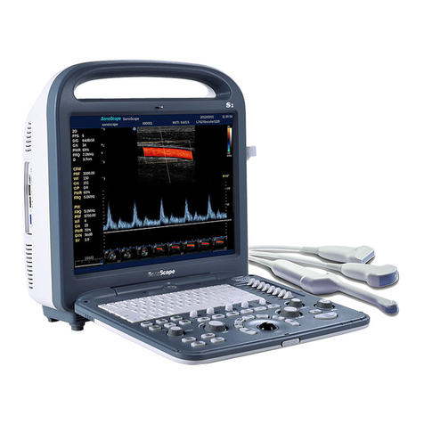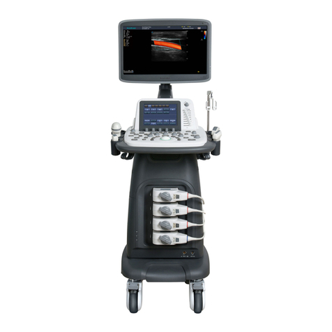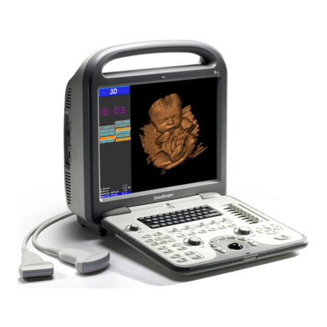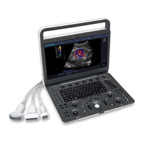
SSI-6000/SSI-5800/SSI-5500/SSI-5500BW
Digital Color Doppler Ultrasound System
When referring to the TI for potential thermal effect, a TI equal to 1 does not mean
the temperature will rise 1 degree C. It only means an increased potential for ther-
mal effects can be expected as the TI increases. A high index does not mean that
bioeffects are occurring, but only that the potential exists and there is no consider-
ation in the TI for the scan duration, so minimizing the overall scan time will reduce
the potential for effects. These operator control and display features shift the safety
responsibility from the manufacturer to the user. So it is very important to have the
Ultrasound systems display the acoustic output indices correctly and the education
of the user to interpret the value appropriately.
RF: De-rating factor
In Situ intensity and pressure cannot currently be measured. Therefore, the acous-
tic power measurement is normally done in the water tank, and when soft tissue
replaces water along the ultrasound path, a decrease in intensity is expected. The
fractional reduction in intensity caused by attenuation is DENOTED by the de-rating
factor RF,
RF=(−.a·f·z)
Where a is the attenuation coefficient in dB cm-1 MHz-1, f is the transducer center
frequency, and z is the distance along the beam axis between the source and the
point of interest.
De-rating factor RF for the various distances and frequencies with attenuation co-
efficient 0.3dB cm-1 MHz-1 in homogeneous soft tissue is listed in the following
table. An example is if the user uses 7.5MHz frequency, the power will be at-
tenuated by .0750 at 5cm, or 0.3x7.5x5=-11.25dB. The De-rated Intensity is also
referred to as ’.3’ at the end (e.g. Ispta.3).
Distance Frequency (MHz)
(cm) 1 3 5 7,5
1 0,9332 0,8128 0,7080 0,5957
2 0,8710 0,6607 0,5012 0,3548
3 0,8128 0,5370 0,3548 0,2113
4 0,7586 0,4365 0,2512 0,1259
5 0,7080 0,3548 0,1778 0,0750
6 0,6607 0,2884 0,1259 0,0447
7 0,6166 0,2344 0,0891 0,0266
8 0,5754 0,1903 0,0631 0,0158
I’=I*RF Where I’ is the intensity in soft tissue, I is
the time-averaged intensity measured in water.
Tissue Model
Tissue temperature elevation depends on power, tissue type, beam width, and
scanning mode. Six models are developed to mimic possible clinical situations.
P/N: 4701-0061-01B
1-7


