CEFLA MyRay Hyperion X9 User manual

97050543
Rev. 17
2018-12
hyperion X9
EN

2
OPERATOR'S MANUAL
EN
ITALIANO

EN
OPERATOR'S MANUAL
3
Contents
1. INTRODUCTION AND INDICATIONS FOR USE .....................................................................................................5
1.1. DESCRIPTION OF THE MANUAL.......................................................................................................................6
1.2. GENERAL WARNINGS........................................................................................................................................6
1.3. REQUIREMENTS (NOT PROVIDED WITH THE PRODUCT) ............................................................................7
1.4. STANDARDS AND REGULATIONS ....................................................................................................................7
1.5. CLASSIFICATIONS..............................................................................................................................................8
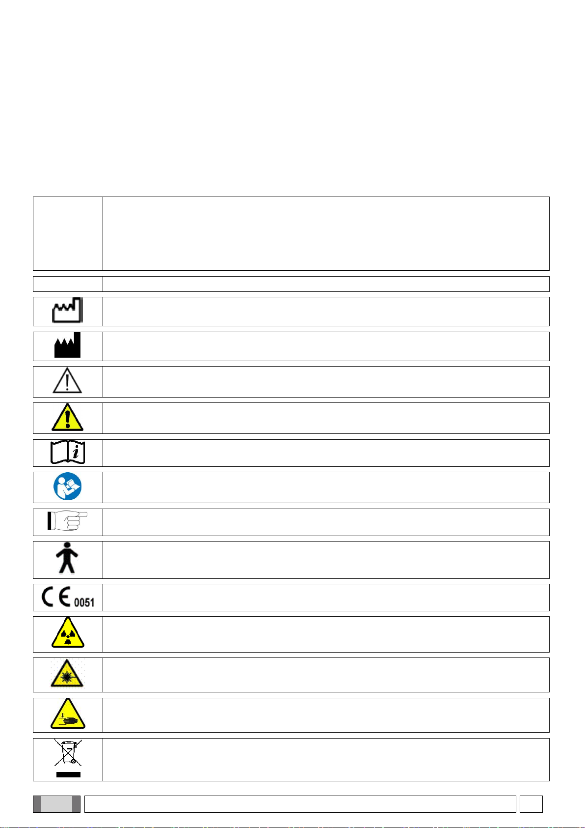
1.6. STYLISTIC CONVENTIONS ................................................................................................................................8
1.7. GENERAL SAFETY WARNINGS.........................................................................................................................9
1.7.1. INSTALLATION CONDITIONS .....................................................................................................................9
1.7.2. CONDITIONS OF USE ...............................................................................................................................10
1.7.3. WARRANTY................................................................................................................................................10
1.7.4. MAINTENANCE AND DISPOSAL ..............................................................................................................11
1.7.5. CLEANING AND DISINFECTION...............................................................................................................12
1.7.6. HYGIENE PROCEDURES FOR PATIENT PROTECTION ........................................................................13
1.8.SAFETY WARNINGS.........................................................................................................................................13
1.8.1. CONDITIONS OF USE ...............................................................................................................................13
1.8.2. GENERAL SAFETY ....................................................................................................................................13
1.8.3. SAFETY DURING X-RAY DEVICE MOVEMENTS ....................................................................................14
1.8.4. EMERGENCY BUTTON .............................................................................................................................15
1.8.5. CONDENSATE FORMATION.....................................................................................................................15
1.8.6. ELECTROSTATIC DISCHARGE ................................................................................................................15
1.8.7. EXPOSURE TO LASER RADIATION.........................................................................................................15
1.8.8. ELECTROMAGNETIC SAFETY .................................................................................................................16
1.8.9. PROTECTION AGAINST RADIATION .......................................................................................................19
1.8.10. APPLIED PARTS...................................................................................................................................19
1.8.11. STRAY RADIATIONS ............................................................................................................................20
2. DESCRIPTION OF OPERATION............................................................................................................................21
3. COMPONENTS .......................................................................................................................................................22
4. CONTROL PANEL ..................................................................................................................................................24
4.1. CONSOLE ONBOARD THE MACHINE.............................................................................................................24
4.2. PUSHBUTTON PANEL ON TELE-X-RAY ARM ................................................................................................24
4.3. X-RAY EMISSION REMOTE CONTROL ...........................................................................................................25
4.4. PERFORM A SIMULATION (DUMMY RUN) .....................................................................................................25
5. PERFORMING A 2D X-RAY EXAMINATION .........................................................................................................26
5.1. STARTING THE SYSTEM .................................................................................................................................26
5.2. SELECTING THE EXAMINATION FROM THE CONTROL CONSOLE............................................................26
5.2.1. 2D EXAMINATIONS AVAILABLE ...............................................................................................................26
5.2.2. SELECTING AN EXAMINATION ................................................................................................................29
5.2.3. SETTING AN EXAMINATION FOR CHILDREN.........................................................................................30
5.2.4. SETTING A CURRENT EXAMINATION AS FAVOURITE .........................................................................30
5.2.5. SETTING THE PROJECTION TYPE..........................................................................................................31
5.2.6. SELECTING A REDUCED ANATOMIC REGION ......................................................................................32
5.2.7. CONFIGURATION OF THE X-RAY TECHNIQUE FACTORS ...................................................................33
5.3. PREPARATION OF THE X-RAY EXAMINATION..............................................................................................34
5.3.1. DEVICES FOR PATIENT POSITIONING ...................................................................................................34
5.3.2. SENSOR POSITIONING.............................................................................................................................35
5.3.3. PATIENT ACCESS STATUS –MINIMUM WAIT STATUS.........................................................................36
5.3.4. EXAMINATION SUMMARY PAGE .............................................................................................................37
5.3.5. DEVICES FOR EDENTULOUS PATIENTS (OPTIONAL)..........................................................................37
5.4. PATIENT POSITIONING....................................................................................................................................38
5.4.1. LASER TRACES .........................................................................................................................................38
5.4.2. PATIENT POSITIONING DESCRIPTION (CRANIOSTAT) ........................................................................39
5.4.3. CRANIOSTAT MOTOR-DRIVEN SUPPORT .............................................................................................40
5.4.4. PAN, DENT AND SIN EXAMINATIONS .....................................................................................................40
5.4.5. TMJ EXAMINATION....................................................................................................................................42
5.4.5.1. LATERAL TMJ ...................................................................................................................................42
5.4.5.2. FRONTAL TMJ ..................................................................................................................................43
5.4.6. TELERADIOGRAPHIC (CEPH) EXAMINATIONS......................................................................................44
5.4.7. POSITIONING FOR DTS EXAMINATION ..................................................................................................45

4
OPERATOR'S MANUAL
EN
5.5. ACQUISITION OF THE EXAM ...........................................................................................................................46
6. 3D TOMOGRAPHIC EXAMINATION (CBCT).........................................................................................................47
6.1. SELECTING THE EXAMINATION FROM THE CONTROL CONSOLE ............................................................47
6.2. POSITIONING THE PATIENT FOR 3D EXAMINATIONS .................................................................................50
6.3. PERFORMING THE EXAMINATION .................................................................................................................50
7. VIEWING AND SAVING ..........................................................................................................................................51
7.1. VIEWING AND SAVING .....................................................................................................................................51
8. CONTROL CONSOLE.............................................................................................................................................52
8.1. LANGUAGE SELECTION ..................................................................................................................................52
8.2. TOUCH SCREEN DISPLAY ICON.....................................................................................................................53
9. PERIODIC CHECKS AND MAINTENANCE ...........................................................................................................56
9.1. PERIODIC INSPECTIONS TO CHECK THE IMAGE ........................................................................................56
10. TECHNICAL DATA..................................................................................................................................................56
10.1. ELECTRICAL CHARACTERISTICS ..............................................................................................................56
10.2. RADIOLOGICAL CHARACTERISTICS .........................................................................................................57
10.3. RADIOLOGICAL FEATURES IN CBCT MODE.............................................................................................58
10.4. ISODOSE CURVES FOR CBCT EXAMINATIONS .......................................................................................59
10.5. ISODOSE CURVES FOR 2D EXAMINATIONS ............................................................................................59
10.6. CTDI (COMPUTED TOMOGRAPHY DOSE INDEX) MEASUREMENTS .....................................................60
10.7. CBCT DETECTOR CHARACTERISTICS......................................................................................................62
10.8. PANORAMIC SENSOR CHARACTERISTICS(PAN) ....................................................................................62
10.9. TELERADIOGRAPHIC (CEPH) SENSOR CHARACTERISTICS (CEPH) ....................................................62
10.10. LASER CHARACTERISTICS.........................................................................................................................62
10.11. DIMENSIONAL CHARACTERISTICS............................................................................................................63
10.12. ENVIRONMENT CHARACTERISTICS..........................................................................................................63
10.13. PC REQUIREMENTS.....................................................................................................................................64
10.14. IDENTIFICATION LABEL POSITION ............................................................................................................67
11. ERROR MESSAGES...............................................................................................................................................68
12. USER’S LICENCE CONTRACT ..............................................................................................................................70
12.1. GENERAL CONDITIONS OF THE IMAGE SOFTWARE LICENCE .............................................................70
12.1.1. USER’S LICENCE .................................................................................................................................70
12.1.2. COPYRIGHT..........................................................................................................................................70
12.1.3. USE OF THE SOFTWARE PRODUCT AND EXPRESS TERMINATION CLAUSE .............................71
12.1.4. GUARANTEE AND EXCLUSIONS FROM THE GUARANTEE ............................................................71
12.1.5. LIMITS OF RESPONSIBILITY...............................................................................................................71
12.1.6. APPLICABLE LAWS, JURISDICTION AND COURT OF JURISDICTION............................................71
12.1.7. WRITTEN FORM ...................................................................................................................................72
12.1.8. TRANSLATION ......................................................................................................................................72
12.1.9. SOFTWARE COMPLIANCE WITH THE REGULATIONS IN FORCE ..................................................72
13. INSPECTION AND MAINTENANCE .......................................................................................................................73
13.1. USER INSPECTION.......................................................................................................................................73
13.2. TECHNICAL MAINTENANCE........................................................................................................................74

EN
OPERATOR'S MANUAL
5
1. INTRODUCTION AND INDICATIONS FOR USE
hyperion X9 is an extraoral X-ray system for digital panoramic exams, tele-X-rays and tomographies, intended to:
(I) produce orthopanoramic images of the maxillofacial region and carry out diagnostic examination on teeth, dental
arches and other structures in the oral cavity;
(II) produce X-ray images of dental arches, cranium parts, carpus, and cephalometric examinations, if equipped with
tele-X-ray arm (CEPH);
(III) produce tomographic images of the oral cavity and maxillofacial structures and carry out diagnostic examination
on teeth, dental arches, structures of the oral cavity and some cranial bones, if equipped with CBCT option.
The system performs tomographic exams with the acquisition of X-ray images through a 360-degree rotating
sequence and the reconstruction of a three-dimensional matrix of the examined volume, thus producing two- and
three-dimensional views of the volume itself. This technique is known as CBCT.
hyperion X9 is a digital X-ray device, suitable for expert professionals, which allows to obtain dental images in a
simple and automatic way. The image is acquired by means of an X-ray detector and a constant potential X-ray
source, powered by a high-voltage and high-frequency generator. Then the image is sent to a computer, in real time
(2D or 3D) or subsequently (2D) according to the needs and choice of the operator.
hyperion X9 allows the following projections:
- standard or panoramic views for paediatric patients (PAN);
- complete or partial views of the teeth, selected by the user (DENT);
- front and side views of maxillary sinuses (SIN);
- side and posteroanterior views of the temporomandibular joints (TMJ), from different angles.
If equipped with tele-X-ray arm (CEPH), hyperion X9 allows the following projections:
- standard or paediatric cephalographies in latero-lateral view;
- cephalographies in anteroposterior and posteroanterior view;
- hand (carpus) X-ray.
If equipped with CBCT option, hyperion X9 also allows to acquire tomographic images.
hyperion X9 is intended for use in dentistry, in the following fields:
- endodontics;
- periodontology;
- dental prosthesis;
- functional diagnosis and therapy of craniomandibular dysfunctions;
- dental surgery;
- dental implants;
- maxillofacial surgery;
- orthodontics.
Contraindications:
- visualisation of cartilaginous structures;
- the CBCT technique has a limited ability to detect soft tissues.
The manufacturer’s website contains a list of authorised agents.

6
OPERATOR'S MANUAL
EN
1.1. DESCRIPTION OF THE MANUAL
This manual is an essential consultation tool and contains important information and instructions for use of
the X-ray system and the relative controls.
These instructions describe how to properly and safely use the digital X-ray system.
Carefully read and familiarise yourself with the entire contents of the manual before attempting to use the
system.
For use of the software, refer to the specific manual.
The manual is only provided in electronic format on a DVD and can be consulted directly on the PC screen
during use.
It is advisable to keep a copy of this manual within reach with the aim of training the operators and as guide
for consultation during use of the device. This manual also contains all the essential information for safety of
the patient, the operator and the device.
It is therefore advisable to particularly carefully read the paragraphs on the safety rules.
The original text of this manual is in Italian.
1.2. GENERAL WARNINGS
The digital X-ray system with its drivers and software have been developed and manufactured by CEFLA s.c. - Via
Selice Prov.le 23/A 40026 Imola (Italia), hereinafter referred to as the Manufacturer, which is the manufacturer and
distributor in compliance with the EC Medical Device Directive.
In order to use the system, when it includes also the optional tomography function, the user must have a Personal
Computer with a suitable software for capturing and saving images; further information about its installation and use
is included in the Software user manual. If the system is used for the acquisition of X-ray and cephalometric images
only, a Personal Computer can be used but is not essential. Carefully read this Manual and the Computer and
Software manuals before using the equipment.
- The contents of this publication are valuable trade secrets and must not be given to third parties, stored, copied,
reproduced, disclosed or transferred in any manner (via computer, photocopies, translations or other means)
without the prior written consent of the Manufacturer.
- The Manufacturer pursues a policy of continual improvement of its products; therefore, some specific instructions
and images contained in this manual may differ from the product purchased.
- The Manufacturer reserves the right to make changes without prior notice.
- The information, technical specifications and illustrations contained in this publication are not binding. The
Manufacturer reserves the right to make technical modifications and improvements without modifying these
instructions.
- All the registered trademarks and the product names mentioned are the property of the respective owners.
- Carefully read the USER LICENSE AGREEMENT before using the product. When installing the program, you will
explicitly be asked to accept the agreement; if you do not accept, the program cannot be installed.

EN
OPERATOR'S MANUAL
7
Please pay particular attention to the sections in the manual where the following symbols appear:
Patient or operator safety-related warnings.
Important information on product use.
For 3D machines only.
In accordance with the privacy laws in force in several countries, all sensitive personal information must be
adequately protected. In addition, patients must sign a consent form before any personal information or
images are transmitted across networks. If required by the laws in force, dentists are obliged to protect data
using a protection password. Refer to the Microsoft® Windows operating system manual for data access
protection methods by means of password.
It is recommended to regularly (at least once a week) make a backup copy of the databases. This will allow
restoring the data in the event of damage to the hard disc of the PC or the databases themselves.
1.3. REQUIREMENTS (NOT PROVIDED WITH THE PRODUCT)
For proper functioning, the device requires connection to a personal computer (PC) and the relative software. For the
minimum requirements of the PC, refer to the paragraph TECHNICAL DATA.
The PC is not included with the device. It is recommended to only use a PC compliant to the IEC 60950-
1:2007 standards for information technology devices.
1.4. STANDARDS AND REGULATIONS
The system has been designed to meet the following standards:
- Directive 93/42/EEC and subsequent amendments and additions
(dir. 2007/47/EC) - Medical Devices Directive;
- Directive 2006/42/EEC - Machinery Directive.
Technical Standards:
IEC 60601-1:2005
IEC 60601-1-2:2014
IEC 60601-1-3:2008
IEC 60601-2-63:2012
IEC 60601-1-6:2010
IEC 62366-1:2015
IEC 62304:2006
The CE marking certifies compliance of the product as described herein with Medical Device Directive
93/42/EEC and subsequent amendments.

8
OPERATOR'S MANUAL
EN
1.5. CLASSIFICATIONS
The system is classified as Class I and Type B as regards safety according to IEC 60601-1.
The system is classified as a Class IIB medical electrical X-ray device in accordance with Medical Device Directive
93/42/EEC and subsequent amendments.
1.6. STYLISTIC CONVENTIONS
The following symbols may be found on the X-ray device and in the Manual:
CEFLA s.c.–
via Selice
Provinciale
23/A - 40026
Imola (BO)
Italia
Name of the Manufacturer and place of production.
SN
Product serial number.
Date of manufacture (month / year).
Manufacturer.
Symbol “Possible hazard: Read the user manual”.
Patient or operator safety-related warnings.
Consult the enclosed documentation before using the relevant part of the equipment.
It is necessary to read the user's manual before using the device.
This symbol in the Manual identifies the paragraphs containing important information on the use of
the product.
Applied part of type B, according to IEC 60601-1.
Equipment compliant with directive 93/42/EEC as amended.
Notified body: IMQ spa.
Ionizing radiation warning symbol.
Class 1 LASER radiation warning symbol.
Crushing hazard.
Disposal symbol in accordance with Directive 2012/19/EU.

EN
OPERATOR'S MANUAL
9
POWER
Power switch.
Product/equipment identification code.
I
Unit ON.
O
Unit OFF.
Ukraine compliance mark.
1.7. GENERAL SAFETY WARNINGS
These instructions describe how to use the system correctly. Please carefully read this manual before using
the device.
The owner or the installation site manager is obliged to verify compliance with the local regulations in force and/or
ask a Qualified Expert for advice. Pay particular attention to fulfilling the obligations of the law regarding the
protection of workers, the population and patients against radiation.
The main REFERENCE STANDARDS are listed in this manual (1.4 - Standards and Regulations).
Do not use the system for uses different from those described in the indications for use (Introduction) and do not use
it if you do not have the necessary knowledge in the dentistry and radiological field.
The law restricts and reserves the sale and use of this device exclusively to physicians, dentists or radiology
specialists.
1.7.1. INSTALLATION CONDITIONS
- The system must not be used if it shows any electrical, mechanical or radiation defect. Like for all medical
electrical systems, this device requires proper installation, use, maintenance and service with the aim of assuring
safe and efficient operation.
- The entire system must be installed by a technician authorised by the Manufacturer under supervision of a
Qualified Expert.
- The room where the system is installed must exclusively be for medical use and designed by an expert in
protection against the risks associated with exposure to radiation in accordance with the regulations in force in
the country of use.
- For Europe, the electric system in the room where the device is installed must comply with the IEC 60364-7-710
standards (requirements for electric systems in rooms used for medical purposes).
- The X-ray system requires special precautions with respect to electromagnetic compliance and must be installed
in accordance with the recommendations given in the paragraph “Electromagnetic safety” in this manual.
- The maximum dimensions reached while the unit is being installed must be taken into consideration to avoid
banging into any objects present in the room. Refer to the dimensional diagram in the service manual.
- Make sure that the operator can communicate verbally and visually with the patient during the examination.
- The system can be installed in the following configurations:
1. wall mounting;
2. on the floor surface with a static baseplate (optional).
- Installation not in compliance with the instructions provided by the Manufacturer might cause increased
electromagnetic emission of the X-ray system and reduce its immunity to disturbances.
- In particular, use a screened cable for connection of the X-ray remote control and make the connection as
specified in the technical manual.

10
OPERATOR'S MANUAL
EN
For further details, refer to the installation template and the detailed instructions given in the service manual.
1.7.2. CONDITIONS OF USE
The equipment may only be used by authorised and adequately trained staff (dentists and paramedics).
Use in compliance with national regulations for protection against ionising radiation, e.g.:
(a) Three-dimensional imaging must not be used for routine screening exams. Three-dimensional imaging
exams must be prescribed according to the patient's needs.
(b) Each exam must be justified by evidence that the benefits outweigh the risks.
(c) All patients must wear leaded aprons with thyroid collar.
(d) Before the exam, ask women of reproductive age if they are pregnant or if there is any possibility that
they might be. If there is, the patient should not undergo the exam, unless a radiologist from a trustworthy
hospital is consulted in order to evaluate, together with the patient and the operator, the benefits and the
risks associated with this procedure, taking into account the possibility of choosing a different kind of exam.
(e) The operator must keep a safe distance, protect him/herself with an adequate shielding and remain
close to the patient in the exam room only if the patient needs assistance. If the operator has to remain in
the exam room, he/she must wear a leaded apron with thyroid collar.
(f) Inform the patient about the risks associated with the exam, acquire his/her informed consent and archive
the document.
In case of claims or need of technical assistance, users in Brazil are required to contact the following email
address: servico.odontologico@cefla.it.
Users in the USA market are required to contact:
Cefla North America Inc.,
6125 Harris Technology Blvd., Charlotte, NC, 28269 United States
1.7.3. WARRANTY
The Manufacturer guarantees the safety, reliability and performance of the device.
The warranty is valid only under the following terms:
-closely observe the conditions specified in the warranty certificate itself;
-the equipment is only to be used as instructed in this manual;
-equipment installation, upgrade and technical support must be performed exclusively by personnel
authorised by the Manufacturer to carry out these operations;
-do not open the device covers: installation, repairs and in general all the operations that require opening
the device must be carried out exclusively by technicians authorised by the Manufacturer;
-the equipment is to be installed in rooms that satisfy the requirements specified in the manual;
-the room where the X-ray unit is installed must be in compliance with the official directives that govern
radiation protection in the country of use.

EN
OPERATOR'S MANUAL
11
1.7.4. MAINTENANCE AND DISPOSAL
Never remove the device covers.
The device does not contain parts that can be repaired directly by the user. In the event of malfunctioning, do not
attempt to carry out any type of maintenance operation. If you find or suspect any kind of system malfunctioning, do
not attempt to carry out any type of maintenance operation and do not use the system on a patient, but directly
contact your local distributor.
The user may not carry out maintenance on any mechanical or electronic part of the x-ray system.
Opening the cases to access the internal circuits may cause device breakage and failure of the electrical safety
devices and will lead to forfeiture of the warranty.
Any maintenance, repairs and modifications of the device must be carried out only by personnel directly authorised
by the Manufacturer or by third parties expressly authorised by the Manufacturer and must be carried out according
to the laws in force and the generally accepted technical standards.
All the system components must be checked and replaced, if necessary, by qualified personnel.
For any maintenance operation, please contact the Manufacturer via the website indicated on the cover of this
manual by filling in the Information Request form.
Further information about the device regular inspection and maintenance is provided in the document “hyperion X9 -
Inspection and Maintenance”.
Should you for any reason need to return the device or its parts to the Manufacturer or a Technical Service centre,
disinfect all the external parts of the device using a specific product (see the paragraph “Cleaning and disinfection”)
and preferably return it in its original packaging.
At the end of its lifetime, dispose of the device in accordance with the regulations in force. It is also advisable to
disinfect all the external parts of the device before disposal and to separate the materials for differentiated waste
collection.
In compliance with Directives 2011/65/EU and 2012/19/EU regarding restriction of the use of certain hazardous
substances in electrical and electronic equipment along with waste electrical and electronic equipment, it is forbidden
to dispose of this equipment in the municipal waste stream as unsorted municipal waste. When purchasing a new
device of an equivalent type, one for one, the device that has come to the end of its lifetime should be returned to the
distributor for disposal. As regards reuse, recycling and other forms of recovery of waste electrical and electronic
equipment, the Manufacturer carries out the functions defined by current local laws. Appropriate differentiated waste
collection for subsequent recycling treatment and environmentally friendly disposal contributes to preventing possible
negative effects on the environment and health and encourages recycling of the materials of which the device is
made up. The crossed-out bin symbol on the device indicates that the product must be collected separately from
other waste at the end of its useful life. Under local legislation, fines can be imposed if the equipment is disposed in
an illegal manner.

12
OPERATOR'S MANUAL
EN
1.7.5. CLEANING AND DISINFECTION
Cleaning is the first step necessary in any disinfection process. The physical action of rubbing a surface with
detergents and surface-active agents and rinsing with water removes a large number of microorganisms. If
a surface is not cleaned first, the disinfection process cannot be successful.
When a surface cannot be adequately cleaned, it should be protected with barriers.
The external parts of the device must be cleaned and disinfected using a product for hospital use with indications for
HIV, HBV and tuberculosis (medium-level disinfectant) specifically for small surfaces.
The various drugs and chemical products used in dentist’s surgeries may damage the painted surfaces and the
plastic parts. The tests and research conducted have demonstrated that the surfaces cannot be fully protected
against attack of all the products found on the market. It is therefore recommended to use barrier protections
whenever possible.
The aggressive effects of chemical products also depend on the time they stay in contact with the surfaces. It is
therefore important not to leave the product on the surfaces of the device for longer than the time indicated by the
manufacturer.
It is recommended to use the specific medium-level disinfectant, STER 1 PLUS (CEFLA S.C.), which is compatible
with the painted surfaces, plastic parts and unpainted metal surfaces. Alternatively, it is recommended to use
products that contain:
-96% ethanol Concentration: maximum 30 g for every 100 g of disinfectant.
-1-propanol (N-propanol, propyl alcohol, N-propyl alcohol). Concentration: maximum 20 g for every 100 g of
disinfectant.
-Combination of ethanol and propanol. Concentration: the combination of the two must be at maximum 40 g for
every 100 g of disinfectant.
-Painted surfaces and plastic parts.
- Incidin Spezial (Henkel Ecolab);
- Omnizid (Omnident);
- Plastisept (Alpro) (not tuberculocide as not an alcohol-based disinfectant);
- RelyOn Virkosept (DuPont);
- Green & Clean SK (Metasys) (not tuberculocide as not an alcohol-based disinfectant).
-Do not use products containing isopropyl alcohol (2-propanol, isopropanol).
-Do not use products containing sodium hypochlorite (bleach).
-Do not use products containing phenols.
-Whatever product you use, follow the instructions given by the manufacturer.
-Do not mix the STER 1 PLUS disinfectant with other products.
-Do not spray the product directly on the device surfaces.
For cleaning and disinfection use disposable, soft, non-abrasive
paper (do not use recycled paper) or sterile gauze.
- It is recommended to turn off the device before cleaning and
disinfecting the external parts.
- Everything used for cleaning and disinfection must be thrown
away after use.

EN
OPERATOR'S MANUAL
13
1.7.6. HYGIENE PROCEDURES FOR PATIENT PROTECTION
Single-use hygienic protections are the main means of protection against transmission of cross-infections
between patients. In order to prevent transmission of infectious diseases from patient to patient, it is
essential to always use the single-use protections provided. The single-use protections are classified as
Class I medical device and may not be replaced with others in a lower class.
The single-use protections must be in compliance with the ISO 10993-1 standards on biocompatibility and
approved by the control bodies where required (e.g. FDA, CE).
Always replace the single-use hygienic protections of the bite piece before positioning a new patient.
The single-use hygienic protections (Cefla code 97901337) must be stored in a dry and clean place without
direct exposure to sunlight or UV rays.
The bite piece and the chinrest can be disinfected by immersing them in a cold sterilizing liquid. For
sterilization of these parts, follow the instructions of the supplier of the sterilizing product.
Cover all the components that will come into contact with the hands of the dental staff with single-use
protections, as they might be contaminated by indirect contact with the patient’s mouth. In particular, be
careful how you handle the control console of the device and the touch screen, mouse and keyboard of the
PC.
Prior to positioning patients for any x-ray exposure, always cover the biteblock with a new clean non-sterile
plastic barrier, to avoid cross contamination.
Note to users in Canada: ask your trusted dental material distributor for any plastic barrier that is suitable in
size and is legally marketed in Canada.
According to Health Canada, bite block covers are Class I devices and are distributed by authorized
establishments only, as listed in the MDEL database.
1.8. SAFETY WARNINGS
1.8.1. CONDITIONS OF USE
In order to use the device in safe conditions, refer to the following paragraphs in the manual.
1.8.2. GENERAL SAFETY
-Do not forget to turn off the main switch on the equipment before leaving the surgery.
-The device is not protected against liquid penetration (Class IPX0 –common protection).
-The equipment is not suitable for use in the presence of a mixture of flammable anaesthetic gas with oxygen or
nitrous oxide.
-Portable telecommunications devices (RF) may interfere with the X-ray device; use in the vicinity of the X-ray
device should therefore be prohibited.
-This equipment must be stored properly so that it is kept in top working order at all times.
-The user must be present at all times when the equipment is turned on or ready for start-up. In particular, never
leave the equipment unattended in the presence of children or other unauthorised personnel in general;
-The Manufacturer shall not be held responsible (under civil and criminal law) for misuse, carelessness or
improper use of the equipment.
-If any person who is not an authorised technician changes the product in any way by replacing parts or
components with other ones not used by the Manufacturer, they shall assume responsibility for the product.
-Any computer, monitor, printer, mouse, keyboard and any other device connected to the X-ray device must be
compliant with ISO, IEC, EN or local standards.
-The Manufacturer is not responsible for problems or malfunction of parts and/or components not approved by
itself, not complying with the regulations and not installed by qualified technical personnel acknowledged by the
Manufacturer.

14
OPERATOR'S MANUAL
EN
-Do not use electronic equipment not compliant with IEC 60601-1-2 in proximity of life support equipment (e.g.
pacemakers or heart stimulators) and hearing aids. Before using any electronic device in health facilities, always
check that it is compatible with the other equipment present.
-The X-ray tube contains insulating mineral oil. This oil is potentially hazardous if ingested or if it comes into
contact with the skin or mucous membranes. In the event of a defect or fault, the oil may leak out. Avoid direct
contact with the oil and do not inhale its vapours.
-Do not eat, drink or smoke near the device.
1.8.3. SAFETY DURING X-RAY DEVICE MOVEMENTS
The X-ray device carries out movements in the vicinity of the patient and the operator.
During the execution of the X-rays, the operator controls the movements by holding down dedicated
buttons.
The reset procedure must be carried out before the patient accesses the device.
The operator must stand at a suitable distance from the moving parts. Movements can be stopped at any
time by pressing the emergency button.
During all the movements of the X-ray device, the operator must:
-closely watch over the patient, and if there is a risk of collision between the X-ray device and the patient,
immediately interrupt the movement by releasing the control button;
-not allow the patient to assume incorrect positions (resting the hands or other parts of the body in inappropriate
areas) or to move away from the examination area.
The operator must be careful not to interfere with the sensor movement during its servo-controlled activation.

EN
OPERATOR'S MANUAL
15
1.8.4. EMERGENCY BUTTON
The system is equipped with an emergency button to stop X-ray device operation, located under the patient support
arm in proximity of the telescopic column. A remote emergency button is connected using the dedicated connector on
the board located at the feet of the lifting column.
1 - Emergency button
These buttons must be activated in the event of danger and emergency, for example, failed interruption of radiation
from the source, in situations of evident danger to persons or when an emergency is signalled.
If the emergency button is pressed, radiation emission is immediately interrupted and all the movements of the
patient support and the rotary arm are stopped, all the linear motors run a distance of less than 10 mm and the rotary
movements a distance of less than 2 degrees, and the button remains locked in safe conditions.
After the emergency, turn the button in the direction indicated by the arrow to resume normal operation.
1.8.5. CONDENSATE FORMATION
Following strong temperature oscillations, condensate may form in the X-ray device. Activate the X-ray device only
after an adequate ambient temperature has been reached. See the chapter “Ambient characteristics”.
1.8.6. ELECTROSTATIC DISCHARGE
Electrostatic discharge (ESD).
Electrostatic discharge from persons may cause damage to the electronic components in the event of contact.
Generally, damaged components must be replaced. The repair must be carried out by qualified technicians.
Do not touch the parts at risk marked with the symbol .
1.8.7. EXPOSURE TO LASER RADIATION
The system contains some Class 1 laser diodes in compliance with IEC 60825-1:2014. Three of them are positioned
on the X-ray generator and one on the chin support base.
Both the patient and the operator may be dazzled by laser traces.
-Do not look directly into the laser beam. Be careful that the laser beam does not hit the patient’s eye.
-Keep a distance of at least 10 cm between the eye and the laser.
The position of the laser sources is indicated with the following symbol .

16
OPERATOR'S MANUAL
EN
1.8.8. ELECTROMAGNETIC SAFETY
The device is intended for use in environments recognised as professional health facilities, as described in IEC
60601-1-2:2014. The device belongs to CISPR 11 Class A Group 1 and complies with immunity test levels specified
by IEC 60601-1-2:2014 for professional health facilities.
Before using any electronic device in health facilities, always check that it is compatible with the other equipment
present.
Even if the device complies with standard IEC 60601-1-2, it is recommended not to use it near life-support
equipment (e.g.: pacemakers or cardiac stimulators). For further information, see the equipment instructions
for use.
Use of this equipment adjacent to or stacked with other equipment should be avoided, because it could
result in improper operation. If such use is necessary, this equipment and the other equipment should be
observed to verify that they are operating normally.
Use of accessories, transducers and cables other than those specified or provided by the manufacturer of
this equipment could result in increased electromagnetic emissions or decreased electromagnetic immunity
of this equipment and result in improper operation.
Portable RF communications equipment (including peripherals such as antenna cables and external
antennas) should be used no closer than 30 cm (12 inches) to any part of the device, including cables
specified by the manufacturer. Otherwise, degradation of the performance of this equipment could result.
Do not subject the device to strong electromagnetic disturbances. These disturbances could degrade the
essential performance of the device.
The emission characteristics of this equipment make it suitable for use in industrial areas and hospitals
(CISPR 11 class A). If it is used in a residential environment (for which CISPR 11 class B is normally
required) this equipment might not offer adequate protection to radio-frequency communication services.
The user might need to take mitigation measures, such as relocating or re-orienting the equipment.

EN
OPERATOR'S MANUAL
17
Guidance and Manufacturer's declaration - Electromagnetic emissions
hyperion X9 is designed to operate in the specified electromagnetic environment. The customer or the user of
hyperion X9 must ensure its use in an electromagnetic environment with the following features:
Emission test
Conformity
Electromagnetic Environment
RF emissions
CISPR 11
Group 1
hyperion X9 uses RF energy only for its internal
operation.
For this, the RF emissions are very low and do not
interfere with the electronic devices nearby.
RF emissions
CISPR 11
Class A
hyperion X9 must be used only by adequately trained
personnel (dentists and paramedics). hyperion X9
may cause radio interferences or disturb the operation
of the nearby equipment. It may be necessary to
adopt countermeasures, such as re-orienting or
moving hyperion X9 or shielding the installation site.
Harmonic emissions
IEC 61000-3-2
Class A
Voltage fluctuations/
flicker emissions
IEC 61000-3-3
Compliant
Guidance and Manufacturer's declaration - Electromagnetic immunity
hyperion X9 is designed to operate in the specified electromagnetic environment. The customer or the user of
hyperion X9 must ensure its use in an electromagnetic environment with the following features:
Immunity test
IEC 60601-1-2
Test level
Level of
conformity
Electromagnetic Environment
Electrostatic
discharge (ESD)
IEC 61000-4-2
± 8 kV contact
± 15 kV air
IEC 60601-1-2
Test level
Floors must be made of wood, concrete or
ceramic tiles. If floors are covered with
synthetic material, the relative humidity
should be at least 30%.
IEC 61000-4-4
fast/burst electric
transients
± 2 kV for electrical
lines
± 1 kV for input/output
lines > 3 m
IEC 60601-1-2
Test level
The power supply line quality should be that
of a typical commercial or hospital
environment.
Overvoltage
IEC 61000-4-5
± 1 kV differential
mode
± 2 kV standard mode
IEC 60601-1-2
Test level
The power supply line quality should be that
of a typical commercial or hospital
environment.
Voltage drops,
short interruptions
and voltage
change on the IEC
61000-4-11 input
electric line
Ut = 0% (a 0, 45°
90°, 135°, 180°, 225°,
270°, 315°) for 0.5
cycles
Ut = 0% per 1 cycle
Ut = 70% (a 0°) per
25/30 cycles
Ut = 0% per 250/300
cycles
IEC 60601-1-2
Test level
The power supply line quality should be that
of a typical commercial or hospital
environment. If the hyperion X9 user
requires a continuous operation also in
case of blackout, it is recommended to
power the hyperion X9 with uninterruptible
power supply or batteries.
Magnetic field at
network frequency
(50/60 Hz)
IEC 61000-4-8
30 A/m
IEC 60601-1-2
Test level
The magnetic fields at network frequency
should feature levels typical of a standard
commercial or hospital environment.

18
OPERATOR'S MANUAL
EN
Guidance and Manufacturer's declaration - Electromagnetic immunity
hyperion X9 is designed to operate in the specified electromagnetic environment. The customer or user of
hyperion X9 must ensure that it is used in such environment.
Immunity test
IEC 60601-1-2
Test level
Level of
conformity
Electromagnetic Environment
The RF communication devices (portable
and mobile) must not be used at a distance
from hyperion X9 and its components,
including cables, lower than the
recommended distance using the
corresponding equation applicable to the
transmitter frequency.
Recommended distance.
Radiated RF
EN 61000-4-3
3 V/m
From 80 MHz to 2.7
GHz
IEC 60601-1-2
Test level
d =
1.2 x √P
80 MHz to 800MHz
d
=
2.3 x √P
800 MHz to 2.7GHz
Conducted RF
EN 61000-4-6
3 V
from 150 kHz to 80
MHz
6V
ISM frequencies
IEC 60601-1-2
Test level
d =
1.2 x √P
Where P is the maximum output power of
the transmitter in Watt (W) according to the
transmitter Manufacturer, and d is the
recommended distance in metres (m).
The field intensity of the fixed RF
transmitters, determined based on an
electromagnetic site, could be lower than
the conformity level in each frequency
interval.
Near the equipment with the following
symbol interferences can be caused:
Recommended distance between the RF portable and mobile communication devices and hyperion X9.
hyperion X9 is intended for use in electromagnetic environment where RF irradiated disturbances are controlled.
The customer or the user of the hyperion X9 can prevent electromagnetic interferences by ensuring a minimum
distance between RF mobile and portable (transmitter) communication devices and hyperion X9 as shown below,
according to maximum power output of the communication devices.
Transmitter maximum nominal
output
(W)
Distance according the transmitter frequency
(m)
150KHz to 80MHz
d = 1.2 x √P
80KHz to 800MHz
d = 1.2 x √P
800KHz to
2.7MHz
d = 2.3 x √P
0.01
0.12
0.12
0.23
0.1
0.38
0.38
0.73
1
1.2
1.2
2.3
10
3.8
3.8
7.3
100
12
12
23
For transmitters having a maximum nominal output power not listed above, the recommended distance d in metres
(m) can be determined using the corresponding equation applicable to the transmitter frequency where P is the
maximum output power of the transmitter in Watt (W) according to the transmitter Manufacturer.
Note:
(1) At 80MHz and 800MHz it is necessary to apply the distance defined for the highest frequency interval.
(2) These guidelines cannot be applicable to all situations. The electromagnetic propagation is influenced by the
absorption and reflection of structures, objects and people.

EN
OPERATOR'S MANUAL
19
1.8.9. PROTECTION AGAINST RADIATION
This system is an X-ray equipment. As such, it exposes patients and operators to risks from radiation. It
must be used in compliance with the safety standards set forth by the radiological protection regulation in
force in the country of use. Following are some of the provisions:
-Control the emission of X-rays exclusively from the control workstation, the exam room must be suitably
shielded (if required by the local regulations in force).
-Make sure that the doors of the X-ray room are closed before starting the exam.
-During the emission, only the patient should stay in the exam room. If it is necessary for the operator to
remain during the exam (e.g. when the patient is not self-sufficient and requires assistance), he/she
must wear protective equipment against scattered radiation and, in any case, no parts of his/her body
should ever be exposed to direct X-ray emission. Patients may not be assisted by pregnant women or
minors.
-During the system's initialisation procedure, nobody is allowed in the exam room and its doors must
remain closed. Authorised personnel must always be present outside the room until the projection is
completed.
-Pay attention not to release the emission button too soon. Please note that during an exposure cycle,
radiation can be emitted several times. Wait for the exposure cycle to be completed.
-Always observe the following points:
-Stay at least 2 metres away from the X-ray source during exposure. For installations in Canada, the
minimum distance is 3 metres.
-All persons that are not directly involved with the patient should stay outside the exam room, or
behind a lead or lead glass shielding during exposure.
-Make sure that the operator is able to communicate with the patient both verbally and visually.
-If required, use a dosimeter for personal monitoring.
-Full use must be made of all radiation protection devices, accessories, and procedures available to
protect the patient and operator from X-ray radiation, especially for child.
1.8.10. APPLIED PARTS
The parts that, during standard use, necessarily come into contact with the patient in order for the device to carry out
its functions correctly, are: chinrest, bite piece and hygienic protections, headrest, handles, ceph nasion and earpiece
protections.
The non-applied parts that may come into contact with patient are the external covers and the patient arm.

20
OPERATOR'S MANUAL
EN
1.8.11. STRAY RADIATIONS
Leaked radiation measurement is highly influenced by ambient conditions, such as wall composition and positioning,
therefore, under certain circumstances, the detected values can be significantly different.
The measurement points used are at 0.5 m, 1.0 m and 2.0 m, respectively, from a central rotation axis.
The circular measurement points are determined based on the patient bite position on the machine.
Leaked radiations are measured in full panoramic mode or in 3D mode, with maximum field (13x13) for well-built
patients, selected by means of a cylindrical PMMA phantom (16 cm Ф x 17 cm height), simulating the patient head.
1) Standard panoramic examination: leaked radiations measured at the maximum usage percentage allowed by the
X-ray generator (corresponding to an average anode power of 42 W).
Distance between rotation axis and measurement point
(Circular measurement points)
Leaked radiations *
0.5 m
40 µGy / h
1.0 m
13 µGy / h
2.0 m
3 µGy / h
3.0 m
1.5 µGy / h
2) Standard panoramic examination: leaked radiations measured at the current average usage percentage or based
on 4 examinations per hour.
Distance between rotation axis and measurement point
(Circular measurement points)
Leaked radiations *
0.5 m
6 µGy / h
1.0 m
1.5 µGy / h
2.0 m
0.5 µGy / h
3.0 m
0.2 µGy / h
3) 3D High Resolution 13x13: leaked radiations measured at the maximum usage percentage allowed by the X-ray
generator (corresponding to an average anode power of 42 W).
Distance between rotation axis and measurement point
(Circular measurement points)
Leaked radiations *
0.5 m
640 µGy / h
1.0 m
160 µGy / h
2.0 m
40 µGy / h
3.0 m
20 µGy / h
4) 3D High Resolution 13x13: leaked radiations measured at the current average usage percentage or based on 4
examinations per hour.
Distance between rotation axis and measurement point
(Circular measurement points)
Leaked radiations *
0.5 m
45 µGy / h
1.0 m
11 µGy / h
2.0 m
3 µGy / h
3.0 m
1.5 µGy / h
* It is the maximum value at 15 cm above the horizontal plane of intersection with patient bite. Other values on the
vertical axis are lower than these values.
Table of contents
Other CEFLA Medical Equipment manuals

CEFLA
CEFLA NewTom VGi evo User manual

CEFLA
CEFLA NEWTOM 5GXL User manual

CEFLA
CEFLA NewTom 5G Series User manual

CEFLA
CEFLA NewTom 5G Series User manual

CEFLA
CEFLA NewTom GiANO HR User manual

CEFLA
CEFLA GiANO HR User manual

CEFLA
CEFLA NewTom 5G User manual

CEFLA
CEFLA NewTom VGi evo User manual

CEFLA
CEFLA NewTom GiANO HR User manual

CEFLA
CEFLA NewTom 5G Series User manual