System Overview / Principles
I. System Overview
I. 1-1
Page
1 System Overview
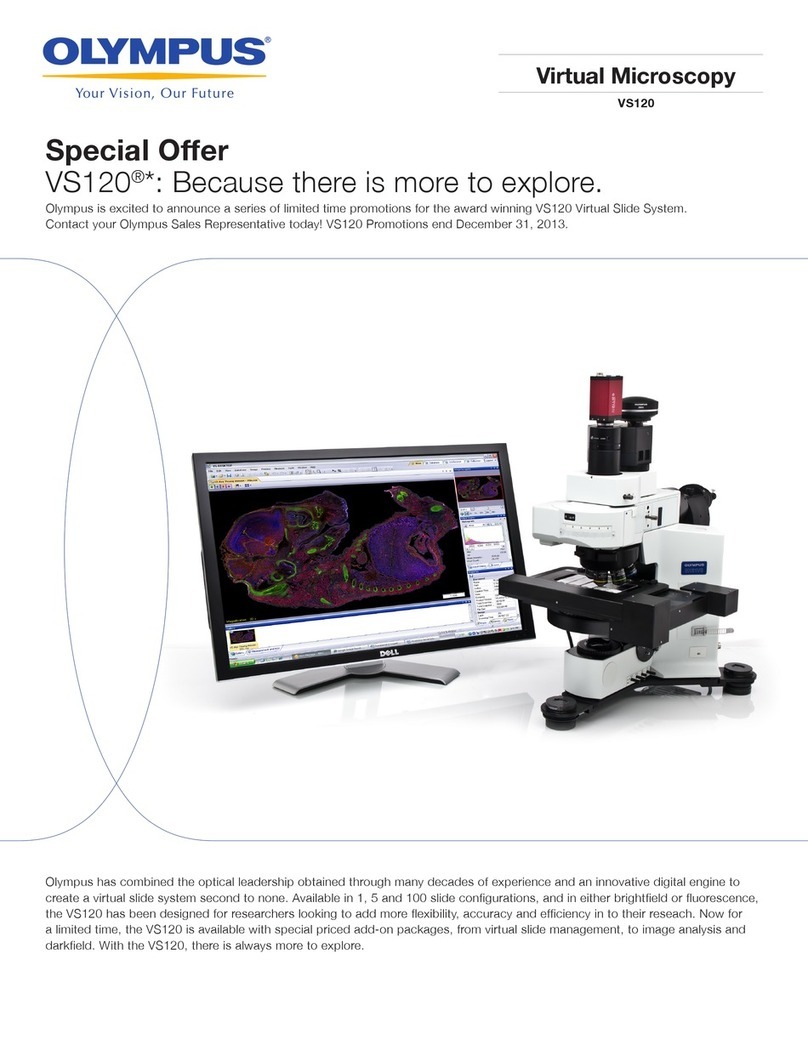
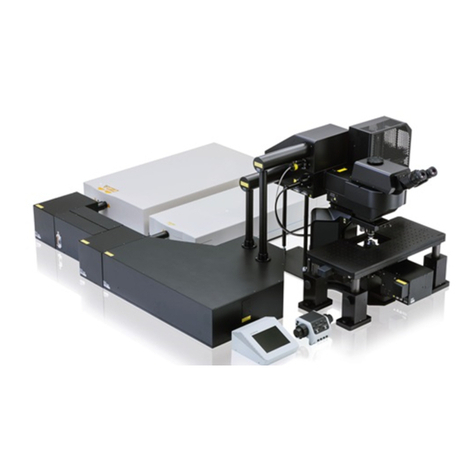
OLYMPUS FV1000 is a confocal laser scanning biological microscope system featuring improved basic
performances (sensor system, scanning system and illumination system performances) by considering the “live
cell observations”, with which long hours of stable measurement of weak fluorescence is required.
This microscope is equipped with 3 fluorescence channels, 3 lasers and AOTF to meet various applications in a
wide range of advanced research fields.
1-1 Principles
A laser scanning microscope converges the laser beam into a small spot using an
objective and scans the specimen in the X-Y direction using the laser beam.
The microscope then captures the fluorescent light and reflected light from the specimen
using light detectors and outputs the specimen image on an image monitor.
As shown in this figure, the confocal
optics incorporates a confocal aperture
on the optically conjugate position
(confocal plane) with the focus position to
eliminate light from other part than the
focus position. This causes the
extraneous light to be viewed as
darkness in the observation image, it is
possible to slice optically a tissue
specimen that has thickness.
On the other hand, an ordinary optical
microscope, the light from other part than
the focus position is overlapped with the
imaging light of the focus position so the
image is blurred in overall.
The laser beam that has transmitted
through the specimen is detected by the
transmitted light detector and provides
the transmitted image, which is not a
confocal image.
However, when the fluorescence images
of the transmitted and confocal images are combined, it is possible to obtain very
important information on the specimen.
Confocal
aperture
Light detector
Objective
Specimen
Light detector
Laser