manuals.online logo
Brands
  1. Home
  2. •
  3. Brands
  4. •
  5. Olympus
  6. •
  7. Microscope
  8. •
  9. Olympus DP80 User manual

Olympus DP80 User manual

DP80
Microscope Digital Camera
A cutting-edge digital microscopy camera equipped with dual CCD sensors,
providing both high sensitivity monochrome and high-quality single-shot color images.
DP80 System Diagram
ø
133
139
DP80
Camera head
To B4-mount
camera adapter
Camera interface cable
External trigger cable
Desktop
PC
PCIe I/F board
cellSens Entry or
cellSens Standard or
cellSens Dimension
* Image acquisition time and speed may be reduced if exposure time increases or several tasks are active in the background.
** "Centering" is a camera function which aligns the positions of the color and monochrome CCD sensors.
* Please use computers that conform to the regulations for your region.
DP80 Camera Head Dimension (Unit: mm)
Weight: approx 2.5 kg
Shinjuku Monolith, 3-1, Nishi Shinjuku 2-chome, Shinjuku-ku, Tokyo, Japan
Wendenstrasse 14-18, 20097 Hamburg, Germany
3500 Corporate Parkway, Center Valley, Pennsylvania 18034-0610, U.S.A.
491B River Vallay Road, #12-01/04 Valley Point Office Tower, Singapore 24837
21 Gilby Road, Mt. Waverley, VIC 3149, Melbourne, Australia
5301 Blue Lagoon Drive, Suite 290 Miami, FL 33126, U.S.A.
A8F, Ping An International Financial Center, No. 1-3, Xinyuan South Road,
Chaoyang District, Beijing, China, 100027
• OLYMPUS CORPORATION is ISO14001 certified.
• OLYMPUS CORPORATION is FM553994/ISO9001 certified.
• All company and product names are registered trademarks and/or trademarks
of their respective owners.
• Images on the PC monitors are simulated.
• Specifications and appearances are subject to change without any notice or
obligation on the part of the manufacturer.
Printed in Japan M1764E-112012
DP80 specifications DP80 system requirement
SpecificationsItem
Color / Monochrome 2CCD camera
Pixel shifting (only for color CCD)
Cooling system: Peltier device (max. Ta -10 degreeC)
[Color]
2/3 inch1.45 mega pixels color CCD, RGB colors on chip filter (Bayer)
[Monochrome]
2/3 inch1.45 mega pixels monochrome CCD
Progressive scan
B4 mount (2/3 inch Bayonet mount)
Auto,SFL-Auto,Manual
±2.0 EV step: 1/3 EV
23 µs to 60 s
Full image, 30%, 1%, 0.1%
2x2,4x4
1360 x 1024 (1x1): 15 fps, 680 x 512 (1x1) : 15 fps
680 x 510 (2x2): 29 fps, 340 x 250 (4x4) : 57 fps
[Centering OFF]
4080 x 3072 (Pixel shift)
2040 x 1536 (Pixel shift)
1360 x 1024 (1x1), 680 x 512 (1x1)
680 x 510 (2x2), 340 x 250 (4x4)
ISO200/400/800/1600 equivalent
14 bit *Number of effective bit: 12 bits@16 bit mode image
4080 x 3072: approx. 3.3 s, 1360 x 1024: approx. 0.3 s
sRGB,AdobeRGB (only for color CCD)
[Centering OFF]
1360 x 1024 (1x1), 680 x 512 (1x1)
680 x 510 (2x2), 340 x 250 (4x4)
x0.5/x1/x2/x4/x8/x16
14 bit *Number of effective bit: 14 bits@16 bit mode image
17000e- (Gain 0.5x)
7e-
2300:1 (Gain 0.5x)
55% (500 nm)
0.4e-/pixel/s (Ta=25degere C)
1360 x 1024: approx. 7.7fps, 680 x 510 (2 x 2): approx. 14.3fps,
340 x 250 (4 x 4): approx. 20fps
File formats supported by cellSens software
133 mm(W) x 130 mm(D) x 139 mm(H) /approx. 2.5 kg
Approx. 2.8 m/approx. 0.23 kg
Approx. 0.2 m/approx. 40 g
Intel Core i5/ Intel Core i7 / Intel Xeon
4 GB or more
(8 GB recommended for high speed image acquisition)
Free space of 1 GB or larger
(at the time of installation)
1280x1024 (min. 1024 x768) monitor resolution with
32-bit-video card with separate graphics memory
(no integrated graphics processor with shared memory)
PCI-Express x1 Rev. 1.0a or later
Compatible with half size or LowProfile
PCIe board (106.7 mm x 174.6 mm)
Windows 7 Professional/Ultimate with SP1 (64 bit)
250 W or more
Unoccupied FDD power cable, HDD
power cable (4-pin size), or Serial ATA
power cable must be available
PC/AT compatible
Camera type
Size
Image sensor
Camera mount
Exposure
control
Metering modes
Binning
Color
Image Resolution
ISO Sensitivity
A/D
Image acquisition time *
Color space
Image Resolution
Gain
A/D
Full well capacity
Readout noise
Dynamic range
Quantum efficiency
Dark current
Image acquisition speed *
Image file format
Dimensions,
weight
Camera head
Interface cable
External trigger cable
Scanning mode
Mode
Adjustment
Exposure time
Monochrome
[Centering ON]**
3648 x 2736 (Pixel shift)
1824 x 1368 (Pixel shift)
1216 x 912 (1x1), 608 x 456 (1x1)
608 x 456 (2x2), 304 x 228 (4x4)
[Centering ON]
1216 x 912 (1x1), 608 x 456 (1x1)
608 x 456 (2x2), 304 x 228 (4x4)
CPU
RAM
HDD
Graphics
Extension slot
OS
Power supply
Live frame rate
Monitor
Olympus-Tower, 114-9 Samseong-Dong, Gangnam-Gu, Seoul, Korea
Low-profile bracket
SATA-to-HDD
power conversion
adapter
HDD-to-FDD
power conversion
cable
1 2
Microscope Digital Camera DP80
Superior bright-field images
at high resolution
A single camera featuring color / monochrome dual CCD sensors.
Clear fluorescence images
with excellent sensitivity
Consider how convenient and easy it would be if both high resolution bright-field
and high-sensitivity fluorescence images could be observed and acquired using
a single microscope camera. The DP80 digital camera fulfills this simple yet
unfulfilled requirement for two cameras in one.
Since it is possible to rapidly switch between the monochrome CCD sensor and
color CCD sensor, it is possible to easily acquire high-quality bright-field and
high-sensitivity fluorescence images of the same field. Without the need to
switch camera ports with a prism, the time required to align the camera sensors
is eliminated.
The DP80 assists you in your research activities from observation to
documentation in a smooth and easy manner.
1 2
Microscope Digital Camera DP80
Superior bright-field images
at high resolution
A simple switching operation allows the use of either a color or monochrome CCD
sensor on the single camera unit.
Clear fluorescence images
with excellent sensitivity
Consider how convenient and easy it would be if both high resolution bright-field
and high-sensitivity fluorescence images could be observed and acquired using
a single microscope camera. The DP80 digital camera fulfills this simple yet
unfulfilled requirement for two cameras in one.
Since it is possible to rapidly switch between the monochrome CCD sensor and
color CCD sensor, it is possible to easily acquire high-quality bright-field and
high-sensitivity fluorescence images of the same field. Without the need to
switch camera ports with a prism, the time required to align the camera sensors
is eliminated.
The DP80 assists you in your research activities from observation to
documentation in a smooth and easy manner.
Live view display
[email protected]
3 4
Microscope Digital Camera DP80
High quality images adapted to observation and
imaging methods can be readily obtained.
The DP80 is equipped with Fine Detail Processing, which reduces
pseudo-colors and moire of ultrastructures and improves resolving
power. Clear imaging of details is achieved by fully extracting the
resolving power of objective lenses.
A monochrome camera that detects and captures dim fluorescence images Color camera provides clear real-time live preview display
High definition uncompressed live images of 1360×1024 pixels
Live view display of high definition RGB 24-bit color images of 1360 ×
1024 pixels at 15 frames per second.Distortion-free focusing or
framing is provided because there is no deterioration of image quality
due to non-compression, and so sample details are sharp and clear
whether the sample is stationary or moving.
Clearly observe weak fluorescence signals with the
DP80's high sensitivity
We significantly improved the DP80's fluorescence imaging
performance by incorporating a separate high dynamic range
monochrome CCD sensor within the body of the camera. Combined
with thermo-electric cooling and high resolution capture, the DP80
meets the demands for low-light fluorescence imaging. With a high
quantum efficiency along a wide spectrum, the DP80 provides
exceptional fluorescence singal detection.
High-sensitivity fluorescence imaging available up to Cy7
wavelengths
The DP80 responds to a wide range of wavelengths from visible to
near infrared. Now sensitive to fluorescence signals with longer
wavelengths, the DP80 captures near-IR wavelengths from samples
stained with Cy7 (767 nm).
Color and monochrome images can be overlayed with
pixel precision
Since the camera is equipped with a centering function, which
minimizes positional differences between color images and
monochrome images, a combined image can be produced by
precisely superimposing the monochrome images and color images
that are acquired for a given observation method. Since positions
accurately fixed morphology and localization can be examined by, for
example, superimposing a bright-field image and a fluorescence image.
Efficient support of observation and experimentation 
High-quality color and multi-channel imaging is automated with the
DP80's switchable CCD sensors and the intuitive Olympus cellSens
software interface. From complex image acquisition to image processing
to report generation, the researcher can focus on research activities
instead of routine labor-intensive acquisition setup and data preparation.
Faithful panoramic imaging, with
high-quality in brightfield or fluorescence
Multiple-region capture of saved images can be
easily recombined and restitched seamless into a
single image. Numerous annotations and
comments can be saved with images for later
retrieval. These features are useful for
standardization and accuracy control of inspection
and research processes.
Fine-detail Processing that suppresses pseudo-colors and
moiré artifacts
Subtle hue differences within colors such as brown, blue, and purple were
difficult to distinguish in the past, but now, such slight differences in color
can be reproduced by incorporating AdobeRGB* color space which
reproduces a wide range of color tones and a new algorithm of color
reproduction. Color images faithful to the original samples can be acquired.
*Color reproduction fidelity depends on monitor specifications. Monitors supporting
AdobeRGB are required to accurately reproduce images recorded in AdobeRGB mode.
Superior color reproducibility captures fine differences in color
High resolution imaging up to 12.5 megapixels
The DP80 uses pixel-shifting technology to reach a maximum recording
image size of 4080 × 3072, a high resolution equivalent to 12.5
megapixels.
Research workflow is improved through
Olympus cellSens imaging software
Conventional processing
Fine-detail processing
cy7サイセブンのアプリ(赤に光る試薬)
Relative Sensitivity
Color image (exposure time: 60 s) Monochrome image (exposure time: 2 s)
Monochrome image
Color image Superimposed image
R
(color)
G
(color)
B
(color)
BW
1
0.8
0.6
0.4
0.2
0
400 500 600 700 800 900 1000 1100
Wavelength
(nm)
+
+
+
+
Higher sensitivity!
Longer wavelength!
5 6
Microscope Digital Camera DP80
High quality bright-field and high-sensitivity multi-channe l fluorescence imaging
The DP80 alone can provide all of the images that are illustrated below.
In the dark field images we can see the borders of the lateral amygdala, a brain region important for fearful emotions. In the fluorescence image are cells and processes expressing a fusion protein of
channelrhodopsin/EYFP. Channelrhodopsin is a blue light activated non specific cation channel that is used in 'optogenetics' experiments. We can express channelrhodpsin in lateral amygdala neurons
and produce emotional fear memories by activating the cells with blue light. These microscope images allow us to verify that expression of channelrhodopsin has occured in the lateral amygdala.
Image data courtesy of : RIKEN BRAIN SCIENCE INSTITUTE Neural Circuitry of Memory Joshua P. Johansen, Ph.D.
Observation of Collagen type
Ⅰ and type Ⅲwith multicolor immuno-fluorescence staining during wound healing process
Bright-field image of total collagen with Elastica van Gieson (EVG) staining (Left; DP80 color mode) and, multi-fluorescent pseudo-color image of collagen type
Ⅰ labeled with Cy7 and collagen type Ⅲ
labeled with Cy5 respectively (Right; DP80 monochrome mode). Location of the collagen type I and III is confirmed clearly by the long-wavelength observation without auto-fluorescence noise such as
erythrocytes and/or other tissue components.
Surface of Drosophila melanogaster expressing fluorescence protein in periphearal sensory cells.
Image data courtesy of : Institute of Molecular and Cellular Biosciences, University of Tokyo Kei Ito, Ph.D.
Caputured by color CCD Caputured by monochrome CCD with pseudo-colors Caputured by color CCD Caputured by monochrome CCD
Caputured by color CCD Caputured by monochrome CCD with pseudo-colors
Image data courtesy of : JAPANESE FOUNDATION FOR CANCER RESEARCH The Cancer Institute, Division of Pathology Noriko Motoi, M.D., Ph.D. Yuichi Ishikawa, M.D., Ph.D.
Fig.1: Histology of lung with EML4-ALK fusion gene. (HE stain).
Fig.3: Histology showing small round cell with many mitoses and nuclear atypia. (HE stain)
Fig.2: Same case as Figure 1. FISH was performed using ALK Split Dual color FISH probe
(green = FITC and red = TexRed) (GSP Laboratory) . The abnormal ALK split signal was
observed as green and red colored signal, in addition to normal yellow-colored signal.
Fig.4: Same case as Figure 3. FISH was performed using EWSR1 (22q12) dual color break
apart rearrangement FISH probe (green = spectrum green and orange = spectrum orange)
(Vysis™ , Abbott Japan). The abnormal EWSR1 split signal was observed as green and
orange colored signal, in addition to normal yellow-colored signal.
Caputured by color CCD Caputured by monochrome CCD with pseudo-colors
Ⅲ

Other manuals for DP80

1

Other Olympus Microscope manuals

Olympus U-CBS User manual

Olympus

Olympus U-CBS User manual

Olympus IX71 User manual

Olympus

Olympus IX71 User manual

Olympus VANOX Operating instructions

Olympus

Olympus VANOX Operating instructions

Olympus IMT-2-11 User manual

Olympus

Olympus IMT-2-11 User manual

Olympus MVX10 User manual

Olympus

Olympus MVX10 User manual

Olympus IX73 User manual

Olympus

Olympus IX73 User manual

Olympus BX60 User manual

Olympus

Olympus BX60 User manual

Olympus U-AW User manual

Olympus

Olympus U-AW User manual

Olympus BX60 User manual

Olympus

Olympus BX60 User manual

Olympus U-UVF248 User manual

Olympus

Olympus U-UVF248 User manual

Olympus CHA User manual

Olympus

Olympus CHA User manual

Olympus BX51 User manual

Olympus

Olympus BX51 User manual

Olympus GX41 User manual

Olympus

Olympus GX41 User manual

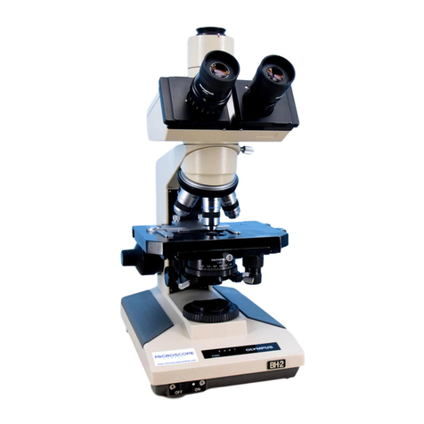
Olympus BH2 Series Installation instructions

Olympus

Olympus BH2 Series Installation instructions

Olympus BX53M User manual

Olympus

Olympus BX53M User manual

Olympus VANOX User manual

Olympus

Olympus VANOX User manual

Olympus SZX-DA User manual

Olympus

Olympus SZX-DA User manual

Olympus SLIDEVIEW  VS200 User manual

Olympus

Olympus SLIDEVIEW VS200 User manual

Olympus FV3000 User manual

Olympus

Olympus FV3000 User manual

Olympus PROVIS AX70 Installation guide

Olympus

Olympus PROVIS AX70 Installation guide

Olympus SZX16 User manual

Olympus

Olympus SZX16 User manual

Olympus FHT-521 User manual

Olympus

Olympus FHT-521 User manual

Olympus JM User manual

Olympus

Olympus JM User manual

Olympus VS120 Owner's manual

Olympus

Olympus VS120 Owner's manual

Popular Microscope manuals by other brands

Leica DI C800 User's manual & installation instructions

Leica

Leica DI C800 User's manual & installation instructions

Atlantis SL 2000 user manual

Atlantis

Atlantis SL 2000 user manual

Ryf AG Schott EasyLED Spotlicht Plus user manual

Ryf AG

Ryf AG Schott EasyLED Spotlicht Plus user manual

VWR VisiScope 300 Series instruction manual

VWR

VWR VisiScope 300 Series instruction manual

Accu-Scope 3012-LED manual

Accu-Scope

Accu-Scope 3012-LED manual

Discovery Kids TDK25 instruction manual

Discovery Kids

Discovery Kids TDK25 instruction manual

Labomed Lx POL user manual

Labomed

Labomed Lx POL user manual

Di-Li 900 Manual instruction

Di-Li

Di-Li 900 Manual instruction

OCULUS BIOM 4 instruction manual

OCULUS

OCULUS BIOM 4 instruction manual

Leica DM750 M instruction manual

Leica

Leica DM750 M instruction manual

KERN OZM 902(UK) User instructions

KERN

KERN OZM 902(UK) User instructions

Dino-Lite 411X Series user manual

Dino-Lite

Dino-Lite 411X Series user manual

Zeiss OPMI Pentero quick guide

Zeiss

Zeiss OPMI Pentero quick guide

ViTiny VT101 user manual

ViTiny

ViTiny VT101 user manual

Labomed STELLA user manual

Labomed

Labomed STELLA user manual

KERN OZO 551 User instructions

KERN

KERN OZO 551 User instructions

Zeiss opmi lumera 700 user manual

Zeiss

Zeiss opmi lumera 700 user manual

Meiji Techno MT-420 Series user manual

Meiji Techno

Meiji Techno MT-420 Series user manual

manuals.online logo
manuals.online logoBrands
  • About & Mission
  • Contact us
  • Privacy Policy
  • Terms and Conditions

Copyright 2025 Manuals.Online. All Rights Reserved.