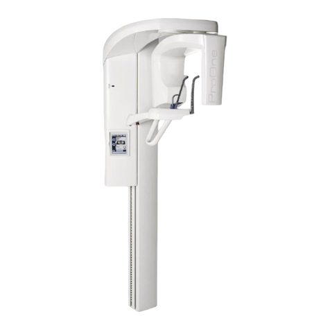
User’s Manual (3D Imaging) Planmeca ProMax 3D s & 3D Classic 1
Table of Contents
1 INTRODUCTION .................................................................................................................1
2 ASSOCIATED DOCUMENTATION ...................................................................................... 2
3 SYMBOLS ON PRODUCT LABELS ..................................................................................... 3
3.1 Symbols on permanently installed X-ray units ........................................................................ 3
3.2 Symbols on X-ray units with detachable power supply cord .................................................. 4
4 SAFETY PRECAUTIONS .................................................................................................... 5
5 SWITCHING X-RAY UNIT ON ............................................................................................. 9
6 MAIN PARTS .................................................................................................................... 10
6.1 General view of X-ray system ...............................................................................................10
6.2 General view of X-ray unit .................................................................................................... 11
6.3 Sensor .................................................................................................................................. 12
6.4 Tube head ............................................................................................................................ 13
6.5 Patient supports ................................................................................................................... 14
6.6 Telescopic column ................................................................................................................ 15
6.7 Exposure switch ................................................................................................................... 16
6.8 Emergency stop button ........................................................................................................ 16
6.9 Touch screen ........................................................................................................................ 17
6.10 ProTouch desktop application ..............................................................................................20
6.11 Patient positioning controls ................................................................................................... 21
7 PLANMECA PROMAX 3D s PROGRAMS .......................................................................... 23
7.1 3D Dental ............................................................................................................................. 23
7.2 3D Models ............................................................................................................................ 23
8 PLANMECA PROMAX 3D CLASSIC PROGRAMS .............................................................. 24
8.1 3D Dental ............................................................................................................................. 24
8.2 3D Models ............................................................................................................................ 24
9 3D PATIENT EXPOSURE ................................................................................................. 25
9.1 Preparing X-ray system ........................................................................................................ 25
9.2 Preparing patient .................................................................................................................. 32
9.3 Selecting exposure settings .................................................................................................. 33
9.4 Patient positioning ................................................................................................................ 37
9.5 Selecting exposure values .................................................................................................... 38
9.6 Selecting patient movement correction ................................................................................ 41
9.7 Selecting 3D face photo (X-ray units with ProFace sensor) ................................................. 41
9.8 Adjusting volume position ..................................................................................................... 42
9.9 Taking a scout image or 2D views (LAT, PA or LAT-PA) ..................................................... 46
9.10 Taking a 3D exposure .......................................................................................................... 49
10 3D FACE PHOTO ............................................................................................................. 51
10.1 Before exposure ................................................................................................................... 51
10.2 Patient positioning ................................................................................................................ 51
10.3 Selecting exposure settings .................................................................................................. 53
10.4 Taking a 3D face photo ........................................................................................................ 54
11 3D MODEL EXPOSURE .................................................................................................... 56
11.1 Calibrating X-ray unit for impression or plaster material ....................................................... 56
11.2 Taking an exposure of an impression or plaster cast ........................................................... 60
