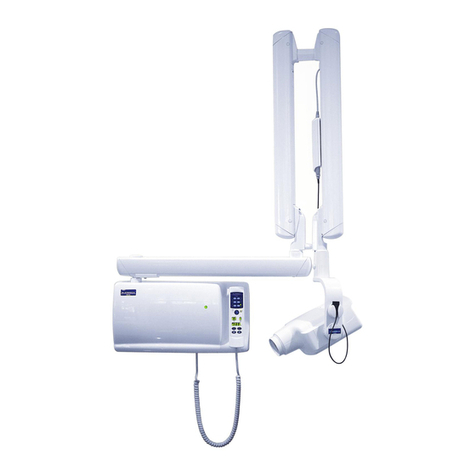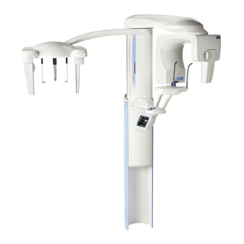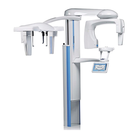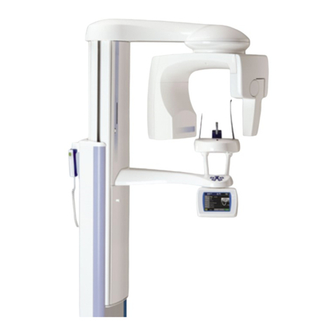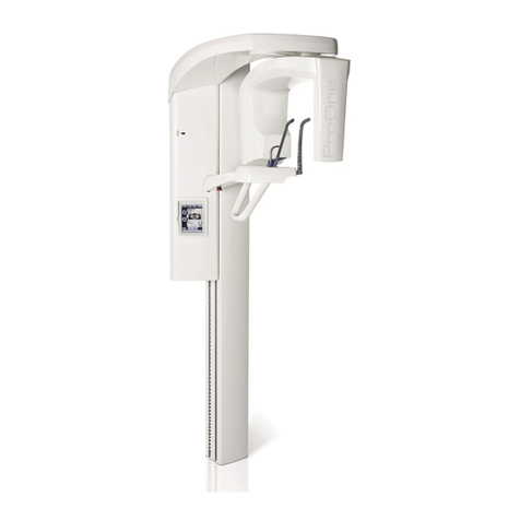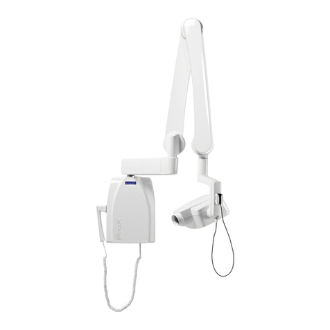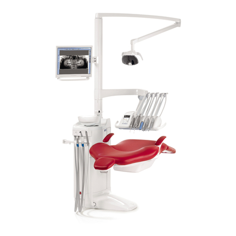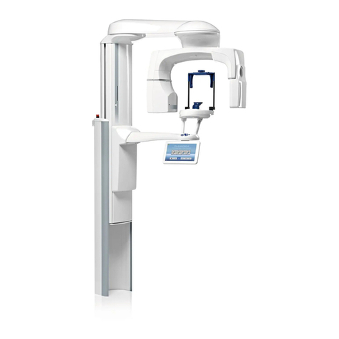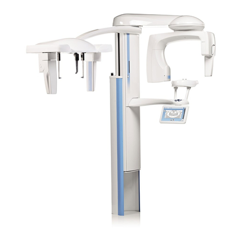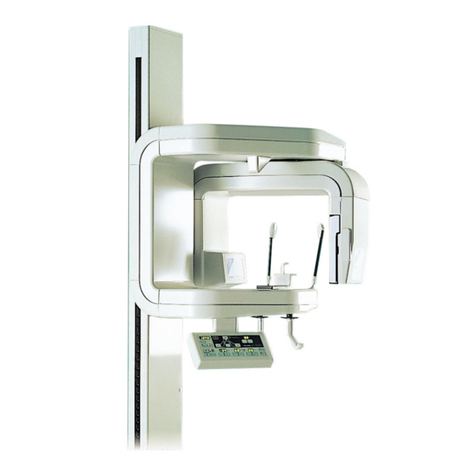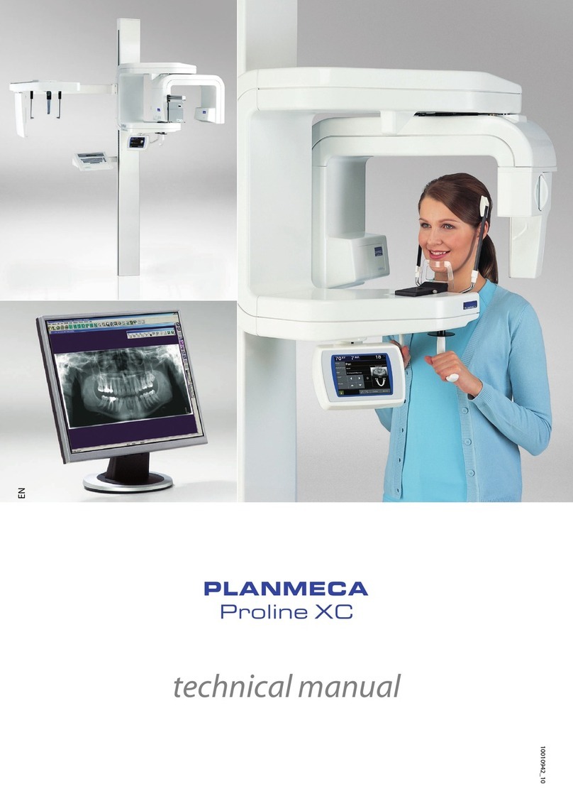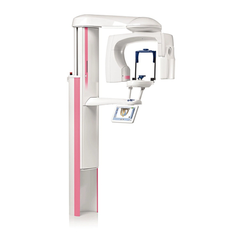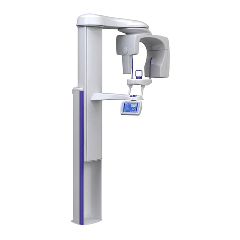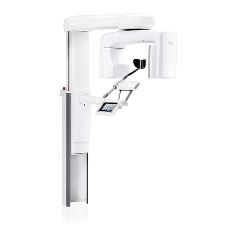
Planmeca ProMax 3D s / 3D 5
SAFETY PRECAUTIONS
User’s Manual (3D)
NOTE Before taking an exposure, ask any female patient of
childbearing age whether she might be pregnant. The
X-ray unit is not intended for use on pregnant women.
NOTE FOR CANADIAN USERS:
All patients must be provided with a shielded apron for
gonad protection and a thyroid shield. The use of a
thyroid shield is especially important in children. The
shielded apron and thyroid shield should have a lead
equivalence of at least 0.25 mm on both sides (front
and back of the patient).
NOTE If the X-ray unit has been stored at temperatures under
+10°C for more than a few hours, time must be allowed
for the unit to reach room temperature before turning
it on.
NOTE Ensure efficient air conditioning in the X-ray room. It is
recommended to keep the room temperature between
+20°C and +25°C at all times.
NOTE If exposures are taken in rapid succession the X-ray
tube may overheat and a cooling time will flash on the
touch screen. The cooling time indicates the delay
before the next exposure can be taken.
NOTE If the X-ray system is not connected to an
Uninterruptible Power Supply (UPS), disconnect the
system from mains during lightning storms.
NOTE FOR US & CANADIAN USERS:
The patient positioning lights are class II laser
products (21 CFR § 1040.10).
NOTE FOR EUROPEAN USERS:
The patient positioning lights are class 1 laser
products (Standard IEC / EN 60825-1: 2007).
NOTE EMC requirements have to be considered, and the
equipment must be installed and put into service
according to the specific EMC information provided in
the accompanying documents.
NOTE Portable and mobile RF communications equipment
can affect the X-ray unit.
NOTE External equipment intended for connection to signal
input, signal output or other connectors, shall comply
with relevant IEC standard (e.g. IEC 60950 for IT
equipment and the IEC 60601 series for medical
electrical equipment). In addition, all such
combinations - systems - shall comply with the
standard IEC 60601-1-1, Safety requirements for
medical electrical systems. Equipment not complying
to IEC 60601 shall be kept outside the patient area
(more than 2m (79 in.) from the X-ray unit).
Any person who connects external equipment to
signal input, signal output or other connectors has
formed a system and is therefore responsible for the
system to comply with the requirements of IEC 60601-
1-1. If in doubt, contact your service technician or local
representative for help.
NOTE Contact your service technician if you notice a
decrease in image quality.
NOTE Contact your service technician if you have taken an
exposure but the image does not appear in the
Planmeca Romexis program. The last ten images can
be manually imported into Romexis.
NOTE Never place or hang any objects on any part of the X-
ray unit.
CAUTION
LASER RADIATION -
DO NOT STARE
INTO BEAM
1mW
635nm
CLASS II
LASER
PRODUCT
LBL-X-099
CLASS 1 LASER PRODUCT
APPAREIL À LASER DE CLASSE 1
IEC 60825-1:2007
