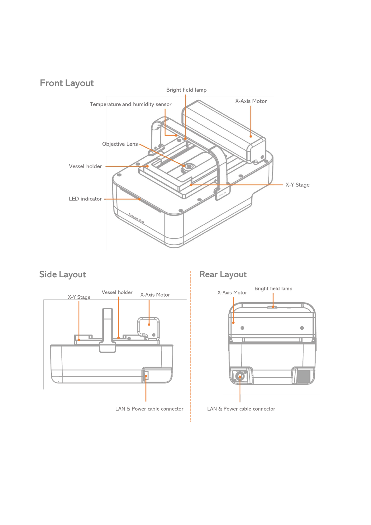
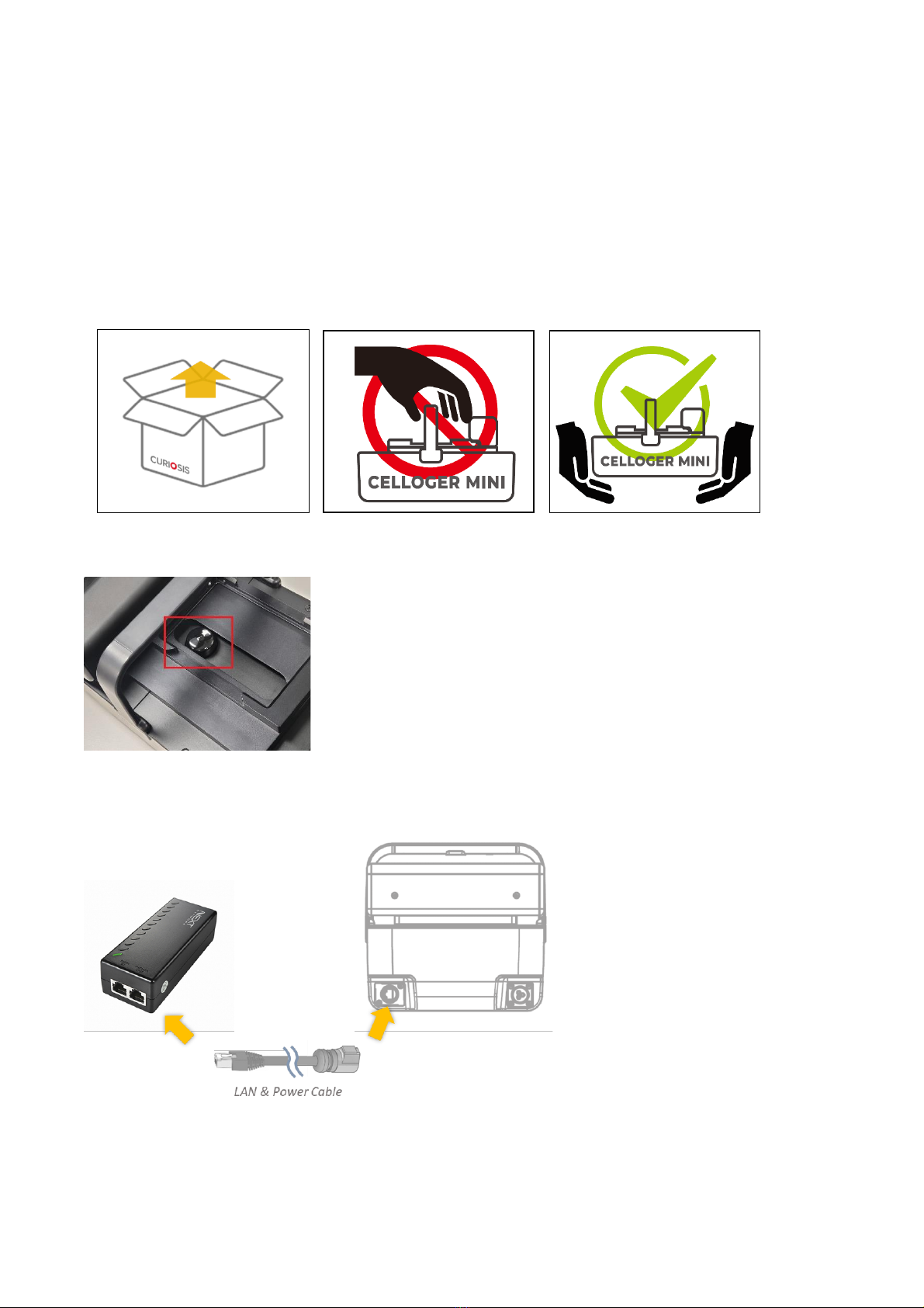
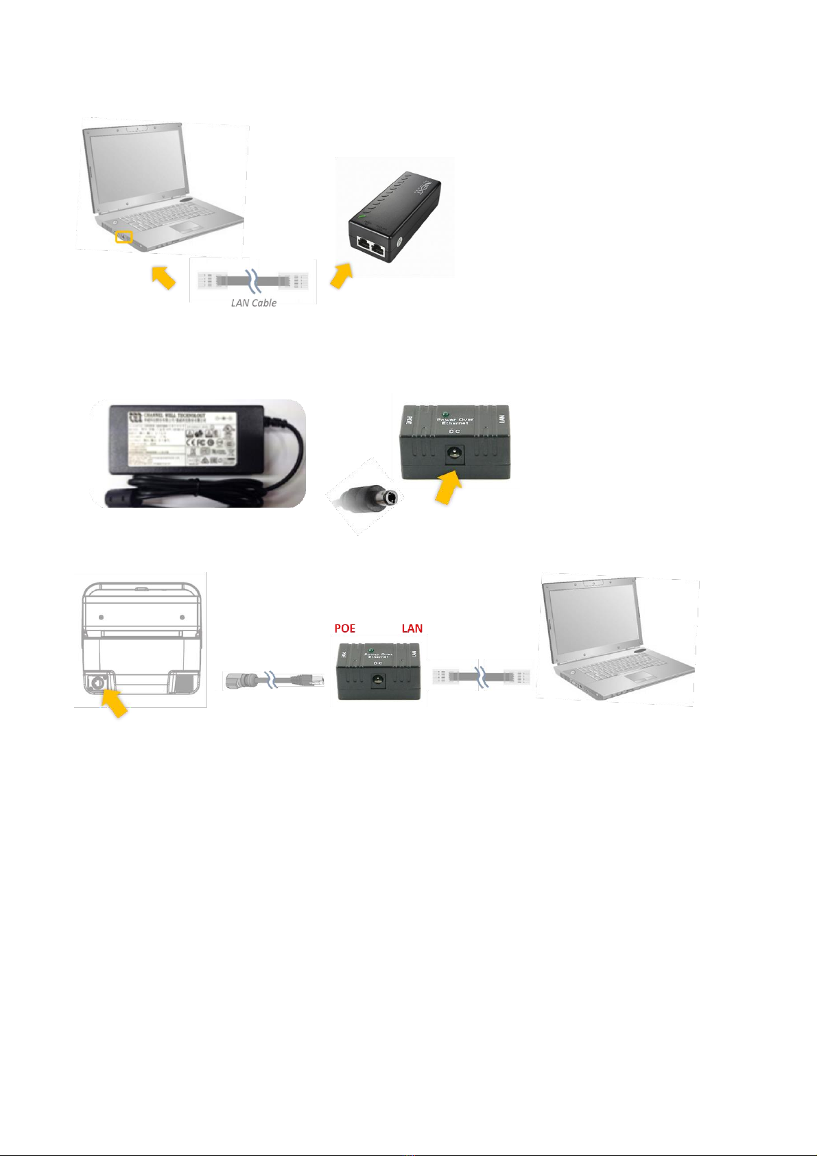
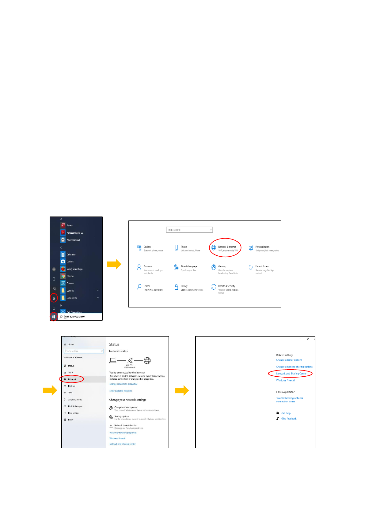
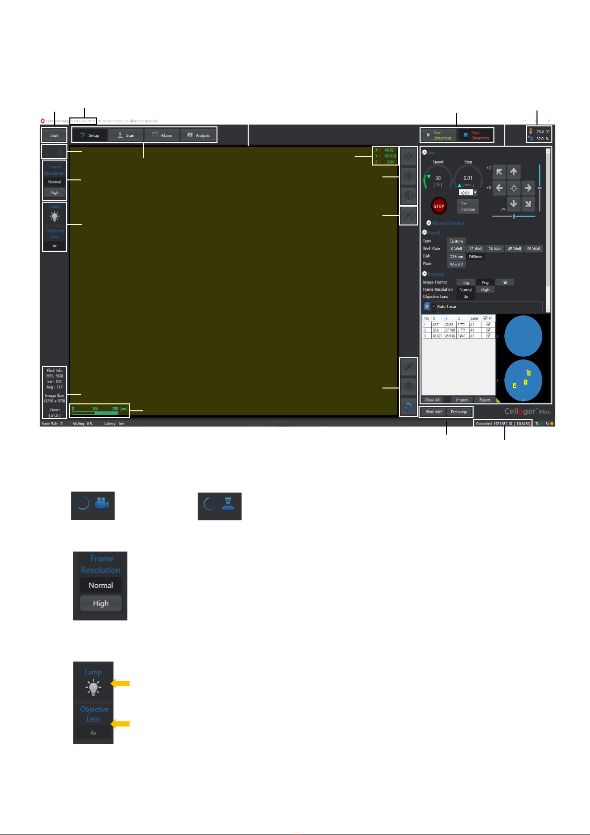
CURIOSIS Celloger Mini User manual
Other CURIOSIS Laboratory Equipment manuals

CURIOSIS
CURIOSIS FACSCOPE B User manual

CURIOSIS
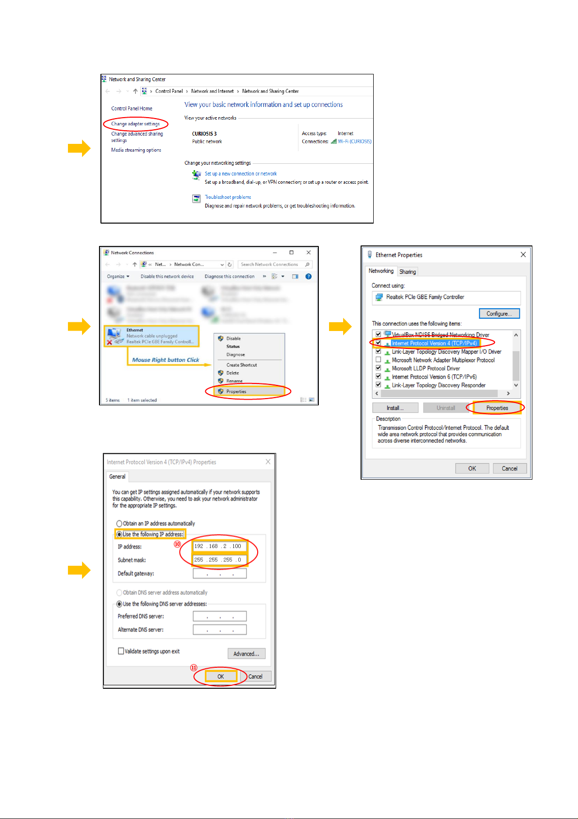
CURIOSIS Celloger Mini Installation guide

CURIOSIS
CURIOSIS Cellpuri Installation guide

CURIOSIS
CURIOSIS Celloger Nano User manual

CURIOSIS
CURIOSIS Celloger Mini Plus User manual

CURIOSIS
CURIOSIS FACSCOPE B User manual

CURIOSIS
CURIOSIS Celloger Mini Installation guide

CURIOSIS
CURIOSIS FACSCOPE B Instruction manual

CURIOSIS
CURIOSIS Celloger Mini Plus Installation guide
Popular Laboratory Equipment manuals by other brands


Agilent Technologies
Agilent Technologies 5800 ICP-OES user guide

Endress+Hauser
Endress+Hauser Cleanfit CPA875 operating instructions

NI
NI PXI-5422 CALIBRATION PROCEDURE

Collomix
Collomix Aqix operating instructions

SPEX SamplePrep
SPEX SamplePrep 6875 Freezer/Mill Series operating manual

Ocean Insight
Ocean Insight FLAME-NIR+ Installation and operation manual

Parker
Parker ALIGN-MG-NA Installation, operation and maintenance manual

BD
BD 644787 user guide

DENTAURUM
DENTAURUM Compact Megaplus Instructions for use

Biuged Laboratory Instruments
Biuged Laboratory Instruments BGD 626 instruction manual

VWR
VWR SAS Super IAQ instruction manual

illumina
illumina MiSeqDx reference guide