Motic PA43 User manual

WWW.MOTIC.COM
MOTIC HONG KONG LIMITED
If the equipment is used in a manner
not specified by the manufacturer,
the protection provided by the
equipment may be impaired.
Note
PA43
Operation Manual

1
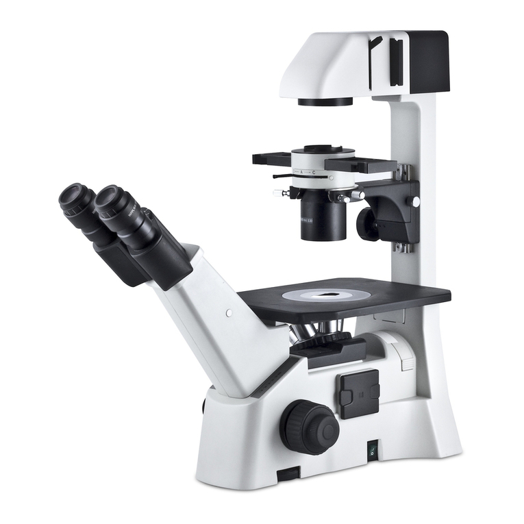
This instruction Manual has been prepared for users of the Motic PA43 Upright
Microscope.
For your own safety, read this manual carefully and thoroughly before using the
product. Do not discard this manual. Always keep it near the product for easy
reference.
We are constantly endeavoring to improve our instruments and to adapt them to the
requirements of modern research techniques and testing methods. This involves
modification to the mechanical structure and optical design of our instruments.
Therefore, all descriptions and illustrations in this instruction manual, including all
specifications are subject to change without notice.

2
TABLE OF CONTENTS
1. General Notes on Instrument Safety 4
1.1 General safety notes and Instruction 4
1.2 Instrument safety, FCC and EMC conformity 5
1.3 Transporting, unpacking, storage of the Instrument 6
1.4 Instrument Disposal 6
1.5 Use of the Instrument 6
1.6 Intended use of the Microscope 8
1.7 Instrument warranty 8
2. Nomenclature 9
2.1 PA43 9
3. Setting up the Instrument 11
3.1 Operating environment 11
4. Assembling the Microscope 12
4.1 Verifying input voltage 12
4.2 Illumination 12
4.2.1 Halogen bulb 12
4.2.2 LED 12
4.3 Rackless Stage 12
4.4 Objectives 13
4.5 Condenser 13
4.6 Eyepiece tube 14
4.7 Eyepieces 14
4.8 Filters 15
4.9 Installing the Polarzier with the filter holder 16
4.10 Installing the Analyzer with the analyzer slot 17
4.11 Power cord 17
5. Usage of Microscope Components 18
5.1 Focusing Block 18
5.1.1 Replacing the Fine Focus Knob 18
5.1.2 Adjusting the Coarse Focus Knob Tension 18
5.1.3 Pre-focusing Lever 19

3
5.2 Interpupillary distance adjustment 19
5.3 Diopter adjustment 20
5.3.1 Selecting the Light Path of the Trinocular Tube 21
5.4 Kohler Illumination alignment procedure: Centering the Condenser 22
5.5 Kohler Illumination alignment procedure: Setting the Aperture Iris Diaphragm 23
5.6 Brightness and contrast adjustment 23
5.7 Illumination brightness adjustment 24
5.7.1 PA43 with light manager 24
5.8 Standby mode 24
6. Photomicrographic Procedure 25
7. Using Oil Immersion Objectives 26
8. Care and Maintenance 27
8.1 Do not disassemble 27
8.2 Cleaning the microscope 27
8.2.1 Lenses and filters 27
8.2.2 Cleaning of painted or plastic components 27
8.3 Disinfecting the Microscope 27
8.4 When not in use 27
8.5 Bulb replacement 28
8.5.1 Replacing the 6V 30W halogen module or the two different LED module 28
8.5.2 Electrical Specifications 29
8.6 Shifting of Instrument 29
9. Specification 30
10. Troubleshooting Table 31
10.1 Optical 31
10.2 Electrical 32
11. Selection of the Power Supply Cord 33
Microscope Terminology 35

4
1. GENERAL NOTES ON INSTRUMENT SAFETY
1.1 General safety notes and Instruction
Please familiarize yourself with this Operation Manual before starting to use the Instrument.
In case additional information or support is needed, please contact Motic after sales Service.
To ensure safe operation and optimal function of the Instrument, strictly observe the precautions and
warnings given in this Operation manual.
Please observe the following indicators:
CAUTION, Risk of Danger
This symbol indicates a possible hazard to the user of the instrument.
CAUTION, Risk of Dangerous heat
This symbol indicates a possible hazard to the user of the instrument.
CAUTION, Risk of Electrical Danger
Disconnect the plug-in power unit from line power before opening the Instrument.
INFO
This symbol refers to a useful information or hint.
Disposal
This symbol refers you a advice that must be observed under all circumstances
Instrument ON
This symbol refers to the switched on (electrical system active) status of the
instrument
Instrument OFF
This symbol refers to the switched off (electrical system inactive) status of the
instrument
Risk of Dangerous Electrical Current
This symbol refers to potential hazardous electrical voltage/current and must be
observed at all circumstances!

5
1.2 Instrument safety, FCC and EMC conformity
This Instrument has been designed, produced and tested in compliance with required safety standards
and regulations. UL, CE, FCC, EMC
“Safety of equipment used for measurement control and laboratory use”
PA43 Microscope Series Products are in conformity with the requirements of the CE
Directive 98/79/EC Annex 1 and carries the CE mark accordingly.
Conform to Class B Noise immunity is in compliance with EN 61326 and EMC and
Radio-noise suppression is in compliance with EN 5501 1
The instruments listed in this document are disposed of in compliance with WEEE Directive
2002/96/EC.
Lamp bulbs, LED Modules, Lampdrawers become very hot during and after a period of
operation. Risk of burn – Do not touch the lamp during or immediately after period of operation.
Don’t pick the microscope up from the bottom during equipment operation.
Proper handling of the microscope will ensure years of trouble free service.
If repair become necessary, please contact your Motic agency or our Technical Service directly.

6
1.3 Transporting, unpacking, storage of the Instrument
Please observe and follow the safety notes for transportation, unpacking and storage
given in this document:
● The microscope is delivered as set, packed into commercial standard plastic and
cardboard packaging; Please re-use the original packaging only for any transportation.
● It is advised to keep and use the original packaging for longer storage or return to the
manufacturer to avoid losing the warranty.
● At receiving and unpacking the equipment, please verify that all parts specified on the
delivery note are present.
● Keep Transport and storage temperatures as specified in this Manual.
● Set the microscope up on a stable worktable with solid and smooth top surface suitable for
Instrument use.
● Do not touch optical surfaces.
1.4 Instrument Disposal
Please observe the following safety notes for the disposal of the microscope:
Defective Instruments, accessories and consumables should be disposed in compliance with
the provisions of the local law.
1.5 Use of the Instrument
The microscope Instrument as well its accessories must not be used for microscopic techniques or
purposes other than those described in this Operating Manual.
Please always observe the following safety notes when using the microscope:
Motic cannot assume any liability for other applications then the intended use, including the
included modules and components. This includes to service or repair work that is not carried
out by authorized Motic service personnel. In case of non-compliance, all warranty claims and
liabilities shall be forfeited.
The microscope Instrument should only be operated by trained personnel who are familiar with
this Operation Manual and therefore aware of the possible dangers involved.


7
This Microscope is a high-precision instrument that can be impaired in its performance or even
be destroyed when handled improperly.
This Instrument is equipped with a build in power supply allowing electric-net voltages to be
used in the range between 100 - 240 V ± 10%, 50/60 Hz. In case of any irregularity on the
electrical system observed, please switch of the Instrument, keep it safe guarded and consult
the Motic after sales service.
Please only use rated Power cords as described in the Appendix of this Document
Always disconnect the power cable, before opening the instrument and changing the lamp or
LED source.
Wait for the lamp to cool down before replacing it and do not touch the new bulb.
The instrument may only be opened by qualified Motic service staff.
The operation of the instrument in explosion-risk environments is not allowed.
Immersion Oil can be harmful. Please only use Motic immersion Oil and follow the safety
Instructions given. Immersion oil is to be used for Microscope application only! Please avoid
skin contact or swallow or any other use then the intended.

8
1.6 Intended use of the Microscope
This Microscope is an optical instrument that has been designed to be used to observe and document
magnified images of specimens in teaching, routine and research applications.
Do not use this instrument for any other purpose than its intended use.
This Instrument complies with the requirements of directive 98/79/EG concerning in-vitro
diagnostic medical devices. CE marking shows the conformity to the directive.
NOTE: This Instrument has been tested and found to comply with the limits for a Class A
digital device, pursuant to Part 15 of the FCC Regulations. These limits are designed to
provide protection against harmful interference during operation in a commercial environment.
This equipment uses and generates frequencies and energy, if not properly installed in
accordance with the instruction manual, those may cause interference to radio
communications. Operation of this Instrument in a residential area is not permitted.
FCC WARNING: Changes or modifications would void the Instruments FCC compliance and is
therefore not permitted.
1.7 Instrument warranty
The Instrument should only be used for microcopy applications mentioned and instructed in this
operation manual. Please note the following information on the instruments warranty.
● If possible defects Motic must be notified immediately and steps be taken to minimize damage.
● If notified of such a defect Motic will evaluate the defect and if within warrant, rectify it at his
discretion, either by repairing the instrument or by delivering a replacement.
● Natural wear, or defects caused by improper use are not covered by the Instrument warranty.
● Motic shall not be liable for damage caused by faulty operation, negligence or tampering with the
Instrument.
● Altering or tampering of the instrument shall lead to a forfeit of all warranty claims

9
2. NOMENCLATURE
2.1 PA43
(Fig. 1)

10
(Fig. 2)

11
3. SETTING UP THE INSTRUMENT
Avoid placing the instrument in locations exposed to direct sunlight, dust, vibration, high temperature,
high humidity and where it is difficult to unplug the power supply cord.
3.1 Operating environment
● Indoor use
● Altitude: Max 2000 meters
● Ambient temperature: 15°C to 35°C
● Maximum relative humidity: 75% for temperature up to 31°C decreasing linearly to 50% relative
humidity at 40°C
● Supply voltage fluctuations: Not to exceed ±10% of the normal voltage.
● Pollution degree: 2 (in according with IEC60664)
● Installation / Overvoltage category: 2 (in according with IEC60664)
● Air pressure of 75kPa to 106 kPa
● Avoid frost, dew, percolating water, and rain.
Please familiarize yourself with the instructions given in this Operation Manual.
In case of unresolved questions, please contact Motic after sales Service or consult Motic
Web services for further instructions.

12
4. ASSEMBLING THE MICROSCOPE
4.1 Verifying input voltage
● The automatic voltage selection works with a broad range of settings, please check the power
rating of your country is admitted before the use of the Instrument under chapter specification.
However, always use a power cord that is rated for the voltage used in your area and that has
been approved to meet local safety standards. Using the wrong power cord could cause fire
or equipment damage.
● If using an extension cord, use only a power supply cord with a protective earth (PE) wire.
● In order to prevent electric shock, always turn the power switch on the power supply off before
connecting the power cord.
4.2 Illumination
4.2.1 Halogen bulb
● The quartz halogen bulb, provides a nearly complete vis light specimen with high luminance and
natural temperature.
● The halogen bulb maintains the same level of brightness and color temperature through its full
lifespan.
4.2.2 LED
● LED is more economical and environmental friendly and combines the advantages of low heat and
long life span. There are two different color temperature available. High contrast blue/warm color
temperature.
4.3 Rackless Stage
● Stage is fitted with a Rackless providing more
clearance for the user and avoid the possibility
to scratch the users hand.
(Fig. 3)

13
4.4 Objectives
● Lower the stage completely. Screw the objectives into the revolving nosepiece so that clockwise
rotation of the nosepiece brings the next higher magnification objective into position.
(Fig. 4)
4.5 Condenser
● Raise the stage by turning the coarse focus knob.
● Completely lower the condenser carrier by turning the condenser focus knob. (Fig. 5-1)
● Insert the condenser into the dovetail mount with aperture scale facing forward towards the user.
Secure it with the condenser clamp screw. (Fig. 5-2)
● Turn the condenser focus knob to raise the condenser as far as it will go. (Fig. 5-1)
Please consider this is the tentative alignment procedure, more detailed and accurate
alignment should refer to Kohler illumination alignment procedure listed at 5.4 and 5.5 of this
manual.
(Fig. 5-1) (Fig. 5-2)

14
4.6 Eyepiece tube
● Loosen the eyepiece clamp screw (Fig. 6-2). Insert the round dovetail mount on the eyepiece tube
into the round dovetail mount on the microscope arm (Fig. 6-1). Tighten the eyepiece tube clamp
screw to secure the eyepiece tube in place. (Fig. 6-2)
(Fig. 6-1) (Fig. 6-2)
4.7 Eyepieces
● Use the same magnification eyepieces for both
the eyes.
● To secure the eyepiece in the eyepiece sleeve,
tighten the clamp screws.
● Twist the eyepiece (anti-clockwise or clockwise)
with 20~30 degree (Fig. 7.1) and pull the eyepieces
gently out when removing the eyepiece. (Fig. 7.2)
(Fig. 7)

15
4.8 Filters
● Remove the collector cover (Fig. 8-1) and place the filter in the filter holder located around the field
lens (Fig. 8-2), screw back the collector cover (Fig. 8-1), taking care that dust, dirt and fingerprints
do not get on the filter and the field lens.
(Fig. 8-1) (Fig. 8-2)
Filter selection:
Filter Function
ND2 (T=50%)
For brightness adjustment in photomicrography
ND4 (T=25%)
ND16 (T=6.25%)
Blue filter (colour balance filter) For routine microscopy and photomicrography
Green interference (546nm) For phase contrast and contrast adjustment with black and
white film
HE (didymium filter) For colour photomicrography of HE stained specimen with
tungsten type film

16
4.9 Installing the Polarzier with the filter holder
● Remove the collector cover (Fig. 9-1) and place the polarzier in the filter holder located around the
field lens (Fig. 9-2), taking care that dust, dirt and fingerprints do not get on the filter and the field
lens.
(Fig. 9-1) (Fig. 9-2)
(Fig. 9-3)

17
4.10 Installing the Analyzer with the analyzer slot
● Remove the dust cover (Fig. 10-1) and insert the Analyzer into the analyzer slot. (Fig. 10-2)
(Fig. 10-1) (Fig. 10-2)
(Fig. 10-3)
4.11 Power cord
● Connect the socket of the of the power cord to the AC inlet on the rear of the base of the
microscope. Plug in the other end of the cord to an AC outlet with ground conductor.

18
5. USAGE OF MICROSCOPE COMPONENTS
5.1 Focusing Block
5.1.1 Replacing the Fine Focus Knob
● The fine focus knob (Fig. 11) is designed detachable to prevent interference with hand during
manipulation of the fine adjustment knob or XY-axis knob.
● Usually attach the knob on the opposite side to the XY-axis knob.
● Using the Allen screwdriver, loosen the clamping screw (Fig. 11) and remove the fine focus knob.
(Fig. 11)
5.1.2 Adjusting the Coarse Focus Knob Tension
● If desired, you can change the coarse adjustment tension using the tension adjustment ring.
(Fig. 12)
● Turing the ring clockwise increase tension, and vice versa.
NOTE
If the tension is too low, the stage drops by itself and
focus is quickly lost after adjustment with the fine
focus knob.
In this case, turn the ring clockwise to increase tension.
(Fig. 12)

19
5.1.3 Pre-focusing Lever (Fig. 13)
● The pre-focusing lever ensures that the objectives does not come in contact with the specimen
and simplifies focusing.
● After focusing on the specimen with the coarse adjustment knob, turn this lever clockwise and lock.
The upper limit on coarse adjustment movement is set at the locked position.
● After changing a specimen, refocusing is easily accomplished by rotating the coarse adjustment
knob to reach the pre-focused position, then making fine adjustment with the fine adjustment knob.
● Turn the lever counterclockwise to unlock.
NOTE
When the pre-focusing lever is locked, a coarse movement stroke is reduced due to the microscope’s
mechanism, causing the stage not to be able to be lowered to the lower limit. To lower the stage to the
lower limit, release the pre-focusing lever.
(Fig. 13)
5.2 Interpupillary distance adjustment
● Before adjusting the interpupillary distance, bring a specimen into focus using the 10x objective.
● Adjust the interpupillary distance so that both the right and left field of view become one.
● This adjustment will enable the user to observe the specimen with both eyes
● Depending on the working environment and its demands, the PA43 has a solution. Each
eyepiece tube allows a 360°swiveling movement (Fig. 14-1) as well as a flexible adjustment of the
interpupillary distance between 48 and 75mm. The “Butterfly mode” increase the viewing height
by 40mm. (Fig. 14-2)
Table of contents
Other Motic Microscope manuals

Motic
Motic S-10 Series User manual

Motic
Motic K500 User manual

Motic
Motic SILVER250 Series User manual

Motic
Motic BA410 Series User manual

Motic
Motic B1-220A User manual

Motic
Motic RED30 Series User manual

Motic
Motic F-11 LED Series User manual

Motic
Motic B1-211 User manual

Motic
Motic MOTICAM T2 User manual

Motic
Motic D-Moticam 1080 User manual

Motic
Motic BA310 Pol User manual

Motic
Motic BA210 series User manual

Motic
Motic DM-143-FBGG Series User manual

Motic
Motic SWIFTLINE MAE-31R User manual

Motic
Motic SILVER3H Series User manual

Motic
Motic BA310E User manual

Motic
Motic SILVER30 Series User manual

Motic
Motic Moticam S Series User manual

Motic
Motic SMZ168 Series User manual

Motic
Motic AE30 User manual